
Acid-Base Disorders
Key points
- Four primary disorders: metabolic acidosis, metabolic alkalosis, respiratory acidosis and respiratory alkalosis - defined by the pH, the PaCO₂ (respiratory component) and the bicarbonate (metabolic component).
- Compensation is never complete: the pH does not return fully to normal by compensation alone. A normal pH with abnormal CO₂ and bicarbonate means a mixed disorder, not perfect compensation.
- The anion gap: Na⁺ − (Cl⁻ + HCO₃⁻), normally 8-12 mmol/L. It divides metabolic acidosis into raised anion gap (added acid) and normal anion gap / hyperchloraemic (bicarbonate loss).
- Correct the gap for albumin: add ~2.5 mmol/L to the anion gap for every 10 g/L the albumin is below 40. Albumin is the main unmeasured anion, so hypoalbuminaemia masks a raised gap.
- Raised anion gap causes: lactate, ketones, urate (renal failure) and toxins - remember GOLDMARK or the classic MUDPILES.
- Normal anion gap causes: diarrhoea, renal tubular acidosis, acetazolamide, ureteric diversion and large-volume 0.9% saline.
- Renal tubular acidosis: type 1 (distal) - cannot acidify urine, urine pH >5.5, low K⁺, stones. Type 2 (proximal) - bicarbonate wasting, low K⁺. Type 4 - hypoaldosteronism, HIGH K⁺.
- Metabolic alkalosis: use the urinary chloride: <20 mmol/L = chloride-responsive (vomiting, diuretics) and corrects with saline; >20 = chloride-resistant (hyperaldosteronism, Bartter, Gitelman).
Introduction
Normal arterial pH is tightly maintained between 7.35 and 7.45, despite the body producing large quantities of acid every day. This is achieved by three lines of defence acting over very different timescales, and understanding them makes the disorders far easier to reason about.1
- Chemical buffers - immediate (seconds). The bicarbonate-carbonic acid system is the most important extracellular buffer, alongside haemoglobin, phosphate and protein
- Respiratory compensation - rapid (minutes to hours). Altering alveolar ventilation changes PaCO₂, and therefore carbonic acid
- Renal compensation - slow (hours to days), but the most powerful. The kidney reabsorbs filtered bicarbonate in the proximal tubule and generates new bicarbonate in the distal nephron by excreting hydrogen ions as ammonium and titratable acid
The relationship is captured by the Henderson-Hasselbalch equation, in which pH depends on the ratio of bicarbonate to PaCO₂. This is why compensation works: a primary change in one component can be partly offset by a change in the other in the same direction, restoring the ratio even though both values are abnormal.
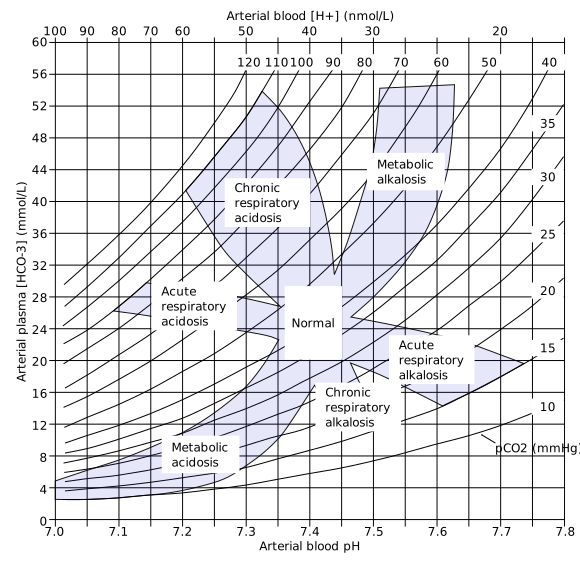
The four primary disorders and expected compensation
| Disorder | Primary change | Compensation | Expected compensation |
|---|---|---|---|
| Metabolic acidosis | ↓ HCO₃⁻ | Hyperventilation → ↓ PaCO₂ | Winter's formula: expected PaCO₂ (kPa) ≈ 0.16 × HCO₃⁻ + 1.1 (in mmHg: 1.5 × HCO₃⁻ + 8 ± 2). A higher CO₂ than expected means a coexisting respiratory acidosis; a lower one a coexisting respiratory alkalosis |
| Metabolic alkalosis | ↑ HCO₃⁻ | Hypoventilation → ↑ PaCO₂ | PaCO₂ rises ~0.09 kPa (0.7 mmHg) per 1 mmol/L rise in HCO₃⁻. Compensation is limited by hypoxia, which eventually drives ventilation |
| Respiratory acidosis | ↑ PaCO₂ | Renal HCO₃⁻ retention | Acute: HCO₃⁻ rises ~1 mmol/L per 1.3 kPa (10 mmHg) rise in PaCO₂. Chronic (2-5 days): rises ~4 mmol/L per 1.3 kPa. The bicarbonate therefore dates the disorder |
| Respiratory alkalosis | ↓ PaCO₂ | Renal HCO₃⁻ excretion | Acute: HCO₃⁻ falls ~2 mmol/L per 1.3 kPa fall in PaCO₂. Chronic: falls ~5 mmol/L per 1.3 kPa |
A practical example of why this matters: a patient with COPD and a PaCO₂ of 8 kPa who has a bicarbonate of 26 has an acute respiratory acidosis (the kidney has not had time to retain bicarbonate) and is decompensating now. The same PaCO₂ with a bicarbonate of 36 indicates chronic CO₂ retention with established renal compensation - a very different clinical situation and a very different urgency.
Metabolic acidosis and the anion gap
Metabolic acidosis arises either because acid has been added to the body or because bicarbonate has been lost. The anion gap distinguishes the two and is the single most useful calculation in acid-base medicine.
Raised anion gap metabolic acidosis
The traditional mnemonic is MUDPILES; the more modern GOLDMARK reflects current practice better.
| Letter | Cause | Clinical notes |
|---|---|---|
| G | Glycols - ethylene glycol, propylene glycol | Antifreeze ingestion; raised osmolar gap, calcium oxalate crystals in urine, AKI |
| O | Oxoproline (5-oxoproline) | Chronic paracetamol use, especially in malnourished women |
| L | L-lactate | Type A - tissue hypoperfusion: sepsis, shock, ischaemia, hypoxia. Type B - without hypoperfusion: metformin, liver failure, malignancy, thiamine deficiency, alcohol |
| D | D-lactate | Short bowel syndrome with bacterial overgrowth; causes encephalopathy and is missed by standard lactate assays |
| M | Methanol | Raised osmolar gap; causes blindness through optic nerve injury |
| A | Aspirin (salicylates) | Classically a mixed picture: respiratory alkalosis (direct respiratory centre stimulation) plus raised anion gap metabolic acidosis |
| R | Renal failure | Accumulation of urate, sulphate and phosphate |
| K | Ketoacidosis | Diabetic, alcoholic, or starvation ketoacidosis |
Normal anion gap (hyperchloraemic) metabolic acidosis
- Diarrhoea - the commonest cause; loss of bicarbonate-rich intestinal fluid
- Renal tubular acidosis - types 1, 2 and 4 (see below)
- Acetazolamide - a carbonic anhydrase inhibitor causing bicarbonate wasting
- Ureteric diversion - ileal conduit or ureterosigmoidostomy, where bowel mucosa absorbs chloride and secretes bicarbonate
- Large-volume 0.9% sodium chloride infusion - a genuinely iatrogenic cause. 'Normal' saline has a chloride concentration of 154 mmol/L, well above plasma, and large volumes produce a dilutional hyperchloraemic acidosis. This is a principal argument for balanced crystalloids such as Hartmann's or Plasma-Lyte in resuscitation
- Pancreatic or biliary fistulae
- Addison's disease and early renal failure
- Ammonium chloride and parenteral nutrition
The renal tubular acidoses
The renal tubular acidoses are a group of disorders in which the kidney fails to acidify the urine or to reabsorb bicarbonate normally, producing a normal anion gap metabolic acidosis with normal or near-normal GFR. They are heavily examined because the pattern of potassium and urine pH distinguishes them cleanly.
| Feature | Type 1 (distal) | Type 2 (proximal) | Type 4 (hyperkalaemic) |
|---|---|---|---|
| Defect | Failure to excrete H⁺ in the distal tubule | Failure to reabsorb HCO₃⁻ in the proximal tubule | Aldosterone deficiency or resistance (hyporeninaemic hypoaldosteronism) |
| Serum potassium | LOW | LOW | HIGH - the key discriminator |
| Urine pH | Always > 5.5 - cannot acidify urine even when systemically acidotic | Variable: >5.5 while bicarbonate is being wasted, but <5.5 once serum bicarbonate falls below the reabsorptive threshold | Usually < 5.5 |
| Serum bicarbonate | Can be very low (often <10) | Moderately low (12-20), stabilises at the new threshold | Mildly low (>17) |
| Causes | Autoimmune - Sjögren syndrome, SLE, rheumatoid arthritis; hypercalciuria; drugs (amphotericin B, lithium); genetic | Fanconi syndrome (with glycosuria, aminoaciduria, phosphaturia); myeloma; Wilson disease; acetazolamide; tenofovir | Diabetic nephropathy (classic); drugs - ACE inhibitors, ARBs, spironolactone, NSAIDs, trimethoprim, heparin, calcineurin inhibitors; Addison's disease; obstructive uropathy |
| Complications | Nephrocalcinosis and renal stones (calcium phosphate, due to alkaline urine and hypocitraturia); rickets/osteomalacia; growth failure | Osteomalacia and rickets; stones are uncommon | Hyperkalaemia and its cardiac consequences |
| Treatment | Oral bicarbonate or potassium citrate | Large doses of bicarbonate plus potassium; thiazides may help | Fludrocortisone; stop culprit drugs; dietary potassium restriction; loop diuretic |
Note that type 3 RTA is a rare hybrid with features of both types 1 and 2 and is not usually required knowledge. A useful shortcut for exams: the RTA with a high potassium is type 4, and the RTA that causes stones is type 1.
Metabolic alkalosis
Metabolic alkalosis requires both a generating process (loss of acid or gain of bicarbonate) and a maintaining process - normally the kidney would simply excrete excess bicarbonate, so alkalosis persists only if something prevents this. The maintaining factor is usually volume depletion, chloride depletion, hypokalaemia or mineralocorticoid excess, and identifying it is what directs treatment.
Respiratory disorders
Respiratory acidosis (raised PaCO₂)
- Reduced respiratory drive - opiates, benzodiazepines, anaesthetics, head injury, stroke, oxygen-induced hypercapnia in chronic type 2 respiratory failure
- Neuromuscular disease - Guillain-Barré syndrome, myasthenia gravis, motor neurone disease, muscular dystrophy, cervical cord injury
- Airway and lung disease - COPD (the commonest cause), severe asthma (a rising CO₂ in acute asthma is an ominous, life-threatening sign), pneumonia, ARDS, pulmonary oedema, obstructive sleep apnoea
- Chest wall and pleural - kyphoscoliosis, obesity hypoventilation, flail chest, large pneumothorax or effusion
- Inadequate mechanical ventilation
Respiratory alkalosis (low PaCO₂)
- Anxiety and hyperventilation - and remember that this lowers ionised calcium, producing perioral paraesthesiae, carpopedal spasm and tetany with a normal total calcium
- Hypoxia - pulmonary embolism, pneumonia, pulmonary oedema, high altitude, anaemia
- Pain and fever
- Sepsis - often an early feature, before lactic acidosis supervenes
- Drugs - salicylates (early), doxapram, progesterone
- Central causes - stroke, meningitis, encephalitis, head injury
- Pregnancy - progesterone-driven hyperventilation causes a chronic compensated respiratory alkalosis, which is normal
- Liver failure, and excessive mechanical ventilation
Management principles
The governing principle is to treat the underlying cause rather than the number. Acid-base abnormalities are almost always a manifestation of a disease process, and correcting the pH without addressing the cause achieves little and may cause harm.2
- Metabolic acidosis - treat the cause: fluid resuscitation and antibiotics for sepsis, insulin and fluids for DKA, stop metformin in metformin-associated lactic acidosis, antidotes and dialysis for toxic alcohols, fluids and steroids in Addison's disease
- Sodium bicarbonate - controversial and not routinely recommended in lactic acidosis or DKA. It may worsen intracellular acidosis (CO₂ crosses membranes more readily than bicarbonate), causes hypernatraemia, volume overload and hypokalaemia, and shifts the oxygen dissociation curve. It has a clearer role in severe hyperchloraemic acidosis, renal tubular acidosis, hyperkalaemia with acidosis, and tricyclic antidepressant overdose (where it is genuinely indicated for cardiotoxicity)
- Renal replacement therapy - for severe refractory acidosis (pH <7.1), particularly with renal failure, fluid overload, hyperkalaemia or a dialysable toxin
- Metabolic alkalosis - 0.9% saline and potassium if chloride-responsive; treat the endocrine cause and use spironolactone if chloride-resistant. Acetazolamide is occasionally used, and dilute hydrochloric acid via a central line very rarely
- Respiratory acidosis - improve ventilation: treat the underlying lung disease, reverse opiates or benzodiazepines, use controlled oxygen therapy targeting 88-92% in those at risk of hypercapnia, and escalate to NIV or invasive ventilation where indicated
- Respiratory alkalosis - treat the underlying cause; reassurance and rebreathing in anxiety-driven hyperventilation, but always exclude hypoxia, PE and sepsis before attributing hyperventilation to anxiety
- Correct associated electrolytes - potassium, magnesium, calcium and phosphate are frequently deranged and interact with the acid-base state
- Recheck the gas after intervention to confirm the direction of travel
Red flags
Prognosis
Acid-base disturbances are almost never the primary determinant of outcome; the underlying disease is. Their prognostic value lies in what they reveal about the severity of that disease, and in this respect they are among the most useful markers available at the bedside. A lactate above 4 mmol/L in suspected sepsis, for example, identifies patients at substantially increased risk of death and mandates aggressive resuscitation - and serial lactate clearance over the first hours is one of the better predictors of survival.
Severe acidaemia is independently harmful once the pH falls below about 7.2. It depresses myocardial contractility, causes systemic vasodilatation with pulmonary vasoconstriction, predisposes to arrhythmia and blunts the response to catecholamines and inotropes - creating a self-reinforcing cycle of shock and worsening acidosis. Recognising and interrupting that cycle by restoring perfusion is far more effective than attempting to correct the pH directly with bicarbonate, which is why routine bicarbonate administration has largely fallen out of favour.
Prognosis by cause varies enormously. Diabetic ketoacidosis has an excellent outlook with prompt treatment, mortality now well under 1% in the UK. Metformin-associated lactic acidosis and toxic alcohol poisoning carry high mortality but are highly responsive to timely dialysis. Chronic compensated disorders - the raised bicarbonate of long-standing COPD, or the chronic respiratory alkalosis of pregnancy - are physiologically appropriate adaptations requiring no correction at all, and attempting to normalise them causes harm. The renal tubular acidoses generally have a good prognosis with alkali therapy, though type 1 carries a lifelong risk of nephrocalcinosis, stones and progressive renal impairment if untreated, and childhood-onset disease can cause growth failure and rickets. In every case, the practical message is the same: the acid-base result is a window onto the underlying process, and it is that process which should be treated.
References
- Berend K, de Vries APJ, Gans ROB. Physiological approach to assessment of acid-base disturbances. New England Journal of Medicine. 2014. Available here
- NICE CG174. Intravenous fluid therapy in adults in hospital. 2013, updated 2017. Available here
- Joint British Diabetes Societies. The Management of Diabetic Ketoacidosis in Adults. Available here
- NICE NG51. Sepsis: recognition, diagnosis and early management. 2016, updated 2024. Available here
- TOXBASE (National Poisons Information Service). Methanol and ethylene glycol poisoning. Available here
- British Thoracic Society. Guideline for oxygen use in healthcare and emergency settings. Available here
- Huckfinne, public domain, via Wikimedia Commons. Available here
This article is written for revision and education. It is not clinical guidance and must not be used to make decisions about the care of a patient. Always check current NICE guidance and local protocols.