
Encephalitis: Diagnosis and Management
Key points
- Encephalitis: inflammation of the brain parenchyma itself, distinct from meningitis (inflammation of the meninges), though the two frequently coexist as meningoencephalitis.
- Classic triad: fever, altered behaviour/cognition and seizures, often with focal neurological signs - distinguishing it from meningitis alone.
- Commonest identifiable cause: herpes simplex virus (HSV-1), which characteristically affects the temporal lobes.
- Start aciclovir immediately: empirically, on clinical suspicion, before results of CSF PCR are available - delayed treatment significantly worsens outcome in HSV encephalitis.
- Key investigation: CSF PCR for HSV and other viruses; MRI showing temporal lobe changes strongly supports HSV encephalitis.
- Autoimmune encephalitis: an increasingly recognised cause, including anti-NMDA receptor encephalitis, which can be paraneoplastic (classically ovarian teratoma) and presents with prominent psychiatric features.
- EEG: in HSV encephalitis, may show characteristic periodic lateralised epileptiform discharges over the affected temporal lobe.
- Prognosis: even with prompt treatment, HSV encephalitis carries significant mortality and a high rate of long-term cognitive and memory impairment in survivors.
Introduction
Encephalitis is inflammation of the brain parenchyma itself, in contrast to meningitis, which involves the meninges - though in practice the two often overlap, producing meningoencephalitis with features of both.1 Involvement of brain tissue is what produces the features that distinguish encephalitis clinically: altered behaviour or personality, cognitive impairment, seizures and focal neurological deficit, rather than headache and neck stiffness alone.
Herpes simplex virus (HSV-1) is the commonest identifiable cause of sporadic encephalitis in immunocompetent adults in the UK, and its recognition is a genuine emergency: outcome is strongly time-dependent, and empirical aciclovir must be started on clinical suspicion, before confirmatory tests return. This single principle - treat first, confirm afterwards - is the most heavily tested point in the whole topic.
In practice, meningitis and encephalitis frequently coexist as meningoencephalitis, and the distinction is one of emphasis rather than a clean dichotomy. What matters clinically is recognising when brain parenchyma is involved, because that changes the differential, the imaging strategy and the drugs given. A patient with headache, fever and neck stiffness alone needs empirical antibiotics; the same patient who is also confused, behaving oddly or having seizures needs empirical aciclovir alongside them, because HSV encephalitis is now in play and the cost of omitting it is severe.
The other reason parenchymal involvement matters is that it produces the diagnostic traps. Temporal lobe inflammation causes memory disturbance, odd olfactory or gustatory sensations and behavioural change - a presentation that can be mistaken for delirium, an acute psychiatric illness, or intoxication, particularly in a patient who is not obviously febrile at the time of assessment.
A practical point on lumbar puncture: CSF in viral encephalitis typically shows a lymphocytic pleocytosis with a mildly raised protein and a normal glucose, but it can be entirely normal very early in the illness. A normal CSF therefore does not exclude the diagnosis, and neither does a negative HSV PCR taken within the first day or two - which is why aciclovir is continued and the PCR repeated if suspicion remains high.
Encephalitis is worth thinking of as three broad groups, because the treatment differs completely. Infectious encephalitis, dominated by HSV, is treated with antivirals started empirically. Autoimmune encephalitis, increasingly recognised, is treated with immunotherapy and, where relevant, removal of an underlying tumour. Post-infectious demyelination (ADEM) follows an infection rather than being caused by ongoing infection, and is treated with corticosteroids.
The three overlap in presentation and can be difficult to separate early, which is why the standard approach is to cover the treatable and time-critical possibility first - aciclovir for HSV - while the investigations that distinguish them, principally CSF PCR, MRI and antibody testing, are processed.
Incidence is low - in the region of 5-10 per 100,000 per year - but the consequences of a delayed diagnosis are severe enough that the threshold for considering it must be correspondingly low. HSV encephalitis in particular has no seasonal pattern, affects all age groups with peaks in the young and the elderly, and occurs in previously healthy, immunocompetent people, so there is no demographic in which it can be safely discounted.
Aetiology
Viral
- Herpes simplex virus (HSV-1) - the commonest identifiable cause in immunocompetent adults; characteristically affects the temporal and inferior frontal lobes, reflecting the virus's route of spread along the olfactory and trigeminal pathways
- Varicella zoster virus - can cause encephalitis, particularly in the immunocompromised or older adults, sometimes with a preceding or concurrent shingles rash
- Enteroviruses
- Arboviruses (e.g. Japanese encephalitis, tick-borne encephalitis, West Nile virus) - relevant with an appropriate travel or exposure history
- HIV - can cause encephalitis directly (seroconversion or advanced disease) or predispose to opportunistic causes
Autoimmune
- Anti-NMDA receptor encephalitis - increasingly recognised, particularly in young women, often (though not always) paraneoplastic in association with an ovarian teratoma; presents with prominent early psychiatric features (agitation, hallucinations, behavioural change) before progressing to seizures, movement disorder (orofacial dyskinesias) and autonomic instability2
- Other antibody-mediated encephalitides (e.g. anti-LGI1, associated with faciobrachial dystonic seizures and hyponatraemia) - a growing and increasingly recognised category
- Post-infectious/parainfectious encephalomyelitis (ADEM) - typically follows a preceding infection or vaccination, more common in children, usually monophasic
Other
- Bacterial (as a complication of bacterial meningitis, or from Listeria, Mycoplasma, or Lyme disease)
- Fungal or parasitic - in the immunocompromised, or with relevant exposure (e.g. toxoplasmosis in advanced HIV)
Clinical features
- Fever
- Altered behaviour, personality change, or confusion - often the earliest and most striking feature, and can be mistaken for a primary psychiatric presentation, particularly in autoimmune encephalitis
- Reduced conscious level, ranging from mild drowsiness to coma
- Seizures - focal or generalised, common and can be the presenting feature
- Focal neurological signs - dysphasia, hemiparesis, depending on the area of brain involved (in HSV encephalitis, often reflecting temporal lobe dysfunction: memory disturbance, olfactory or gustatory hallucinations, behavioural change)
- Headache and meningism - may coexist if there is a meningitic component
- Movement disorder and autonomic instability - particularly characteristic of anti-NMDA receptor encephalitis, alongside its early psychiatric presentation
- Preceding or concurrent rash - in varicella zoster or other viral causes with a characteristic exanthem
Clinical examination
- Glasgow Coma Scale and cognitive assessment - looking specifically for memory disturbance and behavioural change
- Full neurological examination for focal deficit, dysphasia, or movement disorder
- Meningism - may or may not be present
- Skin examination for a rash suggesting varicella zoster or another specific viral cause
- Psychiatric assessment - for the prominent early psychiatric features of autoimmune encephalitis, which can otherwise be misattributed to a primary psychiatric disorder
- Abdominal/pelvic examination or imaging consideration - to look for an ovarian teratoma if anti-NMDA receptor encephalitis is suspected
Differential diagnosis
- Meningitis alone - without significant behavioural change, seizures, or focal deficit
- Primary psychiatric presentation (particularly first-episode psychosis) - can closely mimic early anti-NMDA receptor encephalitis; new-onset seizures, movement disorder, or autonomic instability alongside psychiatric symptoms should prompt reconsideration
- Metabolic encephalopathy - hepatic, uraemic, or hypoglycaemic causes of confusion, generally without fever or focal signs unless coincidental
- Non-convulsive status epilepticus
- Cerebral abscess - focal deficit and fever, but typically with a more localised, mass-effect picture on imaging rather than the diffuse/temporal changes of HSV encephalitis
- Toxic/drug-related delirium
- Stroke - acute focal deficit, but typically without fever or the behavioural/seizure prominence of encephalitis
Investigations
- Lumbar puncture with CSF PCR - the key diagnostic test; HSV PCR is highly sensitive and specific, though it can occasionally be falsely negative very early in the illness, so a single negative result soon after onset does not exclude the diagnosis if clinical suspicion remains high
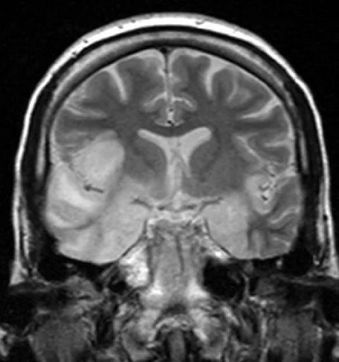
- MRI brain - the imaging investigation of choice; in HSV encephalitis, characteristically shows T2/FLAIR hyperintensity in the temporal lobes and inferior frontal lobes, sometimes with haemorrhagic change, often asymmetric
- EEG - may show focal slowing or, characteristically in HSV encephalitis, periodic lateralised epileptiform discharges (PLEDs) over the affected temporal lobe
- Blood tests - FBC, U&Es, LFTs, glucose, blood cultures, and specific viral/autoimmune serology as indicated
- Autoimmune/paraneoplastic antibody panel (serum and CSF) - if autoimmune encephalitis is suspected, particularly anti-NMDA receptor antibodies
- Imaging for an underlying tumour (pelvic ultrasound/MRI for ovarian teratoma, CT chest for other malignancy) - if autoimmune/paraneoplastic encephalitis is confirmed or strongly suspected
- HIV test - relevant given both direct and opportunistic associations with encephalitis
Management
- IV aciclovir - typically continued for 14-21 days in confirmed HSV encephalitis, with the duration extended and CSF PCR sometimes repeated towards the end of treatment in immunocompromised patients or where response is incomplete
- Supportive care - management of raised intracranial pressure if present, seizure control, and general intensive supportive care for reduced consciousness
- Broad empirical cover alongside aciclovir if bacterial meningitis cannot be excluded, until the diagnosis is clarified
- Immunotherapy - for confirmed autoimmune encephalitis: first-line options include corticosteroids, IV immunoglobulin, or plasma exchange, with second-line agents (rituximab, cyclophosphamide) for refractory cases
- Tumour removal - resection of an identified ovarian teratoma in anti-NMDA receptor encephalitis, alongside immunotherapy, since this addresses the underlying trigger
- Multidisciplinary rehabilitation - given the frequent and sometimes severe cognitive sequelae, particularly memory impairment after HSV encephalitis
Complications
- Persistent memory impairment and other cognitive deficits, particularly after HSV encephalitis affecting the temporal lobes (which contain the hippocampus)
- Epilepsy
- Personality and behavioural change
- Focal neurological deficit
- Relapse - a recognised phenomenon after HSV encephalitis, sometimes representing an autoimmune (anti-NMDA receptor) encephalitis triggered by the preceding HSV infection, rather than viral relapse itself, and requiring a different treatment approach (immunotherapy rather than further aciclovir)
- Death, particularly if treatment is delayed
Red flags
The recurring theme in delayed diagnosis is that encephalitis is mistaken for something more common - delirium, intoxication, or a first psychiatric presentation. The features below should shift the balance towards treating empirically while investigations are processed.
Prognosis
Even with prompt aciclovir treatment, HSV encephalitis carries significant mortality, and a substantial proportion of survivors are left with lasting cognitive impairment - particularly memory difficulty, given the predilection for the temporal lobes and hippocampus - as well as behavioural change or epilepsy. Outcome is strongly related to conscious level at the time treatment is started, reinforcing the importance of early empirical treatment. Autoimmune encephalitis, including anti-NMDA receptor encephalitis, often responds well to prompt immunotherapy (and tumour removal where relevant), though recovery can be slow and some patients are left with residual cognitive or behavioural deficits.
Because the deficits after encephalitis are predominantly cognitive and behavioural rather than physical, recovery is easily underestimated in its difficulty. A patient may walk out of hospital appearing physically well while being unable to retain new information, regulate emotion or return to work. Formal neuropsychological assessment, rather than a bedside cognitive screen, is what characterises these deficits properly, and referral to neurorehabilitation and to epilepsy services where seizures have occurred should be arranged before discharge rather than left to primary care to initiate.
Relatives should be given clear information about what to expect, since behavioural change and memory impairment are frequently more distressing to families than to the patient, who may have limited insight into the deficits.
Driving is an important practical issue that is easily overlooked at discharge. A seizure occurring during the acute illness carries DVLA implications in the same way as any other seizure, and patients should be given clear written advice rather than a verbal comment made in passing. Employment and return to study also need addressing, since cognitive fatigue and reduced processing speed frequently make a phased return necessary even in patients who appear physically recovered.
References
- NICE CKS and British Infection Association. Management of suspected viral encephalitis in adults. Available here
- Dalmau J, Graus F. Antibody-mediated encephalitis. New England Journal of Medicine. 2018. Available here
- Solomon T, Michael BD, Smith PE et al. Management of suspected viral encephalitis in adults - Association of British Neurologists and British Infection Association National Guidelines. Journal of Infection. 2012. Available here
This article is written for revision and education. It is not clinical guidance and must not be used to make decisions about the care of a patient. Always check current NICE guidance and local protocols.