
Hyponatraemia
Key points
- Hyponatraemia: serum sodium below 135 mmol/L - the commonest electrolyte abnormality in hospital practice. Mild 130-134, moderate 125-129, severe below 125.
- It is a water problem, not a salt problem: hyponatraemia almost always reflects excess water relative to sodium, driven by ADH. Total body sodium may be low, normal or high.
- The two-step approach: first measure serum osmolality to exclude pseudo- and hypertonic hyponatraemia; then assess volume status to classify true hypotonic hyponatraemia as hypovolaemic, euvolaemic or hypervolaemic.1
- SIADH: euvolaemic, hypotonic hyponatraemia with inappropriately concentrated urine (osmolality >100) and urinary sodium >30 mmol/L, with normal thyroid and adrenal function and no diuretics.
- Key exclusions before diagnosing SIADH: hypothyroidism and adrenal insufficiency must be excluded - both cause euvolaemic hyponatraemia and are readily treatable.
- Severe symptomatic hyponatraemia: seizures, coma or reduced GCS need hypertonic 3% saline in boluses, aiming to raise sodium by 4-6 mmol/L to stop symptoms - not to normalise it.
- The correction limit: do not exceed 10 mmol/L in 24 hours (8 mmol/L if high risk). Over-rapid correction causes osmotic demyelination syndrome.
- Chronic versus acute: chronic hyponatraemia (>48 hours) carries the demyelination risk; acute hyponatraemia (<48 hours) risks cerebral oedema and can be corrected faster.
Introduction
Hyponatraemia is defined as a serum sodium below 135 mmol/L and is the commonest electrolyte disturbance in clinical practice, present in up to 30% of hospital inpatients. It is independently associated with increased mortality, longer admissions and, even when mild and chronic, with falls, fractures and cognitive impairment.1
Severity is graded mild (130-134 mmol/L), moderate (125-129 mmol/L) and severe (below 125 mmol/L) - but as with potassium, symptoms and the rate of fall matter more than the absolute number. A sodium of 125 developing over hours is far more dangerous than 118 developing over weeks.
Classification and causes
Step 1 - measure the serum osmolality
| Category | Serum osmolality | Causes |
|---|---|---|
| Hypotonic (true) hyponatraemia | Low (<275 mOsm/kg) | The vast majority. Proceed to assess volume status |
| Isotonic ('pseudohyponatraemia') | Normal | A laboratory artefact from severe hyperlipidaemia or hyperproteinaemia (e.g. myeloma, paraproteinaemia) displacing plasma water. The true sodium is normal - modern direct ion-selective electrodes avoid this |
| Hypertonic hyponatraemia | High (>295 mOsm/kg) | Another osmole draws water out of cells and dilutes the sodium - severe hyperglycaemia (the commonest), mannitol, or glycine irrigation (TURP syndrome) |
Step 2 - assess volume status
| Volume status | Clinical signs | Causes | Urinary sodium |
|---|---|---|---|
| Hypovolaemic | Dry mucous membranes, reduced skin turgor, postural hypotension, tachycardia, low JVP, oliguria | Renal losses: diuretics (especially thiazides), adrenal insufficiency, salt-wasting nephropathy, cerebral salt wasting. Extrarenal losses: vomiting, diarrhoea, burns, pancreatitis, third-space losses | Renal cause: >30 mmol/L. Extrarenal cause: <20 mmol/L (kidneys appropriately conserving sodium) |
| Euvolaemic | No oedema, no signs of dehydration - clinically normal volume | SIADH, hypothyroidism, adrenal insufficiency (glucocorticoid deficiency), primary polydipsia, low solute intake ('beer potomania', tea-and-toast diet), exercise-associated hyponatraemia | >30 mmol/L in SIADH; low in primary polydipsia |
| Hypervolaemic | Peripheral oedema, raised JVP, ascites, pulmonary oedema, weight gain | Heart failure, liver cirrhosis, nephrotic syndrome, advanced chronic kidney disease | <20 mmol/L in heart failure and cirrhosis (avid sodium retention); >30 in renal failure |
Syndrome of inappropriate ADH secretion (SIADH)
SIADH is a diagnosis of exclusion and the commonest cause of euvolaemic hyponatraemia. ADH is secreted despite a low plasma osmolality, causing inappropriate water retention.
- Diagnostic criteria: hypotonic hyponatraemia (serum osmolality <275), inappropriately concentrated urine (urine osmolality >100 mOsm/kg), urinary sodium >30 mmol/L, clinical euvolaemia, and normal thyroid, adrenal and renal function with no recent diuretics
- Malignancy - classically small cell lung cancer; also pancreatic, prostate and lymphoma
- CNS disease - stroke, subarachnoid haemorrhage, meningitis, encephalitis, head injury, tumours
- Pulmonary disease - pneumonia, tuberculosis, lung abscess, and positive-pressure ventilation
- Drugs - SSRIs and other antidepressants, carbamazepine, antipsychotics, NSAIDs, opiates, chemotherapy (cyclophosphamide, vincristine), PPIs, MDMA
- Surgery and pain - postoperative ADH release is very common and compounded by hypotonic IV fluids
- Other - HIV, porphyria, and idiopathic (particularly in older people)
Clinical features
Symptoms result from water moving into brain cells down the osmotic gradient, causing cerebral oedema. Their severity depends far more on the rate of fall than on the absolute sodium, because the brain adapts to slowly developing hyponatraemia by extruding organic osmolytes - an adaptation that is also what makes rapid correction dangerous.
By severity
- Mild / chronic: often asymptomatic, or non-specific - nausea, headache, malaise, lethargy, poor concentration. Importantly, even 'asymptomatic' chronic hyponatraemia is associated with gait instability, falls, fractures and osteoporosis in older people, and with subtle cognitive impairment
- Moderate: confusion, disorientation, vomiting, muscle cramps, weakness, restlessness
- Severe: seizures, reduced GCS, coma, respiratory arrest, and features of raised intracranial pressure from cerebral oedema - this constitutes a medical emergency
Assessing the patient
- Volume status is the central clinical task - JVP, skin turgor, mucous membranes, postural blood pressure, capillary refill, oedema, ascites, and crucially the fluid balance chart and daily weights
- Duration - establish whether this is acute (<48 hours) or chronic (>48 hours), using previous results where available. This determines how fast correction may proceed
- Drug history - thiazides, SSRIs, carbamazepine, antipsychotics, PPIs, NSAIDs, desmopressin; drugs are among the commonest causes
- Fluid intake history - including recent IV fluids (especially hypotonic), excessive water or beer intake, and endurance exercise
- Look for the underlying cause - respiratory examination and smoking history (small cell lung cancer), neurological examination, signs of heart failure or chronic liver disease, and features of hypothyroidism or Addison's disease (pigmentation, postural hypotension)
Investigations
- Serum osmolality - the essential first step, to exclude pseudo- and hypertonic hyponatraemia
- Urine osmolality - <100 mOsm/kg indicates appropriately dilute urine and suggests primary polydipsia or low solute intake; >100 indicates ADH is acting, appropriately or otherwise
- Urinary sodium - <20 mmol/L suggests hypovolaemia with appropriate renal conservation, or an oedematous state (heart failure, cirrhosis); >30 mmol/L suggests renal salt loss, diuretics, adrenal insufficiency or SIADH
- U&Es, creatinine and eGFR
- Glucose - to correct for hyperglycaemia and exclude a hypertonic cause
- Thyroid function tests - hypothyroidism must be excluded before diagnosing SIADH
- 9 am cortisol, and a short Synacthen test if indicated - adrenal insufficiency must be excluded before diagnosing SIADH; it is readily treatable and easily missed
- Lipid profile and total protein / paraproteins - if pseudohyponatraemia is suspected
- Liver function tests and albumin - cirrhosis
- Chest X-ray - essential if SIADH is diagnosed, looking for small cell lung cancer, pneumonia or tuberculosis; consider CT chest if suspicion is high
- CT head - if there are neurological signs or a CNS cause is suspected
- Daily weights and accurate fluid balance - often more informative than any single blood test
Management
Severe symptomatic hyponatraemia - a medical emergency
- Indications: seizures, reduced consciousness, coma, or severe vomiting attributable to hyponatraemia - regardless of the absolute level
- Give hypertonic 3% sodium chloride: typically 150 mL over 20 minutes, repeated up to two or three times, with sodium checked after each bolus
- The target is symptom relief, not normalisation - aim to raise sodium by 4-6 mmol/L, which is usually enough to stop seizures and reverse cerebral oedema
- Manage in a high-dependency or critical care setting with close monitoring, and involve senior, critical care and nephrology input early
- Once symptoms resolve, stop the hypertonic saline and revert to treating the underlying cause within the 24-hour correction limit
- Protect the airway, treat seizures, and correct coexisting hypokalaemia (which itself raises sodium as it is corrected, contributing to over-rapid rise)
Treatment by category
| Category | Management |
|---|---|
| Hypovolaemic | Volume replacement with 0.9% sodium chloride, and treat the cause. Stop diuretics. Sodium often rises briskly once ADH release is switched off by volume repletion - monitor closely for over-rapid correction |
| Euvolaemic (SIADH) | Fluid restriction - typically 800-1000 mL/day, and less than urine output. Treat the underlying cause and stop culprit drugs. If restriction fails: tolvaptan (a vasopressin V2 receptor antagonist, started in hospital with close monitoring), demeclocycline, or oral urea. Increasing dietary salt and protein intake can also help |
| Hypervolaemic | Fluid AND sodium restriction, plus loop diuretics. Treat the underlying condition - heart failure, cirrhosis or nephrotic syndrome. Tolvaptan may be used in selected patients |
| Adrenal insufficiency | Hydrocortisone - this corrects the hyponatraemia and is life-saving; do not simply fluid restrict |
| Hypothyroidism | Levothyroxine (with hydrocortisone first if adrenal insufficiency is also possible) |
| Primary polydipsia / beer potomania | Reduce water intake and improve solute intake. High risk of very rapid autocorrection once free water intake stops - monitor sodium closely |
Complications
Of the hyponatraemia itself
- Cerebral oedema, seizures, coma and death - particularly with acute severe hyponatraemia
- Brainstem herniation and respiratory arrest in extreme cases
- Falls, gait instability and fractures - even mild chronic hyponatraemia impairs balance and attention, and is associated with osteoporosis
- Cognitive impairment and delirium
- Prolonged hospital stay and increased mortality - hyponatraemia is an independent marker of poor outcome across many conditions
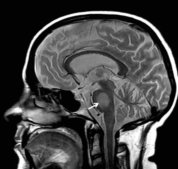
Of over-rapid correction - osmotic demyelination syndrome
- Central pontine myelinolysis and extrapontine myelinolysis - demyelination caused by rapid osmotic shift out of adapted brain cells
- Classically biphasic: the patient initially improves as sodium rises, then deteriorates 2 to 6 days later
- Clinical features: dysarthria, dysphagia, spastic quadriparesis, pseudobulbar palsy, behavioural change, and in the worst cases the 'locked-in' syndrome
- MRI changes lag behind the clinical picture by days - a normal early scan does not exclude the diagnosis
- Often irreversible, though partial or occasionally substantial recovery does occur with supportive care and rehabilitation
- Risk factors: chronic hyponatraemia, sodium below 105 mmol/L, alcohol dependence, malnutrition, liver disease and transplantation, hypokalaemia, and advanced age
- Entirely preventable by respecting correction limits - which is why they are worth memorising
Red flags
Prognosis
Prognosis is determined largely by the underlying cause rather than by the sodium concentration itself. Hyponatraemia from a thiazide, an SSRI or a self-limiting episode of vomiting resolves completely once the cause is removed, with no lasting consequence. By contrast, hyponatraemia complicating advanced heart failure, cirrhosis or malignancy is a well-established marker of disease severity and an independent predictor of mortality - the low sodium is a signal of how unwell the patient is, and correcting the number without addressing the disease achieves little.
Even mild chronic hyponatraemia is not benign, which is a change in thinking worth appreciating. Long regarded as an incidental finding to be tolerated, it is now recognised to cause subtle cognitive impairment, unsteadiness, falls, fractures and osteoporosis, particularly in older people. There is therefore a reasonable argument for investigating and treating persistent mild hyponatraemia rather than simply noting it.
The outcome of treatment depends almost entirely on the rate of correction. Appropriately managed hyponatraemia, even when severe, generally resolves without neurological sequelae, and hypertonic saline given for seizures or coma is highly effective. Osmotic demyelination syndrome is the feared exception: it is frequently irreversible, can leave patients with devastating deficits including a locked-in state, and is almost entirely iatrogenic and preventable. Some recovery is possible with supportive care and rehabilitation, but the prognosis is poor once established. The practical conclusion is a slightly counterintuitive one that examiners like to test: in chronic hyponatraemia, the danger lies less in the low sodium than in correcting it too enthusiastically - the aim is to relieve symptoms and then to proceed slowly, deliberately and with frequent monitoring.
References
- Spasovski G, Vanholder R, Allolio B et al. Clinical practice guideline on diagnosis and treatment of hyponatraemia. European Journal of Endocrinology / Nephrology Dialysis Transplantation. 2014. Available here
- NICE Clinical Knowledge Summaries. Hyponatraemia. Available here
- Society for Endocrinology. Emergency Endocrine Guidance: Acute hyponatraemia in adults. Available here
- NICE CG174. Intravenous fluid therapy in adults in hospital. 2013, updated 2017. Available here
- NICE TA. Tolvaptan for hyponatraemia secondary to SIADH. Available here
- Sterns RH. Disorders of plasma sodium - causes, consequences, and correction. New England Journal of Medicine. 2015. Available here
- MBq, CC0, via Wikimedia Commons. Available here
This article is written for revision and education. It is not clinical guidance and must not be used to make decisions about the care of a patient. Always check current NICE guidance and local protocols.