
Lung Cancer: Recognition, Staging and Treatment by Cell Type
Key points
- Lung cancer: the third commonest cancer in the UK and the commonest cause of cancer death, with around 48,000 new diagnoses a year.
- Smoking: causes around 85% of cases and raises risk 15 to 30 fold. Radon is the second commonest cause, and asbestos multiplies the risk in smokers.
- Non-small cell lung cancer: around 85% of cases - adenocarcinoma (peripheral, commonest overall and in non-smokers), squamous cell (central, cavitates, causes hypercalcaemia) and large cell.
- Small cell lung cancer: around 15%, arising from neuroendocrine cells. Central, strongly smoking-related, grows fast, metastasises early, and causes most paraneoplastic endocrine syndromes.
- Referral: urgent chest X-ray within 2 weeks under NICE NG12 for anyone over 40 with two unexplained symptoms, or one if they have ever smoked.
- Staging: contrast CT of the chest, abdomen and pelvis, then PET-CT and often brain imaging where treatment with curative intent is being considered.
- NSCLC treatment: surgery or stereotactic radiotherapy for early disease, chemoradiotherapy for stage III, and targeted therapy or immunotherapy guided by molecular testing in stage IV.
- SCLC treatment: chemotherapy with platinum and etoposide plus immunotherapy, with concurrent radiotherapy in limited-stage disease. Surgery has almost no role.
Introduction
Lung cancer is the third commonest cancer in the UK and by a wide margin the commonest cause of cancer death, accounting for around 35,000 deaths a year - more than breast, prostate and bowel cancer combined.3 Around 48,000 people are diagnosed each year.
The dominant clinical problem is late presentation. Symptoms are non-specific and easily attributed to smoking, COPD or ageing, and by the time most patients present the disease is advanced: fewer than a third are diagnosed at a stage where curative treatment is possible. This is why the referral thresholds are deliberately low and why a national screening programme has been introduced.
Risk factors
- Smoking - responsible for around 85% of cases, with a 15 to 30-fold increase in risk. Risk is related to duration more than intensity, falls after cessation but never returns to that of a never-smoker.
- Second-hand smoke - raising risk by around 25% in non-smoking partners of smokers
- Radon - naturally occurring radioactive gas seeping from granite bedrock, and the second commonest cause overall and the commonest in never-smokers. Highest exposures in the UK are in Devon, Cornwall and parts of Scotland.
- Asbestos - and note that asbestos and smoking act multiplicatively rather than additively
- Other occupational carcinogens - arsenic, chromium, nickel, cadmium, silica, diesel exhaust and polycyclic aromatic hydrocarbons
- Air pollution - particulate matter, now classified as a definite human carcinogen
- Pre-existing lung disease - COPD and idiopathic pulmonary fibrosis both increase risk independently of smoking
- Previous radiotherapy to the chest, for example for lymphoma or breast cancer
- Family history and, increasingly recognised, inherited susceptibility
- HIV infection - roughly doubling the risk
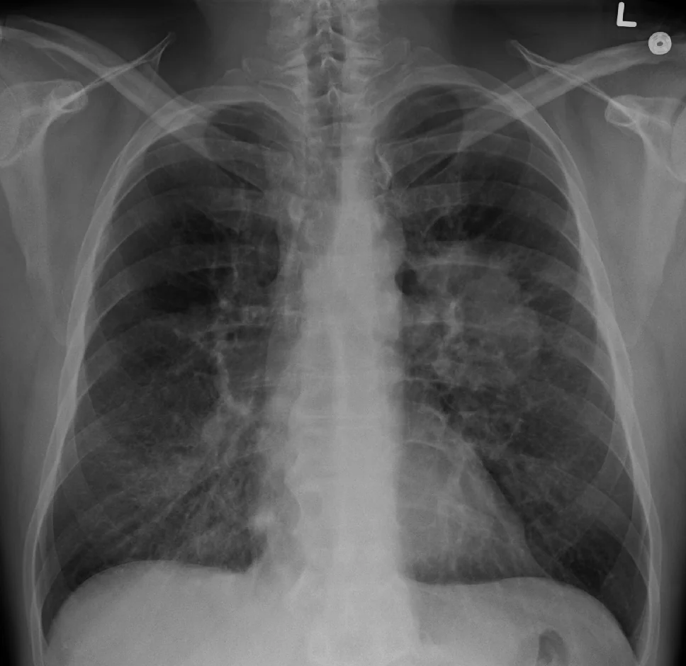
Types of lung cancer
The primary division is between small cell and non-small cell lung cancer, because it determines the entire treatment approach.
| Type | Proportion | Features |
|---|---|---|
| Adenocarcinoma | About 40% | The commonest type overall, and the commonest in never-smokers and women. Arises from mucus-secreting glandular cells and is typically peripheral. Most likely to carry a targetable driver mutation such as EGFR or ALK. Associated with hypertrophic pulmonary osteoarthropathy. |
| Squamous cell carcinoma | About 25-30% | Arises from bronchial epithelium after squamous metaplasia and is typically central. Cavitates more than any other type. Secretes PTH-related peptide, causing hypercalcaemia. Strongly smoking-related. |
| Large cell carcinoma | About 10% | Poorly differentiated, with no glandular or squamous features. Grows rapidly and metastasises early; poor prognosis. |
| Small cell carcinoma | About 15% | Arises from neuroendocrine (Kulchitsky) cells and is typically central. Almost exclusively in smokers. Very rapid doubling time with early widespread metastasis - around two thirds have metastases at diagnosis. Responsible for most paraneoplastic endocrine and neurological syndromes. |
| Carcinoid tumour | 1-2% | A low-grade neuroendocrine tumour, often in younger non-smokers. Slow growing, may cause recurrent infection through bronchial obstruction, and occasionally carcinoid syndrome. |
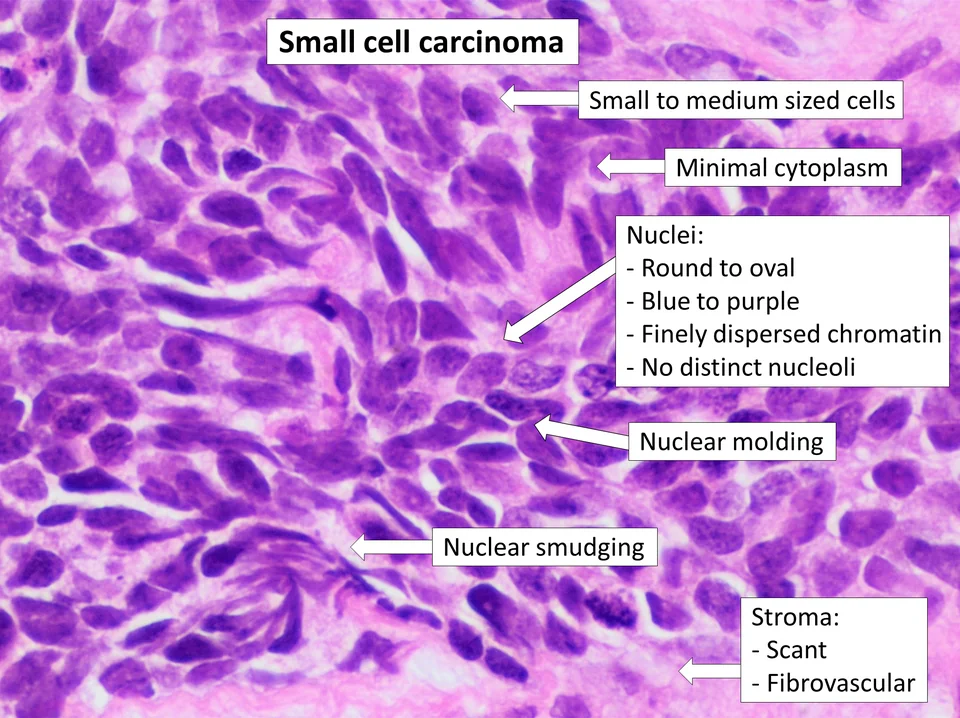
Clinical features
Local effects of the tumour
- Cough - the commonest presenting symptom, or a change in a longstanding smoker's cough
- Haemoptysis - usually small volume, and a symptom that should always be investigated
- Breathlessness - from airway obstruction, collapse, effusion or lymphangitis
- Chest pain - dull and persistent, or pleuritic with chest wall invasion
- Recurrent or slowly resolving chest infection, especially in the same lobe - suggesting post-obstructive pneumonia
- Monophonic wheeze or stridor - a fixed sound from a single obstructed airway, quite unlike the polyphonic wheeze of asthma
Local invasion and regional spread
| Structure | Consequence |
|---|---|
| Recurrent laryngeal nerve | Hoarse voice and a bovine cough - most often the left nerve, which loops under the aortic arch |
| Phrenic nerve | Diaphragmatic paralysis with a raised hemidiaphragm and breathlessness |
| Superior vena cava | SVC obstruction - facial and arm swelling, distended non-pulsatile neck veins, headache worse on bending forward, and a positive Pemberton's sign |
| Sympathetic chain (Pancoast tumour) | Horner syndrome - ptosis, miosis, anhidrosis and apparent enophthalmos |
| Brachial plexus (T1 root) | Pain in the shoulder and inner arm with wasting of the small muscles of the hand |
| Oesophagus | Dysphagia |
| Pericardium | Pericardial effusion and tamponade |
| Pleura and chest wall | Pleural effusion and localised chest wall pain |
| Lymph nodes | Supraclavicular and cervical lymphadenopathy - always palpate for these |
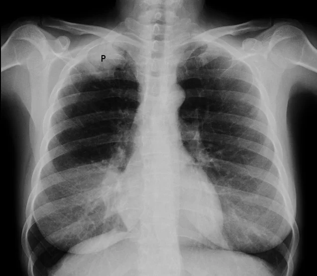
Metastatic disease
- Bone - pain, pathological fracture, spinal cord compression and hypercalcaemia
- Brain - headache, seizures, focal neurological deficit, personality change and confusion
- Liver - hepatomegaly, right upper quadrant pain, jaundice and deranged liver function
- Adrenal glands - usually asymptomatic and found on staging CT
- Skin and subcutaneous nodules
Constitutional and general signs
- Weight loss, anorexia and fatigue
- Finger clubbing - present in around a third, most often with non-small cell disease
- Hypertrophic pulmonary osteoarthropathy - clubbing with painful, swollen wrists and ankles from periosteal new bone formation; a classic association with adenocarcinoma
- Cachexia, anaemia and thrombocytosis
- Thrombophlebitis migrans (Trousseau sign) and unprovoked venous thromboembolism
Paraneoplastic syndromes
These are effects of the tumour that are not caused by its mass or by metastasis, and they may be the presenting feature. They are examined frequently because each points to a particular cell type.
| Syndrome | Mechanism | Cell type |
|---|---|---|
| SIADH | Ectopic ADH secretion causing euvolaemic hyponatraemia with concentrated urine and a high urinary sodium | Small cell |
| Ectopic ACTH (Cushing syndrome) | ACTH secretion causing hypokalaemic metabolic alkalosis, hyperglycaemia, hypertension, muscle weakness and pigmentation. Onset is too rapid for the classical Cushingoid habitus to develop, which catches people out. | Small cell |
| Lambert-Eaton myasthenic syndrome | Antibodies to presynaptic voltage-gated calcium channels. Proximal weakness that improves with repeated use, hyporeflexia with post-tetanic potentiation, and autonomic features such as dry mouth. The reverse of myasthenia gravis, where activity worsens weakness. | Small cell |
| Cerebellar degeneration and limbic encephalitis | Anti-Hu, anti-Yo and related antineuronal antibodies | Small cell |
| Hypercalcaemia | PTH-related peptide secretion. Distinguished from bone metastases by a suppressed PTH with a raised PTHrP. | Squamous cell |
| Hypertrophic pulmonary osteoarthropathy | Periosteal new bone formation with clubbing and painful swollen wrists and ankles | Adenocarcinoma and squamous |
| Dermatomyositis and polymyositis | Proximal myopathy with a heliotrope rash and Gottron papules - a recognised marker of underlying malignancy | Any |
| Thrombophlebitis migrans | Hypercoagulable state | Adenocarcinoma |
Referral and investigation
Who to refer, and how fast
NICE NG12 sets deliberately low thresholds, because early symptoms are so easily dismissed.1
- Offer an urgent chest X-ray (within 2 weeks) to people aged 40 and over with two or more of the following unexplained symptoms, or one or more if they have ever smoked: cough, fatigue, breathlessness, chest pain, weight loss or appetite loss
- Consider an urgent chest X-ray in people aged 40 and over with any of: persistent or recurrent chest infection, finger clubbing, supraclavicular or persistent cervical lymphadenopathy, chest signs consistent with lung cancer, or thrombocytosis
- Refer on a suspected cancer pathway (seen within 2 weeks) if the chest X-ray suggests lung cancer, or if the person is aged 40 or over with unexplained haemoptysis
- A normal chest X-ray does not exclude lung cancer. Around one in five is missed on the plain film. If clinical suspicion remains, refer or arrange a CT anyway - this is one of the most common causes of delayed diagnosis.
Establishing the diagnosis and stage
- Chest X-ray - looking for a mass, hilar enlargement, collapse, effusion, cavitation or a raised hemidiaphragm
- Contrast-enhanced CT of the chest, abdomen and pelvis - the standard staging investigation, covering the liver and adrenals
- PET-CT - where treatment with curative intent is being considered, to detect occult nodal and distant disease that would change the plan
- Brain imaging (contrast MRI preferred) - for stage III disease, for all patients with small cell lung cancer, and where there are neurological symptoms
- Tissue diagnosis - by the least invasive route that gives adequate material and stages the patient at the same time
| Site of lesion | Technique |
|---|---|
| Central airway lesion | Bronchoscopy with biopsy, brushings and washings |
| Mediastinal or hilar nodes | EBUS-TBNA - endobronchial ultrasound-guided transbronchial needle aspiration. Diagnoses and stages simultaneously, and is now the first-line nodal test. |
| Peripheral lesion | CT-guided percutaneous needle biopsy - carries a pneumothorax risk of around 20% |
| Pleural effusion | Pleural fluid cytology, then local anaesthetic thoracoscopy if cytology is negative |
| Accessible metastasis | Biopsy the metastasis - it gives tissue and confirms stage IV in one step |
Other investigations
- Bloods - FBC (anaemia, thrombocytosis), U&Es (hyponatraemia of SIADH), calcium, LFTs including ALP, and clotting
- Lung function tests - FEV1 and TLCO to assess fitness for surgery or radical radiotherapy, with a cardiopulmonary exercise test in borderline cases
- Performance status (WHO or ECOG, 0 to 4) - as important as stage in determining what treatment a patient can tolerate
- Molecular and immunohistochemical testing on NSCLC tissue - EGFR, ALK, ROS1, BRAF, KRAS G12C and other drivers, plus PD-L1 expression. This is now essential rather than optional, because it determines first-line treatment in advanced disease.
Staging
Non-small cell lung cancer is staged with the TNM system, which drives the treatment decision:
- T - size of the primary and what it invades
- N - nodal involvement, from N0 (none) through N1 (ipsilateral hilar), N2 (ipsilateral mediastinal) to N3 (contralateral or supraclavicular)
- M - metastases, including separate tumour nodules and malignant pleural or pericardial effusion
- Stages I and II are potentially curable by surgery; stage III is locally advanced and usually treated with chemoradiotherapy; stage IV is metastatic and treated systemically
Small cell lung cancer is also formally staged by TNM, but the practical division remains:
- Limited stage - confined to one hemithorax and encompassable within a single tolerable radiotherapy field. Treated with concurrent chemoradiotherapy with curative intent.
- Extensive stage - beyond that, in around two thirds of patients at diagnosis. Treated with chemotherapy plus immunotherapy.
Management
Every patient should be discussed at a lung cancer multidisciplinary team meeting and allocated a lung cancer nurse specialist, who is consistently one of the most valued parts of care from the patient's point of view.
Non-small cell lung cancer
- Surgery - lobectomy is the standard operation for stage I and II disease in a patient fit enough, with pneumonectomy, segmentectomy or wedge resection in selected cases. Increasingly performed by VATS or robotically. Fitness is assessed with lung function, exercise testing and cardiac risk.
- Stereotactic ablative radiotherapy (SABR) - highly conformal radiotherapy delivered in a few fractions, for early-stage disease in patients unfit for surgery. Local control rates approach those of surgery.
- Adjuvant chemotherapy after resection of stage II or III disease, and adjuvant targeted therapy or immunotherapy in selected molecular subgroups
- Concurrent chemoradiotherapy for stage III disease, followed by durvalumab consolidation
- Stage IV disease - guided by molecular testing: a targeted tyrosine kinase inhibitor where a driver mutation is present (for example osimertinib for EGFR, alectinib for ALK), immunotherapy such as pembrolizumab alone where PD-L1 expression is high, or chemotherapy combined with immunotherapy where it is not
Small cell lung cancer
- Chemotherapy is the backbone - platinum (cisplatin or carboplatin) with etoposide. SCLC is strikingly chemosensitive initially, with high response rates, but almost always relapses.
- Concurrent chemoradiotherapy for limited-stage disease, given with curative intent
- Chemotherapy plus immunotherapy (atezolizumab or durvalumab) for extensive-stage disease
- Prophylactic cranial irradiation - offered to patients responding to treatment, because brain metastases are so common and the blood-brain barrier limits chemotherapy penetration
- Surgery has almost no role, because the disease has usually disseminated by diagnosis
Palliative and supportive care
- Early specialist palliative care referral - in advanced lung cancer this improves quality of life and mood, and in a landmark trial was associated with longer survival despite less aggressive end-of-life treatment
- Palliative radiotherapy - for haemoptysis, chest pain, bone pain, SVC obstruction and brain metastases
- Endobronchial treatment - stenting, debulking or brachytherapy for major airway obstruction
- Pleural procedures - talc pleurodesis or an indwelling pleural catheter for a recurrent malignant effusion
- Symptom control - opioids for pain and for breathlessness, steroids for raised intracranial pressure and SVC obstruction, and antiemetics
- Smoking cessation - beneficial even after diagnosis, improving treatment tolerance, surgical outcomes and survival
- Nutritional and psychological support, and attention to benefits and financial advice
Complications
- Malignant pleural effusion - occurring in around 15% and defining stage IV disease
- Post-obstructive pneumonia and lung abscess
- Superior vena cava obstruction
- Metastatic spinal cord compression and pathological fracture
- Brain metastases - present in around 10% at diagnosis in NSCLC and far more in SCLC
- Hypercalcaemia and hyponatraemia
- Venous thromboembolism - substantially increased, and often the first clue to an occult cancer
- Massive haemoptysis
- Cachexia and malnutrition
- Treatment complications - neutropenic sepsis, radiation pneumonitis and oesophagitis, and the immune-related toxicities of checkpoint inhibitors, which can affect any organ and include colitis, hepatitis, pneumonitis, thyroiditis and hypophysitis
- Depression and anxiety - very common, and compounded by the stigma many patients feel about a smoking-related cancer
Red flags
Prognosis
Overall survival remains poor. Around 40% of patients survive one year and only about 16 to 20% survive five years, largely because of late presentation - but the figures conceal an enormous range.
- Stage I NSCLC treated surgically - five-year survival of around 65 to 80%
- Stage III NSCLC - around 15 to 25% at five years with chemoradiotherapy
- Stage IV NSCLC - historically a median survival of under a year, but transformed in molecularly selected subgroups, where targeted therapy has produced median survivals of several years and durable responses to immunotherapy are now well documented
- Limited-stage SCLC - median survival around 15 to 20 months, with a minority cured by chemoradiotherapy
- Extensive-stage SCLC - median survival around 8 to 13 months, improved somewhat by the addition of immunotherapy
Prognosis depends on stage, histology, molecular subtype, performance status and weight loss. Performance status in particular is often decisive: a patient with stage IV disease who is fully active may tolerate treatment that would be unthinkable for someone confined to bed for more than half the day, whatever the stage.
The single most important message remains preventive. Smoking cessation reduces risk substantially within 10 to 15 years and improves outcomes even after diagnosis, and the Targeted Lung Health Check programme offers the first realistic prospect of shifting the stage distribution at diagnosis, which is what would change these survival figures most.
References
- NICE NG12. Suspected cancer: recognition and referral. 2015, updated 2023. Available here
- NICE NG122. Lung cancer: diagnosis and management. 2019, updated 2024. Available here
- Cancer Research UK. Lung cancer statistics. Available here
- NHS England. Targeted Lung Health Check programme. Available here
- de Koning HJ, van der Aalst CM, de Jong PA et al. Reduced lung-cancer mortality with volume CT screening (NELSON). NEJM. 2020. Available here
- Temel JS, Greer JA, Muzikansky A et al. Early palliative care for patients with metastatic non-small-cell lung cancer. NEJM. 2010. Available here
- NICE NG234. Metastatic spinal cord compression in adults. 2023. Available here
- Royal College of Physicians. National Lung Cancer Audit. Available here
- James Heilman MD, CC BY-SA 4.0, via Wikimedia Commons. Available here
- Mikael Haggstrom MD, CC0, via Wikimedia Commons. Available here
- Jmarchn, CC BY-SA 3.0, via Wikimedia Commons. Available here
This article is written for revision and education. It is not clinical guidance and must not be used to make decisions about the care of a patient. Always check current NICE guidance and local protocols.