
Radiotherapy and its Side Effects
Key points
- Mechanism: ionising radiation causes DNA double-strand breaks, directly and through free radicals generated from water, killing cells as they attempt to divide.
- Fractionation: the total dose is split into small daily fractions because normal tissue repairs sublethal damage between fractions better than tumour does.
- Dose: measured in gray (Gy). Radical treatment is typically 60 to 70 Gy in 30 to 35 fractions; a single 8 Gy fraction is standard for painful bone metastases.
- Acute toxicity: affects rapidly dividing tissue in the treated field - skin, mucosa, gut - starts in week 2 to 3, peaks at the end, and resolves within weeks.
- Late toxicity: fibrosis, ischaemia and second malignancy appearing months to years later. It is irreversible and is what limits the dose that can be given.
- In the field only: with the exception of fatigue, side effects occur only in the irradiated volume - a patient having pelvic radiotherapy does not lose scalp hair.
- Not radioactive: patients treated with external beam radiotherapy are not radioactive and are safe to be around, including with children and pregnant women.
- Palliative role: single fractions give rapid relief of bone pain, haemoptysis and bleeding, and radiotherapy is central to treating cord compression and brain metastases.
Introduction
About four in ten people cured of cancer have had radiotherapy as part of their treatment, and roughly half of all patients with cancer receive it at some point.1,6 It is used with curative intent as the sole treatment in some tumours, as adjuvant or neoadjuvant treatment around surgery, and very commonly for palliation.
Students consistently underestimate it, partly because it happens in a physically separate department and partly because the physics feels forbidding. What is actually examined is straightforward: why it is given in fractions, what it is used for, the difference between acute and late toxicity, and how to recognise the complications you will meet on a general ward.
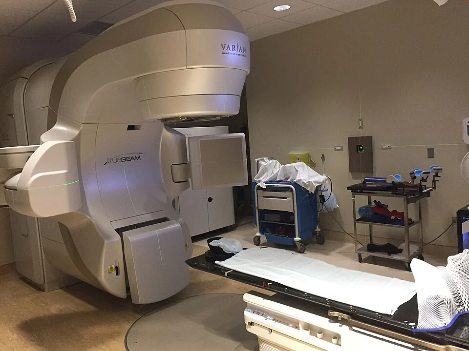
How radiotherapy works
Ionising radiation deposits energy in tissue, damaging DNA in two ways. A small proportion of damage is direct ionisation of the DNA molecule. Most is indirect: radiation ionises water to produce free radicals, principally the hydroxyl radical, which then attack DNA. The lethal lesion is the double-strand break, which is difficult to repair accurately.
Because free radical formation requires oxygen, well-oxygenated tissue is around three times more radiosensitive than hypoxic tissue. This oxygen effect explains why large tumours with necrotic hypoxic centres respond poorly, and why anaemia is corrected before radical treatment. Cells die when they next attempt mitosis, which is why the response is not immediate and why rapidly dividing tissues show effects first.
Why the dose is fractionated
Giving the whole dose at once would kill the tumour but also destroy the normal tissue in the beam. Splitting it into daily fractions exploits four biological differences between normal and malignant tissue, traditionally called the four Rs:
- Repair - normal cells repair sublethal DNA damage between fractions more efficiently than tumour cells, so the gap is worth more to them than to the tumour
- Reassortment (redistribution) - cells surviving one fraction redistribute through the cell cycle into the more radiosensitive G2 and M phases before the next
- Repopulation - normal tissue proliferates to replace losses during a course; this is also why prolonging a course by missing fractions allows tumour repopulation and worsens outcome
- Reoxygenation - as the tumour shrinks, previously hypoxic cells gain a blood supply and become radiosensitive
The practical consequence is that a radical course runs daily on weekdays for several weeks, and that gaps in treatment are avoided wherever possible. If a patient misses fractions through illness, the department will usually compensate rather than simply extend the course.
Techniques
| Technique | How it works | Typical use |
|---|---|---|
| External beam radiotherapy (EBRT) | High-energy photons from a linear accelerator directed from outside the body, from multiple angles | The great majority of treatments, curative and palliative |
| IMRT and VMAT | The beam intensity is modulated and the gantry rotates continuously, shaping the high-dose volume around concave targets | Head and neck (sparing the parotids), prostate, anywhere the target wraps around a critical structure |
| Image-guided radiotherapy (IGRT) | Imaging immediately before or during each fraction to correct for daily positioning and organ movement | Prostate, lung, anywhere the target moves |
| Stereotactic ablative radiotherapy (SABR/SRS) | Very high dose in 1 to 5 fractions with steep dose fall-off | Early-stage lung cancer in patients unfit for surgery, oligometastatic disease, brain metastases |
| Brachytherapy | Radioactive sources placed inside or adjacent to the tumour, giving a very high local dose with rapid fall-off | Cervical cancer (an essential component of curative treatment), prostate, oesophagus, skin |
| Proton beam therapy | Protons deposit most energy at a defined depth (the Bragg peak) with almost no exit dose | Paediatric tumours and base of skull tumours, where reducing late effects and second cancers matters most |
| Radioisotope therapy | A radionuclide given orally or intravenously and taken up by the target tissue | Radioiodine for thyroid cancer, radium-223 for prostate bone metastases, lutetium-177 for neuroendocrine tumours |
Planning
Radical treatment involves a planning process the patient should be prepared for. They attend for a planning CT in the exact position they will be treated in, immobilised - often with a custom thermoplastic shell for head and neck treatment, which some patients find claustrophobic and should be warned about. Small permanent skin tattoos are used to reproduce the position daily.
The oncologist then outlines volumes on the planning scan: the GTV (gross tumour volume, what can be seen), the CTV (clinical target volume, adding a margin for microscopic spread), and the PTV (planning target volume, adding a further margin for daily set-up variation and organ motion). Organs at risk such as the spinal cord, parotids, lungs, heart, rectum and bowel are also outlined, and the plan is optimised to keep each within an accepted dose constraint. Much of the art of radiotherapy lies in these constraints - the spinal cord tolerance is what limits re-treatment of a previously irradiated site.
Indications
Curative and adjuvant
- Radical radiotherapy as definitive treatment - prostate cancer, early laryngeal cancer (preserving the voice), cervical cancer with chemotherapy and brachytherapy, anal cancer (chemoradiotherapy avoids a permanent stoma), and early lung cancer with SABR
- Adjuvant after surgery - whole breast radiotherapy after breast-conserving surgery reduces local recurrence substantially and improves survival; also used after resection of head and neck, sarcoma and some brain tumours
- Neoadjuvant before surgery - long-course chemoradiotherapy or short-course radiotherapy in rectal cancer, to downstage the tumour and improve the chance of a clear circumferential margin
- Chemoradiotherapy - concurrent platinum or 5-FU-based chemotherapy acts as a radiosensitiser, improving control at the cost of considerably more acute toxicity
- Total body irradiation - as part of conditioning before allogeneic stem cell transplantation
Palliative
Palliative radiotherapy uses far fewer fractions, causes much less toxicity, and often works within days to weeks. It is one of the most effective and underused palliative interventions available.
- Painful bone metastases - a single 8 Gy fraction gives useful pain relief in around 60% of patients, can be repeated, and is as effective as longer courses for pain4
- Metastatic spinal cord compression - after dexamethasone, and after considering surgical decompression
- Brain metastases - stereotactic radiosurgery for limited disease; whole brain radiotherapy is used more selectively than it once was, since it adds cognitive toxicity for limited survival benefit
- Haemoptysis, cough and breathlessness from an endobronchial tumour
- Bleeding from bladder, rectal, gynaecological or fungating skin tumours
- Superior vena cava obstruction, though stenting is usually faster
- Dysphagia from oesophageal cancer, sometimes with brachytherapy
Side effects: the general rules
Three principles let you predict almost any radiotherapy side effect without memorising lists.
- Side effects occur in the treated volume. Ask what is in the field. Pelvic radiotherapy causes diarrhoea and cystitis, not oral mucositis. The only reliable exception is fatigue, which is systemic.
- Acute effects hit rapidly dividing tissue. Skin, mucosa, gut epithelium and bone marrow. They begin in week 2 or 3, worsen to a peak at the end of the course or shortly after, and then settle over a few weeks.
- Late effects are fibrosis and ischaemia. They arise from damage to fibroblasts, small vessels and slowly dividing parenchyma, appear months to years afterwards, and are essentially irreversible. They set the maximum dose that can safely be given.
| Acute | Late | |
|---|---|---|
| Timing | During treatment and up to 3 months after | Months to many years after |
| Mechanism | Depletion of rapidly dividing stem cells | Fibrosis, small vessel damage, parenchymal loss, carcinogenesis |
| Reversibility | Usually resolves completely | Progressive and irreversible |
| Relationship to dose | Related to total dose and how quickly it is given | Related to dose per fraction - large fractions cause disproportionately more late effect |
| Examples | Erythema, desquamation, mucositis, oesophagitis, diarrhoea, cystitis, myelosuppression | Fibrosis, lymphoedema, xerostomia, infertility, osteoradionecrosis, second malignancy, myelopathy |
Site-specific toxicity
Skin, and general effects
Radiation dermatitis progresses from erythema through dry desquamation (dry, itchy, flaking skin) to moist desquamation, where the epidermis is lost and the dermis weeps - most likely in skin folds such as the inframammary fold, axilla and groin. Advise gentle washing with unperfumed soap, a simple aqueous moisturiser, loose cotton clothing, avoidance of shaving, deodorant on broken skin, and sun protection to the treated area permanently. Fatigue is near-universal, worsens through the course, and can take months to resolve.
Head and neck
- Mucositis - severe, peaking in the final week, often requiring strong opioids and a nasogastric or gastrostomy tube for nutrition
- Xerostomia - permanent dry mouth from parotid damage, greatly reduced but not eliminated by IMRT. It causes difficulty eating and speaking and accelerates dental disease.
- Dental caries and osteoradionecrosis of the mandible - the reason every patient must have a dental assessment and any necessary extractions before treatment starts. Extractions afterwards in an irradiated mandible risk non-healing necrosis.
- Loss of taste, usually partially recovering, and hypothyroidism if the thyroid is in the field
Thorax
Radiation pneumonitis typically develops 1 to 6 months after treatment with a dry cough, breathlessness and low-grade fever, and radiological change confined to the treated volume rather than following anatomical lobes. It responds to corticosteroids and is an important differential for infection or progressive cancer in a breathless patient after thoracic radiotherapy. Later, pulmonary fibrosis, oesophageal stricture and - after mediastinal treatment for lymphoma in young people - premature coronary disease, valve disease and second cancers including breast cancer.
Abdomen and pelvis
- Acute - nausea, diarrhoea from radiation enteritis, cystitis with frequency and dysuria, and proctitis with tenesmus and mucus
- Late bowel - chronic radiation enteritis with malabsorption, bile acid diarrhoea, strictures, fistulae and occasionally obstruction
- Late bladder - reduced capacity, haemorrhagic cystitis with painless haematuria years later
- Sexual and reproductive - infertility in both sexes, vaginal stenosis and dryness (dilators are prescribed and should be discussed frankly), erectile dysfunction after prostate treatment, and premature menopause
- Pelvic insufficiency fractures of the sacrum, a commonly missed cause of pain after pelvic radiotherapy
Central nervous system
Acutely, raised intracranial pressure from oedema, managed with dexamethasone; alopecia in the treated area; and fatigue. Somnolence syndrome may occur several weeks afterwards. Late effects include cognitive impairment (a substantial concern after whole brain radiotherapy), hypopituitarism, cataract and, rarely, radiation necrosis. Lhermitte's sign - an electric shock down the spine on neck flexion - appears a few months after spinal or neck irradiation and is usually benign and self-limiting, but radiation myelopathy is a devastating and irreversible late complication and is the reason cord dose constraints are treated as absolute.
Practical counselling
Several patient concerns come up so consistently that they are worth having a prepared answer for, and they appear in communication stations.
- "Will I be radioactive?" - No. External beam radiotherapy leaves nothing radioactive in the body, and there is no risk to family members, children or pregnant women at any point.
- The exceptions - patients treated with permanent brachytherapy seed implants or radioisotopes such as radioiodine are given specific, temporary restrictions on close contact, particularly with children and pregnant women, and written instructions to follow
- "Does it hurt?" - Delivery is painless and each fraction takes only a few minutes, most of which is spent positioning. The machine does not touch the patient.
- "Will I lose my hair?" - Only in the treated area, and it may be permanent at radical doses
- Contraception - required during and for a period after treatment, and fertility preservation must be discussed before treatment where the gonads are in or near the field
- Travel and attendance - daily attendance for several weeks is a major practical burden, and transport support should be arranged early
Red flags
Outcomes
Radiotherapy is curative on its own in a range of tumours and contributes to cure in many more; the meta-analyses of adjuvant breast radiotherapy showed that preventing roughly four local recurrences avoids one breast cancer death at 15 years, which is one of the clearest demonstrations of its value.3 In the palliative setting, it is among the most reliable ways to relieve bone pain and to control bleeding.
Its main limitations are the tolerance of surrounding normal tissue, which caps the deliverable dose and often prevents re-treatment of the same site; tumour hypoxia and intrinsic radioresistance; and the practical burden of daily attendance. Modern technique - IMRT, image guidance, SABR and protons - is aimed almost entirely at improving the ratio between dose to tumour and dose to normal tissue.2
The late effect that most influences practice in younger patients is second malignancy, which arises in the irradiated volume typically 10 or more years later. It is the reason proton therapy is prioritised for children, and the reason that survivors of Hodgkin lymphoma treated with mediastinal radiotherapy enter breast screening and cardiac surveillance far earlier than the general population.
References
- Royal College of Radiologists. Radiotherapy dose fractionation, fourth edition. 2024. Available here
- NICE NG191 and related cancer service guidance on radiotherapy. Available here
- Early Breast Cancer Trialists' Collaborative Group. Effect of radiotherapy after breast-conserving surgery on 10-year recurrence and 15-year breast cancer death. The Lancet. 2011. Available here
- Chow E, Zeng L, Salvo N et al. Update on the systematic review of palliative radiotherapy trials for bone metastases. Clinical Oncology. 2012. Available here
- Haviland JS, Owen JR, Dewar JA et al. The UK START trials of hypofractionated radiotherapy for early breast cancer. The Lancet Oncology. 2013. Available here
- Cancer Research UK. Radiotherapy for cancer. Available here
This article is written for revision and education. It is not clinical guidance and must not be used to make decisions about the care of a patient. Always check current NICE guidance and local protocols.