
Malabsorption: Causes, Investigation and Management
Key points
- Malabsorption: impaired absorption of nutrients across the small bowel mucosa, causing nutritional deficiency despite adequate intake.
- Maldigestion: failure to break food down into absorbable units, chiefly from pancreatic exocrine insufficiency or bile salt deficiency - often grouped with malabsorption clinically.
- Cardinal features: steatorrhoea (pale, bulky, offensive stools that are hard to flush), weight loss despite adequate intake, bloating and fatigue.
- Commonest causes: coeliac disease, chronic pancreatitis, Crohn's disease, small intestinal bacterial overgrowth and bile acid malabsorption.
- Key first-line tests: coeliac serology, full blood count and haematinics, faecal elastase for pancreatic function, and faecal calprotectin.
- Deficiency pattern helps localise: iron and folate suggest proximal small bowel disease; B12 suggests terminal ileal disease or bacterial overgrowth.
- Management: treat the underlying cause, replace deficiencies, and involve a dietitian; pancreatic enzyme replacement for exocrine insufficiency.
- Red flags: significant weight loss, iron-deficiency anaemia, and new steatorrhoea in an older patient - exclude pancreatic and GI malignancy.
Introduction
Malabsorption describes the impaired absorption of one or more nutrients across the small intestinal mucosa. It is helpful to distinguish it from maldigestion, the failure to break food down into absorbable components, although the two frequently coexist and produce a similar clinical picture, so they are usually considered together.1
Normal absorption requires three things to work: adequate luminal digestion (pancreatic enzymes and bile salts), an intact mucosal absorptive surface, and functioning transport away from the gut via lymphatics and portal blood. Malabsorption results from failure at any of these three stages, and this framework is the most useful way to structure both the differential diagnosis and the investigation.
Causes
| Stage | Mechanism | Examples |
|---|---|---|
| Luminal (maldigestion) | Insufficient pancreatic enzymes | Chronic pancreatitis, pancreatic cancer, cystic fibrosis, pancreatic resection |
| Luminal (maldigestion) | Insufficient or ineffective bile salts | Cholestasis and biliary obstruction, primary biliary cholangitis, terminal ileal disease or resection, small intestinal bacterial overgrowth (bacteria deconjugate bile salts) |
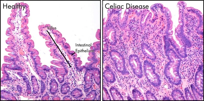
| Mucosal | Loss of absorptive surface or damaged enterocytes | Coeliac disease, Crohn's disease, tropical sprue, Whipple disease, giardiasis, radiation enteritis, autoimmune enteropathy, cow's milk protein enteropathy |
| Mucosal | Specific enzyme or transporter defects | Lactase deficiency (primary or secondary), abetalipoproteinaemia |
| Structural | Reduced length or abnormal transit | Short bowel syndrome after resection, gastric bypass and other bariatric surgery, fistulae, blind loops, motility disorders (systemic sclerosis, diabetic autonomic neuropathy) |
| Post-mucosal | Impaired lymphatic or venous drainage | Intestinal lymphangiectasia, lymphoma, tuberculosis, constrictive pericarditis |
In UK practice, coeliac disease, chronic pancreatitis, Crohn's disease, small intestinal bacterial overgrowth and bile acid malabsorption account for the majority of cases presenting to gastroenterology.
Clinical features
Gastrointestinal features
The classic presentation is steatorrhoea: pale, bulky, greasy, offensive-smelling stools that float and are difficult to flush, reflecting unabsorbed fat. Other features include chronic diarrhoea, abdominal bloating and distension, excessive flatus, and abdominal discomfort.
Weight loss despite an adequate or even increased appetite and food intake is characteristic and an important discriminator from simple reduced intake.
Features of specific deficiencies
Much of the clinical picture comes from the resulting deficiency states, and the pattern helps localise the lesion:
| Deficiency | Clinical features | Localising value |
|---|---|---|
| Iron | Microcytic anaemia, fatigue, koilonychia, angular stomatitis | Absorbed in duodenum and proximal jejunum - suggests proximal disease such as coeliac |
| Folate | Macrocytic anaemia, glossitis | Absorbed in proximal jejunum |
| Vitamin B12 | Macrocytic anaemia, peripheral neuropathy, subacute combined degeneration of the cord | Absorbed in terminal ileum - suggests ileal Crohn's, ileal resection or bacterial overgrowth |
| Vitamin A | Night blindness, dry eyes, hyperkeratosis | Fat-soluble - suggests fat malabsorption |
| Vitamin D and calcium | Osteomalacia, bone pain, proximal myopathy, tetany, osteoporosis | Fat-soluble |
| Vitamin E | Ataxia, peripheral neuropathy, haemolysis | Fat-soluble |
| Vitamin K | Bruising, prolonged prothrombin time | Fat-soluble |
| Protein | Oedema from hypoalbuminaemia, muscle wasting | Generalised |
| Zinc | Poor wound healing, acrodermatitis, altered taste, hair loss | Generalised |
Additional clues in the history include a family history of coeliac disease, a history of alcohol excess or recurrent pancreatitis, previous bowel or bariatric surgery, foreign travel (tropical sprue, giardiasis), and coexisting autoimmune disease.
Investigations
Establishing that malabsorption is present
- Full blood count and blood film: anaemia, which may be microcytic, macrocytic or dimorphic
- Haematinics: ferritin, vitamin B12 and folate
- Urea and electrolytes, calcium, magnesium, phosphate and vitamin D
- Liver function tests and albumin: hypoalbuminaemia reflects protein loss or malnutrition
- Coagulation screen: a prolonged prothrombin time reflects vitamin K deficiency
- Faecal fat estimation is rarely performed now, having been superseded by more practical tests
Identifying the cause
- Coeliac serology: IgA tissue transglutaminase antibodies with total IgA, in every patient - and remember the patient must be eating gluten for the test to be valid
- Faecal elastase: the practical test for pancreatic exocrine insufficiency; a low level indicates reduced enzyme output
- Faecal calprotectin: raised in inflammatory bowel disease, normal in coeliac disease and functional disorders
- Stool microscopy and culture, including for Giardia, particularly with relevant travel history
- Hydrogen breath testing: for small intestinal bacterial overgrowth (glucose or lactulose breath test) and for lactose intolerance
- SeHCAT scan: for bile acid malabsorption, particularly with watery diarrhoea after cholecystectomy or ileal resection
- Thyroid function, HIV testing and immunoglobulins where clinically indicated
Endoscopy and imaging
- Upper GI endoscopy with duodenal biopsies: for coeliac disease and other causes of villous atrophy, and to sample duodenal aspirate for Giardia
- Colonoscopy with ileal intubation and biopsies: for Crohn's disease and microscopic colitis
- MR enterography: for small bowel Crohn's disease, strictures and fistulae
- CT abdomen: for pancreatic disease, including calcification of chronic pancreatitis and pancreatic malignancy
- Capsule endoscopy: for suspected small bowel pathology not identified by other means
Management
Treat the underlying cause
Definitive management is directed at the specific diagnosis, and effective treatment of the cause usually reverses the malabsorption:
- Coeliac disease: lifelong strict gluten-free diet
- Chronic pancreatitis or pancreatic insufficiency: pancreatic enzyme replacement therapy (e.g. Creon) taken with meals and snacks, together with a proton pump inhibitor to reduce acid degradation of the enzymes2
- Crohn's disease: induction and maintenance of remission with steroids, immunomodulators and biologics
- Small intestinal bacterial overgrowth: a course of antibiotics such as rifaximin, and treatment of any underlying motility disorder or blind loop
- Bile acid malabsorption: a bile acid sequestrant such as colestyramine or colesevelam
- Giardiasis: metronidazole or tinidazole
- Lactose intolerance: lactose restriction, or lactase supplementation
Nutritional support and replacement
Replace identified deficiencies: iron, vitamin B12 (parenterally if ileal absorption is impaired), folate, calcium and vitamin D, and the other fat-soluble vitamins A, E and K. Magnesium and zinc are replaced where deficient. Bone density should be assessed in patients with prolonged fat malabsorption, given the risk of osteomalacia and osteoporosis.
Dietitian involvement is essential. Depending on the cause, this may involve a gluten-free diet, a low-fat diet with medium-chain triglyceride supplementation (which is absorbed directly into the portal circulation without requiring bile salts or lymphatics), oral nutritional supplements, or in severe cases enteral or parenteral nutrition. Patients with short bowel syndrome require specialist intestinal failure input and may need long-term parenteral nutrition.
Complications
- Malnutrition, cachexia and sarcopenia, with impaired wound healing and immunity
- Anaemia, which may be microcytic, macrocytic or mixed
- Metabolic bone disease: osteomalacia and osteoporosis with fragility fractures
- Neurological sequelae: peripheral neuropathy and subacute combined degeneration from B12 deficiency, ataxia from vitamin E deficiency
- Coagulopathy from vitamin K deficiency
- Oedema and ascites from hypoalbuminaemia
- Growth failure and delayed puberty in children
- Renal oxalate stones in fat malabsorption, where unabsorbed fatty acids bind calcium, leaving oxalate free to be absorbed and excreted
- Gallstones, particularly with terminal ileal disease and bile salt loss
- Refeeding syndrome if nutrition is reintroduced too rapidly in a severely malnourished patient - monitor and replace phosphate, potassium and magnesium
Red flags
Prognosis
Prognosis depends almost entirely on the underlying cause. Where the cause is treatable - coeliac disease, giardiasis, bacterial overgrowth, bile acid malabsorption, or pancreatic insufficiency managed with enzyme replacement - symptoms and nutritional status usually improve substantially, deficiencies correct with replacement, and the long-term outlook is good.1
Outcomes are less favourable where malabsorption results from irreversible structural loss, such as extensive short bowel syndrome or advanced chronic pancreatitis, in which case management becomes long-term nutritional support rather than cure. Malabsorption secondary to malignancy carries the prognosis of the underlying tumour. Across all causes, early recognition matters, because prolonged deficiency produces complications - particularly metabolic bone disease and neurological damage from B12 deficiency - that may not fully reverse even after the underlying cause is treated.
References
- Zuvarox T, Belletieri C. Malabsorption Syndromes. StatPearls. 2023. Available here
- NICE NG104. Pancreatitis. 2018 (updated 2020). Available here
- NICE NG20. Coeliac disease: recognition, assessment and management. 2015 (updated 2020). Available here
- NICE CG32. Nutrition support for adults. 2006 (updated 2017). Available here
- GeneFood, CC BY 4.0, via Wikimedia Commons. Available here
- NICE DG44. SeHCAT (tauroselcholic acid) for diagnosing bile acid diarrhoea. 2021. Available here
- British Society of Gastroenterology. Guidelines for the investigation of chronic diarrhoea in adults. Gut. 2018. Available here
This article is written for revision and education. It is not clinical guidance and must not be used to make decisions about the care of a patient. Always check current NICE guidance and local protocols.