
Tuberculosis: Diagnosis and Management
Key points
- Mycobacterium tuberculosis: an acid-fast bacillus spread by inhaled droplet nuclei, causing granulomatous infection that is either contained (latent TB) or progresses to active disease.
- Latent TB: viable bacteria contained by cell-mediated immunity - no symptoms, not infectious, but a lifelong risk of reactivation, highest in the first 2 years after infection.
- Reactivation: classically apical, post-primary pulmonary disease when immunity wanes - HIV is the single biggest risk factor.
- Diagnosis: sputum for AFB smear, culture (the gold standard, also giving drug sensitivities) and NAAT (rapid, and detects rifampicin resistance); culture or histology from any suspected extrapulmonary site.
- Standard treatment: the RIPE regimen - rifampicin, isoniazid, pyrazinamide and ethambutol for 2 months, then rifampicin and isoniazid for a further 4 months.
- Pyridoxine: given alongside isoniazid to prevent peripheral neuropathy.
- Public health duty: TB is notifiable, and contact tracing plus screening and treatment of latent infection follows every case.
Introduction
Tuberculosis (TB) is caused by Mycobacterium tuberculosis, an acid-fast, slow-growing bacillus spread person-to-person by inhaled droplet nuclei. It remains one of the world's leading infectious causes of death, and although UK incidence is low by global standards, it is concentrated in specific groups - people born outside the UK in high-incidence countries, those experiencing homelessness, people in prison, and people living with HIV - which makes a good social and travel history central to recognising it.1
TB is a heavily examined topic precisely because it tests several skills at once: recognising a chronic, systemic presentation that mimics malignancy; understanding the difference between latent and active infection; interpreting a battery of different tests correctly; and remembering a multi-drug regimen with genuinely dangerous side effects.
Pathophysiology
Inhaled droplet nuclei reach the alveoli, where the bacilli are phagocytosed by macrophages. In most people, cell-mediated immunity contains the infection within a granuloma, forming a Ghon focus; the combination of this focus with the associated hilar lymph node is the Ghon (Ranke) complex, often visible incidentally on a chest X-ray years later as a healed, calcified nodule.
- Primary TB - infection at first exposure; usually contained, but can progress directly to active disease in young children or the significantly immunosuppressed, who lack the mature cell-mediated response needed to wall it off
- Latent TB - viable bacteria persist within the granuloma, contained but not eliminated; the person has no symptoms and is not infectious, but carries a lifelong risk of reactivation, highest in the first 2 years after infection
- Post-primary (reactivation) TB - the classic adult presentation, occurring when waning immunity allows contained bacteria to multiply again, typically in the apex of the upper lobe, where higher oxygen tension favours this obligate aerobe
- Miliary TB - haematogenous dissemination producing widespread, millet-seed-sized (1-2 mm) granulomas across multiple organs; a feature of primary progressive disease or reactivation in a severely immunosuppressed host
Risk factors for progression to active disease
Most infected people never develop active disease. The risk of progression, whether from primary infection or from reactivation of latent infection, is driven overwhelmingly by the state of the host's cell-mediated immunity.
- HIV infection - by far the single largest risk factor, and TB is an AIDS-defining illness
- Immunosuppressive treatment - long-term corticosteroids, and particularly anti-TNF-alpha biologic therapy, which is why latent TB screening is mandatory before starting these drugs
- Diabetes mellitus
- Chronic kidney disease, especially on dialysis
- Malnutrition and low body weight
- Silicosis and other chronic lung disease
- Malignancy, particularly haematological
- Young children and older adults
- Alcohol excess, smoking and IV drug use
- Close, prolonged contact with a person with active pulmonary TB
Clinical features
Pulmonary TB
- Chronic cough, classically for more than 2-3 weeks, sometimes productive
- Haemoptysis
- Weight loss and anorexia
- Night sweats
- Low-grade fever and malaise
The systemic, gradual, weeks-to-months tempo of these symptoms - rather than any single feature - is what should raise suspicion, especially alongside a relevant travel, migration or exposure history.
Extrapulmonary TB
Occurs in a significant minority of cases overall, and considerably more often in people with HIV. Recognising the range of presentations matters because extrapulmonary disease is easy to misattribute to something else entirely.
| Site | Typical presentation |
|---|---|
| Lymph node ("scrofula") | Chronic, painless cervical lymphadenopathy, sometimes discharging (a "cold abscess" - little surrounding erythema or warmth despite active infection) |
| Pleura | Pleuritic pain and breathlessness from a lymphocyte-predominant exudative effusion, low glucose, high adenosine deaminase (ADA) |
| CNS (TB meningitis) | A subacute, days-to-weeks history of headache, fever and behavioural change, with cranial nerve palsies common; CSF shows a raised lymphocyte count, high protein and low glucose |
| Spine (Pott's disease) | Vertebral destruction causing back pain and progressive kyphosis, with a paravertebral or psoas "cold abscess" and a risk of spinal cord compression |
| Genitourinary | Sterile pyuria (white cells on dipstick/microscopy with a negative routine culture), epididymitis, and a recognised cause of infertility |
| Gastrointestinal/peritoneal | Ascites, abdominal pain and adhesions, which can mimic peritoneal malignancy |
| Pericardium | Pericardial effusion, and a recognised cause of later constrictive pericarditis |
| Miliary/disseminated | Multi-organ involvement with non-specific fever and weight loss; look for choroidal tubercles on fundoscopy |
Investigations
Testing for latent infection
- Mantoux (tuberculin skin test) - intradermal injection with induration measured at 48-72 hours; simple and cheap, but cross-reacts with prior BCG vaccination, causing false positives
- Interferon-gamma release assay (IGRA), for example Quantiferon - a blood test unaffected by BCG status, so more specific; used for contact tracing, new-entrant screening, and before starting immunosuppressive or biologic therapy
Neither test distinguishes latent from active infection, nor confirms the diagnosis of active TB on its own - a positive result in a symptomatic patient prompts investigation for active disease, not treatment for latent infection.
Testing for active disease
- Chest X-ray - upper lobe cavitation, patchy or nodular consolidation, fibrosis, hilar lymphadenopathy, pleural effusion, or a diffuse miliary pattern
- Sputum samples (at least three, ideally including an early morning sample) for AFB smear microscopy, culture and nucleic acid amplification testing (NAAT)
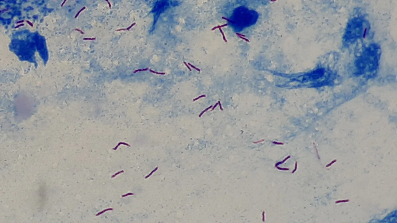
- AFB smear (Ziehl-Neelsen stain) - rapid and cheap but only moderately sensitive; a positive smear indicates a higher bacterial load and greater infectiousness
- Culture - the gold standard, since it confirms viability and allows full drug sensitivity testing, but slow (typically 2-6 weeks on solid or liquid media)
- NAAT (e.g. GeneXpert MTB/RIF) - rapid (hours rather than weeks) and also detects rifampicin resistance, an important early flag for possible multidrug-resistant disease
- Bronchoscopy with bronchoalveolar lavage, or gastric aspirates in young children who cannot expectorate, if sputum cannot be obtained
- Biopsy and histology of an affected extrapulmonary site - caseating granulomas are characteristic, and tissue should also be sent for culture and NAAT
- HIV test, offered to everyone diagnosed with TB
- Baseline bloods before treatment - LFTs (all four first-line drugs can be hepatotoxic), U&Es and FBC, plus visual acuity and colour vision before starting ethambutol
Differential diagnosis
- Lung cancer - shares weight loss, haemoptysis and a chronic cough, and the two can coexist
- Lymphoma - lymphadenopathy and B symptoms overlap closely with TB lymphadenitis
- Sarcoidosis - also produces non-caseating granulomas and hilar lymphadenopathy, but without the caseation, systemic infectious features or acid-fast organisms of TB
- Non-tuberculous mycobacterial infection - can produce a similar clinical and radiological picture, distinguished on culture speciation
- Fungal infection (histoplasmosis, coccidioidomycosis) in the relevant geographic and travel context
- Community-acquired pneumonia and lung abscess - usually more acute in onset than TB
Management
Standard treatment: the RIPE regimen
Standard treatment for fully sensitive pulmonary (and most extrapulmonary) TB is six months in total, in two phases.2
| Phase | Duration | Drugs |
|---|---|---|
| Intensive | 2 months | Rifampicin, Isoniazid, Pyrazinamide, Ethambutol |
| Continuation | 4 months | Rifampicin, Isoniazid |
TB meningitis and bone/joint TB are treated for longer - typically 12 months - reflecting poorer drug penetration and the consequences of under-treatment in these sites. Corticosteroids are added in TB meningitis and TB pericarditis, to reduce the inflammatory damage caused by the immune response as much as the organism itself.
| Drug | Notable side effects |
|---|---|
| Rifampicin | Hepatotoxicity; orange-red discolouration of urine, tears and other secretions (harmless but worth warning patients about); a potent CYP450 inducer, reducing the efficacy of the combined oral contraceptive pill, warfarin and other interacting drugs |
| Isoniazid | Hepatotoxicity; peripheral neuropathy, caused by interference with pyridoxine (vitamin B6) metabolism - pyridoxine is co-prescribed to prevent it |
| Pyrazinamide | Hepatotoxicity (the most hepatotoxic of the four); hyperuricaemia, which can precipitate gout |
| Ethambutol | Optic neuritis, causing reduced visual acuity and impaired colour vision - visual acuity and colour vision are checked before starting and monitored throughout, and the drug is stopped immediately if visual symptoms develop |
Adherence and infection control
Directly observed therapy (DOT) - a health worker witnessing each dose - is used for patients at higher risk of non-adherence, given the consequences of an incomplete course: relapse and the development of drug resistance. Patients with smear-positive pulmonary or laryngeal TB are considered infectious and are isolated (in a negative-pressure side room if admitted) until they have completed at least 2 weeks of effective treatment and are showing clinical improvement, with a lower threshold and longer isolation for suspected multidrug-resistant disease.
Drug-resistant TB
Multidrug-resistant TB (MDR-TB) is resistant to at least rifampicin and isoniazid; extensively drug-resistant TB (XDR-TB) adds resistance to key second-line agents. Both need longer, more complex and more toxic regimens under specialist infectious diseases and microbiology input, which is why rapid NAAT-based detection of rifampicin resistance at diagnosis matters so much.
Latent TB treatment
Treating latent infection substantially reduces the lifetime risk of reactivation, and is offered to those at higher risk of progression - including new HIV diagnoses, people about to start biologic or other significant immunosuppression, and close child contacts. Options include isoniazid and rifampicin for 3 months, isoniazid alone for 6 months, or rifampicin alone for 4 months, chosen according to interactions, hepatotoxicity risk and adherence.
BCG vaccination
The BCG is a live attenuated vaccine, no longer given universally in the UK (targeted at higher-risk neonates and infants since 2005). It reduces the risk of severe, disseminated childhood disease (miliary TB, TB meningitis) more reliably than it prevents pulmonary TB in adults, and it causes false-positive Mantoux results, which is why IGRA testing is preferred where BCG history is uncertain.
Public health duties
- TB is a notifiable disease - notify UKHSA on clinical suspicion, without waiting for microbiological confirmation
- Contact tracing - close contacts are screened with Mantoux and/or IGRA, with a chest X-ray where indicated, and offered treatment for latent or active disease as appropriate
- Isolation for infectious pulmonary or laryngeal disease, as above
- Directly observed therapy for patients at risk of poor adherence, given the public health as well as individual consequences of an incomplete course
Complications
- Massive haemoptysis from cavitary disease eroding into a vessel
- Bronchiectasis and post-TB lung fibrosis, causing long-term respiratory impairment
- Aspergilloma forming within an old, healed TB cavity
- Pneumothorax
- Constrictive pericarditis after TB pericarditis
- Spinal deformity and cord compression in Pott's disease
- Infertility from genitourinary TB
- Historically, adrenal (Addisonian) failure from TB destruction of the adrenal glands
- Death, particularly with delayed diagnosis, drug resistance or HIV coinfection
Prognosis
Fully sensitive TB treated with a complete course has an excellent cure rate. Outcomes worsen significantly with delayed diagnosis, drug resistance, poor adherence, and HIV coinfection or other significant immunosuppression, and CNS and disseminated disease carry a materially higher mortality and rate of long-term disability than uncomplicated pulmonary disease. Completion of treatment, not simply starting it, is what determines cure and prevents the emergence of resistance - which is the underlying rationale for directly observed therapy and close public health follow-up.
References
- NICE NG33. Tuberculosis. 2016, updated 2019. Available here
- World Health Organization. Global tuberculosis report. Available here
- UK Health Security Agency. Tuberculosis: guidance, data and analysis. Available here
- NICE Clinical Knowledge Summaries. Tuberculosis. Available here
- British Thoracic Society. Guidelines on the management of tuberculosis. Available here
This article is written for revision and education. It is not clinical guidance and must not be used to make decisions about the care of a patient. Always check current NICE guidance and local protocols.