
Pleural Effusion: Transudate or Exudate, and What the Fluid Tells You
Key points
- Pleural effusion: an abnormal accumulation of fluid in the pleural space, which normally contains only 10 to 20 ml of lubricating fluid.
- The first question: transudate or exudate. Transudates result from altered hydrostatic or oncotic pressures with normal pleura; exudates from pleural inflammation or increased capillary permeability.
- Light's criteria: the fluid is an exudate if pleural to serum protein ratio is above 0.5, pleural to serum LDH ratio is above 0.6, or pleural LDH exceeds two thirds of the upper limit of normal serum LDH.
- Commonest causes: heart failure is the commonest transudate and the commonest cause overall. Pneumonia and malignancy are the commonest exudates.
- Signs: stony dull percussion, reduced or absent breath sounds, reduced vocal resonance and reduced expansion, with tracheal deviation away from the side if the effusion is massive.
- Ultrasound is mandatory: every pleural procedure must be guided or sited by thoracic ultrasound. Blind aspiration causes avoidable serious harm and is no longer acceptable practice.
- A pH below 7.2: in a parapneumonic effusion indicates a complicated effusion or empyema and mandates chest drain insertion.
- Drainage limit: remove no more than 1.5 litres at a time, or in a single episode, to avoid re-expansion pulmonary oedema.
Introduction
The pleural space normally contains only 10 to 20 ml of fluid, continuously produced by the parietal pleura and reabsorbed by parietal pleural lymphatics. A pleural effusion develops when production exceeds reabsorption, and around 50,000 people are diagnosed with one in the UK each year.1
An effusion is a sign, not a diagnosis, and the whole of its management is directed at answering one question: why is the fluid there? The single most useful step in reaching an answer is dividing effusions into transudates and exudates, because that division separates two entirely different lists of causes and two entirely different management approaches.
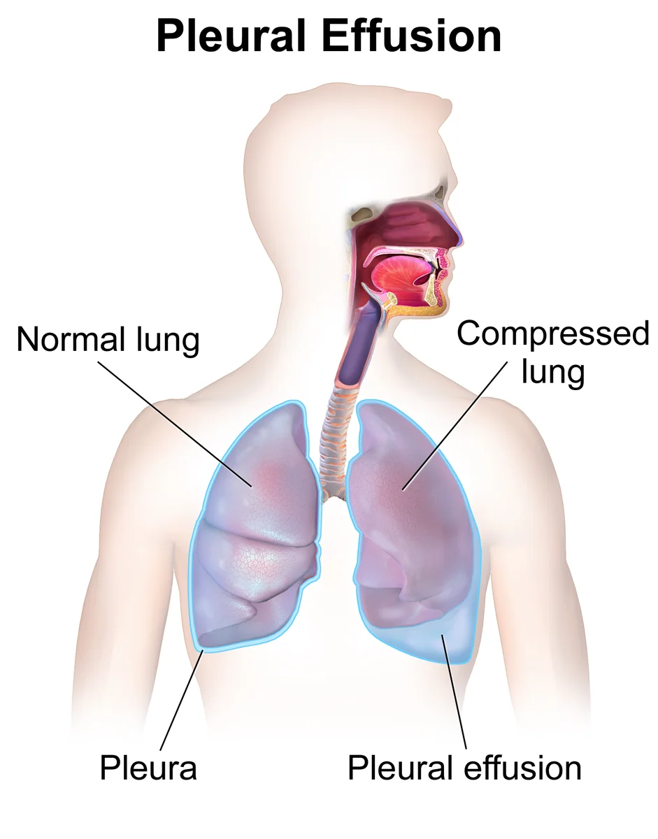
Transudate or exudate
- Transudate - the pleura is normal. Fluid moves out because of raised hydrostatic pressure or reduced oncotic pressure. Protein content is low (typically under 25 g/L), and the fluid is usually clear and straw-coloured. Transudates are commonly bilateral.
- Exudate - the pleura itself is diseased. Inflammation or malignancy increases capillary permeability, or lymphatic drainage is obstructed, allowing protein-rich fluid to leak into the space. Protein content is high (typically above 35 g/L), and the effusion is more often unilateral.
| Transudates | Exudates |
|---|---|
| Heart failure - the commonest cause of an effusion overall, usually bilateral and larger on the right | Parapneumonic effusion and empyema - the commonest exudate |
| Liver cirrhosis with hypoalbuminaemia | Malignancy - lung, breast, lymphoma, mesothelioma and metastases from any primary |
| Nephrotic syndrome | Tuberculosis - typically lymphocyte-predominant with a high adenosine deaminase |
| Hypoalbuminaemia from any cause, including malnutrition and protein-losing enteropathy | Pulmonary embolism - usually small and blood-stained |
| Peritoneal dialysis | Rheumatoid arthritis - characteristically a very low glucose and low pH |
| Hypothyroidism | Systemic lupus erythematosus |
| Constrictive pericarditis | Pancreatitis - left-sided, with a very high amylase |
| Meigs syndrome - benign ovarian fibroma with ascites and a right pleural effusion | Oesophageal rupture - high amylase, low pH, and a surgical emergency |
| Urinothorax | Post-cardiac injury (Dressler) syndrome and post-CABG effusions |
| Asbestos-related benign pleural effusion | |
| Drugs - methotrexate, amiodarone, nitrofurantoin, phenytoin | |
| Chylothorax - thoracic duct disruption from trauma, surgery or lymphoma |
Clinical features
Symptoms
- Breathlessness - the dominant symptom, worsening as the effusion enlarges and often worse lying on the unaffected side
- Pleuritic chest pain - suggests pleural inflammation and therefore an exudate; transudates are typically painless
- Dry cough
- Symptoms of the underlying cause - orthopnoea and oedema in heart failure, fever and purulent sputum in pneumonia, weight loss and haemoptysis in malignancy, night sweats in tuberculosis
- Small effusions may be entirely asymptomatic and found incidentally
Signs
- Stony dull percussion note - the classic and most useful sign, and distinctly duller than the dullness of consolidation
- Reduced or absent breath sounds over the effusion
- Reduced vocal resonance and tactile vocal fremitus
- Reduced chest expansion on the affected side
- Bronchial breathing and egophony at the upper border of the effusion, where the underlying lung is compressed
- Tracheal and mediastinal deviation away from the side of the effusion if it is massive - a large effusion pushes, unlike collapse which pulls
- Signs of the cause - raised JVP and peripheral oedema in heart failure, clubbing and cachexia in malignancy, a rheumatoid hand, ascites and spider naevi in cirrhosis
| Feature | Effusion | Consolidation | Collapse |
|---|---|---|---|
| Percussion | Stony dull | Dull | Dull |
| Breath sounds | Absent or reduced | Bronchial | Reduced |
| Vocal resonance | Reduced | Increased | Reduced |
| Trachea | Pushed away if large | Central | Pulled towards |
| Added sounds | None, or a rub | Coarse crackles | None |
Investigations
Imaging
- Erect chest radiograph - blunting of the costophrenic angle appears once around 200 ml has accumulated on a PA film (only 50 ml is needed on a lateral). Larger effusions show a meniscus curving upwards laterally, and a massive effusion produces a complete white-out with mediastinal shift away from the affected side.
- Thoracic ultrasound - more sensitive than radiography, distinguishes fluid from consolidation and pleural thickening, identifies septations that predict the need for drainage, and is used to select the safest site. It is mandatory before every pleural procedure.
- CT thorax with contrast - for undiagnosed exudative effusions. It should ideally be performed before complete drainage, since fluid provides contrast against which pleural nodularity, thickening and mediastinal pleural involvement - all features suggesting malignancy - are more easily seen.
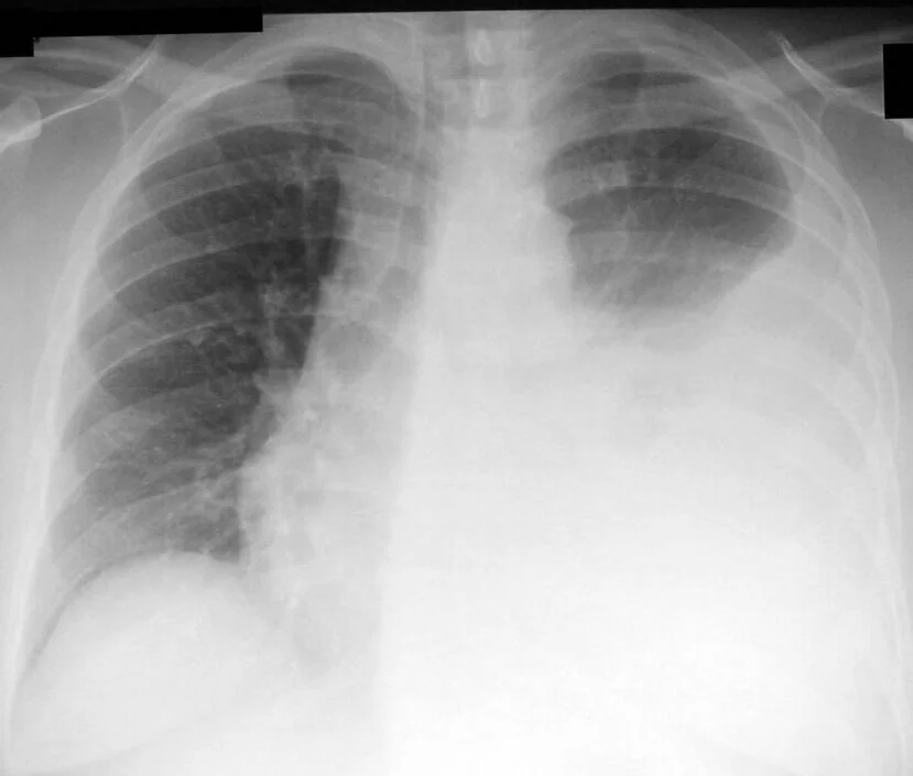
Pleural fluid analysis
Aspirate 20 to 50 ml under ultrasound guidance and send it for the following, taking paired serum protein and LDH at the same time.
| Test | Interpretation |
|---|---|
| Protein and LDH | Apply Light's criteria to classify as transudate or exudate |
| pH | Send in a blood gas syringe. Below 7.2 in a parapneumonic effusion indicates a complicated effusion or empyema and mandates chest drain insertion. Also low in malignancy, tuberculosis, rheumatoid disease and oesophageal rupture. |
| Glucose | Low (below 3.3 mmol/L) in empyema, rheumatoid arthritis (often strikingly low), tuberculosis, malignancy, lupus and oesophageal rupture |
| Gram stain, culture and sensitivities | Including cultures in blood culture bottles, which improve yield. Send AFB smear and culture if tuberculosis is suspected. |
| Cytology | For malignant cells. Sensitivity is only around 60% on a first sample, so send a second if the first is negative and suspicion remains. |
| Amylase | Raised in pancreatitis, oesophageal rupture and some malignancies |
| Triglycerides and chylomicrons | Triglycerides above 1.24 mmol/L indicate a chylothorax |
| Haematocrit | A pleural fluid haematocrit greater than half the blood haematocrit defines a haemothorax, which requires drainage |
| Differential cell count | Neutrophils suggest an acute process; lymphocytes suggest tuberculosis, malignancy, lymphoma or a chronic effusion; eosinophils suggest air or blood in the pleural space, drugs or parasitic infection |
| Adenosine deaminase | Raised in tuberculous effusions and useful where culture is slow or negative |
The undiagnosed exudative effusion
Around a quarter of exudative effusions remain undiagnosed after initial fluid analysis, and the concern in almost all of them is malignancy. The pathway is:
- CT thorax with contrast, before full drainage
- Repeat cytology on a second sample
- Local anaesthetic thoracoscopy - the investigation of choice, giving a diagnostic yield above 90% and allowing talc pleurodesis at the same sitting
- Image-guided pleural biopsy where thoracoscopy is not available or the patient is unfit
- Blind (Abrams) pleural biopsy is now reserved for suspected tuberculosis, where the pleural involvement is diffuse
Other tests
- Bloods - FBC, U&Es, LFTs including albumin, CRP, clotting, serum protein and LDH, and NT-proBNP where heart failure is suspected
- Echocardiogram for suspected heart failure
- Thyroid function, and urine protein-creatinine ratio for nephrotic syndrome
- Autoimmune serology - rheumatoid factor, anti-CCP and ANA where a connective tissue disease is suspected
- Occupational history - a careful asbestos exposure history in every unexplained unilateral effusion
Management
General principles
- Treat the underlying cause. A transudative effusion from heart failure, cirrhosis or nephrotic syndrome should be managed by treating that condition - diuretics, albumin, sodium restriction - and does not usually need drainage.
- Therapeutic aspiration for symptomatic relief in a breathless patient
- Remove no more than 1.5 litres at a time, and stop earlier if the patient develops chest discomfort or persistent cough, to avoid re-expansion pulmonary oedema
- Repeat the chest radiograph after drainage to assess re-expansion and exclude a pneumothorax
Parapneumonic effusion
- Simple parapneumonic effusion - clear fluid, pH above 7.2, negative Gram stain and culture. Treat the pneumonia with antibiotics; the effusion resolves.
- Complicated parapneumonic effusion - pH below 7.2, low glucose, or a positive Gram stain or culture. Requires chest drain insertion in addition to antibiotics.
- Empyema - frank pus. Requires chest drainage, prolonged antibiotics and, if drainage fails, intrapleural fibrinolytics or surgical referral
Malignant pleural effusion
A malignant effusion indicates advanced disease, and treatment is directed at relieving breathlessness rather than at cure. The choice is guided by prognosis, whether the lung re-expands after drainage, and above all by patient preference.
- Therapeutic aspiration - simple and immediate, but the fluid usually recurs within days to weeks. Reasonable in patients with a very short prognosis.
- Chest drain with talc pleurodesis - instilling talc to obliterate the pleural space. Requires the lung to re-expand fully and an inpatient stay of several days. Success is around 70 to 80%.
- Indwelling pleural catheter - a tunnelled catheter drained at home by the patient or a carer, allowing outpatient management. This is the preferred option where the lung fails to re-expand (trapped lung), since pleurodesis cannot work in that situation, and is increasingly chosen by patients who wish to avoid admission.
- Thoracoscopy with talc poudrage - combines diagnosis and pleurodesis in one procedure
- Systemic anti-cancer therapy where the tumour is responsive - for example, effusions from small cell lung cancer or lymphoma may resolve with chemotherapy alone
Complications
- Empyema - infection of the pleural fluid, requiring drainage
- Trapped lung and pleural thickening - causing persistent breathlessness and restriction
- Respiratory failure from a large or bilateral effusion
- Re-expansion pulmonary oedema - after removing too much fluid too quickly, presenting with cough, breathlessness and unilateral pulmonary oedema
- Pneumothorax - from aspiration or drain insertion
- Haemothorax and organ injury - laceration of intercostal vessels, liver or spleen during a poorly sited procedure
- Tumour seeding along the needle or drain tract - a particular problem in mesothelioma, where prophylactic radiotherapy to the tract is sometimes considered
- Infection at the drain site, and catheter-related infection with indwelling catheters
- Recurrence - the norm in malignant effusions without definitive treatment
Red flags
Prognosis
Prognosis is entirely determined by the underlying cause. Transudative effusions from heart failure, cirrhosis or nephrotic syndrome resolve as the cause is treated, and their prognostic significance is that of the underlying organ failure rather than of the fluid itself.
Parapneumonic effusions resolve with adequate treatment of the pneumonia, though a complicated effusion or empyema carries a mortality of around 20% and much longer hospital stays.
Malignant pleural effusion indicates advanced disease, with a median survival of around 4 to 6 months, though this varies considerably by tumour type - measured in a few months for lung cancer and often much longer for breast cancer, ovarian cancer, lymphoma and mesothelioma. The LENT score (pleural fluid LDH, ECOG performance status, neutrophil-to-lymphocyte ratio and tumour type) is used to estimate survival and to guide whether an invasive definitive procedure or a simpler palliative approach is more appropriate. In practice, choosing between talc pleurodesis and an indwelling catheter is as much a conversation about how the patient wants to spend their remaining time as it is a technical decision.
References
- British Thoracic Society. Pleural disease guideline. 2023. Available here
- Light RW, Macgregor MI, Luchsinger PC, Ball WC. Pleural effusions: the diagnostic separation of transudates and exudates. Annals of Internal Medicine. 1972. Available here
- NICE Clinical Knowledge Summaries. Palliative care - dyspnoea and malignant pleural effusion. Available here
- NHS England. Patient safety alert: risks of chest drain insertion. Available here
- Clive AO, Kahan BC, Hooper CE et al. Predicting survival in malignant pleural effusion: the LENT prognostic score. Thorax. 2014. Available here
- NICE NG122. Lung cancer: diagnosis and management. 2019, updated 2024. Available here
- BruceBlaus, CC BY-SA 4.0, via Wikimedia Commons. Available here
- Clinical Cases, CC BY-SA 2.5, via Wikimedia Commons. Available here
This article is written for revision and education. It is not clinical guidance and must not be used to make decisions about the care of a patient. Always check current NICE guidance and local protocols.