
Malignant Pleural Effusion
Key points
- Definition: a pleural effusion containing malignant cells, or occurring as a direct consequence of malignancy involving the pleura.
- Causes: lung and breast cancer account for around half; then lymphoma, ovarian and gastrointestinal cancer, mesothelioma, and cancer of unknown primary.
- Biochemistry: almost always an exudate by Light's criteria; a malignant transudate is rare and should prompt a search for another explanation.
- Cytology: sensitivity of a first pleural fluid sample is only around 60%, and much lower in mesothelioma - a negative result does not exclude malignancy.
- Ultrasound: all pleural procedures are performed under ultrasound guidance, which reduces the risk of pneumothorax and organ injury.
- Symptom-driven: an asymptomatic effusion does not need draining; treat the breathlessness, not the radiograph.
- Drain slowly: remove no more than about 1.5 litres at a time, and stop for chest pain or cough, to avoid re-expansion pulmonary oedema.
- Definitive options: talc pleurodesis or an indwelling pleural catheter - equally effective for breathlessness, so the choice depends on the lung re-expanding and on the patient's priorities.
Introduction
Malignant pleural effusion is one of the commonest reasons a patient with cancer becomes breathless, affecting around 15% of patients with malignancy at some point and being the presenting feature in a significant minority.1 It is almost always a marker of advanced, incurable disease, and the aim of treatment is therefore to relieve breathlessness with the fewest hospital visits and the least discomfort.
The management of these effusions has changed considerably. Repeated therapeutic aspirations, which used to be the default, are now regarded as a holding measure rather than a plan, because the fluid reaccumulates within days to weeks and each aspiration carries risk. The modern choice is between talc pleurodesis and an indwelling pleural catheter, and the trials that compared them are worth knowing.
Aetiology and pathophysiology
The pleural space normally contains only 5 to 15 mL of fluid, produced by the parietal pleura and absorbed largely through parietal lymphatics. An effusion forms when production exceeds absorption, and malignancy interferes with both sides of that balance.
- Increased permeability - tumour deposits on the pleura release VEGF and inflammatory mediators, making the pleural capillaries leaky
- Impaired lymphatic drainage - obstruction of parietal lymphatics or mediastinal nodes by tumour prevents reabsorption. This is often the dominant mechanism, and explains effusions with little visible pleural disease.
- Direct tumour invasion of the pleura
- Paramalignant effusions - fluid caused by the cancer but without malignant cells in the pleura: post-obstructive pneumonia, atelectasis from bronchial obstruction, pulmonary embolism, chylothorax from thoracic duct obstruction, hypoalbuminaemia, or pericardial involvement. These matter because their management differs.
| Primary | Approximate share | Note |
|---|---|---|
| Lung cancer | About a third | The commonest cause overall; usually ipsilateral. Makes the tumour stage M1a and therefore incurable by surgery. |
| Breast cancer | About a fifth | Often occurs years after the primary; frequently responsive to systemic treatment |
| Lymphoma | About 10% | May respond well to chemotherapy, so effusion control alone is not the whole answer |
| Ovarian and gastrointestinal cancer | About 10% combined | Often with ascites; consider a transdiaphragmatic route |
| Mesothelioma | Less common but important | Asbestos exposure; cytology is particularly insensitive, and pleural biopsy is usually needed |
| Unknown primary | Around 10% | Immunohistochemistry on the pleural fluid or biopsy directs the search |
Clinical features
- Breathlessness - the dominant symptom, typically progressive over weeks, worse on exertion and on lying on the unaffected side. Its severity correlates poorly with the size of the effusion, since it also depends on lung reserve, pleural elastance and diaphragm mechanics.
- Chest discomfort - a dull ache or a sense of heaviness. Sharp pleuritic pain suggests parietal pleural involvement and is characteristic of mesothelioma.
- Dry cough, from compression of the lung
- Constitutional symptoms - weight loss, anorexia, fatigue
- Sometimes asymptomatic - found incidentally on a staging scan, in which case it does not require drainage
Examination
The classic signs of an effusion are reduced chest expansion, stony dull percussion note, reduced or absent breath sounds and reduced vocal resonance over the fluid, sometimes with bronchial breathing and egophony at the upper level where the underlying lung is compressed. Tracheal deviation away from the side of the effusion indicates a large effusion; deviation towards it suggests underlying collapse from an obstructing tumour, which changes the management entirely.
Look also for the features that point to the primary and to the stage: clubbing, a supraclavicular node, a breast mass or mastectomy scar, hepatomegaly, cachexia, and signs of superior vena cava obstruction.
Investigations
Imaging
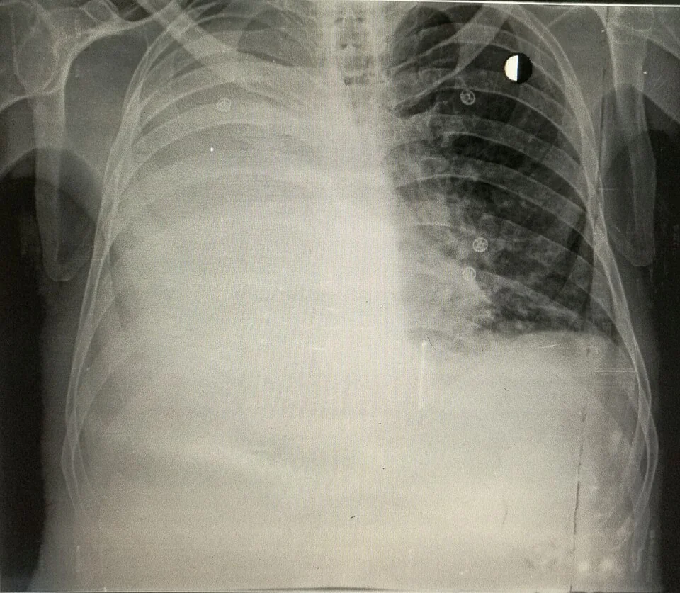
- Chest radiograph - blunting of the costophrenic angle appears with about 200 mL of fluid on a PA film; a meniscus with a concave upper border rising laterally indicates a moderate to large effusion
- Thoracic ultrasound - now mandatory before any pleural procedure. It confirms fluid, measures depth, identifies septations, marks a safe site, and is far more sensitive than a radiograph. Complex septated fluid or visible pleural nodules strongly suggest malignancy or empyema.
- CT of the thorax with contrast, performed before complete drainage - the fluid provides contrast against the pleura, so imaging before drainage shows pleural nodularity, circumferential or mediastinal pleural thickening, and parietal pleural thickening over 1 cm, all of which suggest malignancy. Drain first and this information is lost.
- PET-CT - occasionally used where the primary is unknown or the pleural findings are equivocal
Pleural fluid analysis
Diagnostic aspiration under ultrasound guidance yields fluid for biochemistry, cytology and microbiology. Malignant fluid is typically straw-coloured, but is blood-stained in around half of cases, and a haemorrhagic effusion in the absence of trauma or infarction is highly suggestive of malignancy.
Malignant effusions are essentially always exudates. Light's criteria are highly sensitive but misclassify around a quarter of transudates as exudates, particularly in patients on diuretics for heart failure - in that situation the serum-to-pleural-fluid albumin gradient (a gradient above 12 g/L indicates a transudate) is more reliable.2
| Test | Interpretation |
|---|---|
| Cytology | Diagnostic when positive. Sensitivity is around 60% on a first sample, rising modestly with a second; below 30% in mesothelioma. Send a large volume (at least 25 to 50 mL). |
| pH | Below 7.30 in malignant effusion predicts a poorer prognosis and a lower success rate for pleurodesis. Below 7.20 in a parapneumonic effusion mandates drainage. |
| Glucose | Low (below 3.3 mmol/L) in malignancy, empyema, tuberculosis and rheumatoid disease |
| Differential cell count | Lymphocyte predominance in malignancy, tuberculosis and lymphoma; neutrophils in parapneumonic effusion |
| Microbiology | Gram stain, culture and acid-fast bacilli - always exclude empyema and tuberculosis |
| Triglycerides | Above 1.24 mmol/L indicates chylothorax, from thoracic duct obstruction by lymphoma or tumour |
| Amylase | Raised in pancreatic disease and oesophageal rupture, and in some malignant effusions |
When cytology is negative
A negative cytology in a patient with a high clinical suspicion requires a pleural biopsy. Image-guided (ultrasound or CT) cutting-needle biopsy of thickened pleura has good sensitivity and is minimally invasive. Local anaesthetic thoracoscopy (medical pleuroscopy) has sensitivity above 90%, allows direct visualisation and biopsy of the parietal pleura, drains the effusion and permits talc poudrage at the same sitting - which makes it the preferred approach where mesothelioma is suspected or where a definitive diagnosis and pleurodesis are both needed. Blind (Abrams) pleural biopsy is now reserved for suspected tuberculosis.
Management
The guiding principle is that treatment is driven by symptoms rather than by radiological size. An asymptomatic effusion found on staging CT should be observed, not drained.1,3
Therapeutic aspiration
Ultrasound-guided aspiration relieves breathlessness quickly and confirms that removing the fluid actually helps, which is useful information before committing to a definitive procedure. It is appropriate as a one-off in a patient with a very short prognosis or where the diagnosis is not yet clear, but repeated aspiration is not a plan: the fluid returns, and each procedure carries risks of pneumothorax, infection and loculation.
Definitive options
| Talc pleurodesis | Indwelling pleural catheter (IPC) | |
|---|---|---|
| How it works | Talc instilled through a chest drain or at thoracoscopy causes an inflammatory reaction that fuses the visceral and parietal pleura, obliterating the space | A tunnelled catheter left in place, drained intermittently at home by the patient or a district nurse |
| Requires the lung to re-expand | Yes - it fails in trapped lung | No - works regardless of re-expansion |
| Hospital stay | Usually several days as an inpatient | Day-case insertion, then managed at home |
| Effectiveness for breathlessness | Comparable to an IPC | Comparable to pleurodesis, with fewer further pleural procedures |
| Main drawbacks | Failure rate of 20 to 30%, pain, fever, and rarely ARDS | Ongoing catheter care, risk of infection and cellulitis, catheter blockage, and living with a tube |
| Best suited to | Fitter patients with a re-expandable lung and a longer prognosis, who want a single definitive procedure | Trapped lung, short prognosis, a wish to avoid inpatient stay, or failed pleurodesis |
The TIME2 and AMPLE trials showed that indwelling catheters and talc pleurodesis give similar relief of breathlessness, with IPCs requiring fewer days in hospital and fewer subsequent pleural interventions but more adverse events related to the catheter itself.4,5 Daily rather than alternate-day drainage of an IPC increases the chance of spontaneous pleurodesis, after which the catheter can be removed.
Other measures
- Systemic anti-cancer therapy - where the tumour is chemosensitive or hormone-sensitive (lymphoma, small cell lung cancer, breast cancer), effective systemic treatment may control the effusion without a pleural procedure
- Surgical options - video-assisted thoracoscopic pleurectomy or a pleuroperitoneal shunt, rarely used and reserved for fit patients with refractory disease
- Symptomatic management of breathlessness - a handheld fan directed at the face, positioning, breathing techniques, pulmonary rehabilitation-style pacing, and low-dose oral morphine, all of which help independently of the fluid
- Palliative care involvement - appropriate at diagnosis, since a malignant effusion signifies incurable disease
Complications
- Procedural - pneumothorax, bleeding including haemothorax, and injury to liver or spleen. All are substantially reduced by ultrasound guidance, which is why non-guided pleural aspiration is no longer acceptable practice.
- Re-expansion pulmonary oedema - as above, related to volume and speed of drainage
- Pleural infection and empyema - a particular concern with an indwelling catheter, though the rate is lower than initially feared and most cases are managed with antibiotics without removing the catheter
- Failed pleurodesis - in 20 to 30%, most often because of trapped lung or incomplete drainage before talc
- Loculation - repeated procedures and inflammation produce septations, making later drainage harder
- Tumour seeding along the tract - characteristic of mesothelioma, and the reason prophylactic tract radiotherapy was traditionally given, though recent trials have not supported its routine use
- Catheter problems - blockage, dislodgement, symptomatic loculation and cellulitis at the exit site
Red flags
Prognosis
A malignant pleural effusion indicates advanced disease, and median survival from diagnosis is in the region of 4 to 12 months, varying substantially by primary. Patients with breast cancer, lymphoma and ovarian cancer do relatively well; those with lung cancer and mesothelioma considerably less so.1
The LENT score is a validated prognostic tool using pleural fluid LDH, ECOG performance status, the neutrophil-to-lymphocyte ratio and tumour type, and it is used precisely to inform the choice of procedure - a patient with a predicted survival of weeks is better served by an indwelling catheter or a single aspiration than by an admission for pleurodesis.6
The message that generalises is that the treatment should be proportionate to the time available. The right question is not "what is the most definitive way to control this effusion" but "what will give this patient the most symptom-free time out of hospital". That framing is also what examiners are looking for in the discussion of these cases.
References
- Roberts ME, Rahman NM, Maskell NA et al. British Thoracic Society guideline for pleural disease. Thorax. 2023. Available here
- Light RW, Macgregor MI, Luchsinger PC, Ball WC. Pleural effusions: the diagnostic separation of transudates and exudates. Annals of Internal Medicine. 1972. Available here
- NICE NG122 and BTS guidance on the management of malignant pleural effusion. Available here
- Davies HE, Mishra EK, Kahan BC et al. Effect of an indwelling pleural catheter vs chest tube and talc pleurodesis for relieving dyspnea in patients with malignant pleural effusion (TIME2). JAMA. 2012. Available here
- Thomas R, Fysh ETH, Smith NA et al. Effect of an indwelling pleural catheter vs talc pleurodesis on hospitalization days in patients with malignant pleural effusion (AMPLE). JAMA. 2017. Available here
- Clive AO, Kahan BC, Hooper CE et al. Predicting survival in malignant pleural effusion: development and validation of the LENT prognostic score. Thorax. 2014. Available here
This article is written for revision and education. It is not clinical guidance and must not be used to make decisions about the care of a patient. Always check current NICE guidance and local protocols.