
Pulmonary Embolism: Wells Scoring, Imaging and Anticoagulation
Key points
- Pulmonary embolism: occlusion of the pulmonary arterial tree, usually by thrombus embolised from a deep vein of the leg or pelvis.
- Presentation: breathlessness, pleuritic chest pain, haemoptysis, tachycardia and tachypnoea. Syncope or hypotension indicates a large embolus with right ventricular strain.
- Wells score: above 4 means PE is likely - go straight to CTPA. Four or less means PE is unlikely - measure a D-dimer, and image only if it is raised.
- D-dimer: highly sensitive but very non-specific. It is used to rule out PE in low-probability patients, never to rule it in.
- Interim anticoagulation: if imaging will be delayed, anticoagulate immediately unless contraindicated. Do not wait for the scan in a patient you believe has a PE.
- First-line treatment: a DOAC - apixaban or rivaroxaban - started as soon as PE is suspected or confirmed.
- Massive PE: haemodynamic instability with a systolic blood pressure below 90 mmHg is the indication for thrombolysis with alteplase.
- Duration: 3 months for a provoked PE, at least 3 to 6 months and often indefinitely for an unprovoked one, and 3 to 6 months or longer in active cancer.
Introduction
Pulmonary embolism is obstruction of the pulmonary arterial circulation, in the great majority of cases by thrombus that has formed in the deep veins of the leg or pelvis and embolised through the right heart. PE and deep vein thrombosis are two presentations of a single disease process, venous thromboembolism (VTE), and around 70% of patients with a confirmed PE have a demonstrable DVT.
It is common, dangerous and frequently missed. VTE causes an estimated 25,000 deaths a year in UK hospitals, a substantial proportion of which are considered preventable, and PE remains one of the commonest causes of avoidable in-hospital death.4 The clinical picture is notoriously non-specific, which is why the diagnosis rests on a structured approach rather than on clinical impression.
Pathophysiology
Thrombus formation follows Virchow's triad: venous stasis, endothelial injury and hypercoagulability. Once the embolus lodges in the pulmonary circulation, three consequences follow:
- Ventilation-perfusion mismatch - lung is ventilated but not perfused, creating alveolar dead space and causing hypoxaemia. Hyperventilation in response blows off CO2, producing the characteristic low PaO2 with a low PaCO2 and a respiratory alkalosis.
- Increased pulmonary vascular resistance - from mechanical obstruction and from vasoconstrictor mediators released by platelets. If more than around 30 to 50% of the vascular bed is occluded, the thin-walled right ventricle cannot generate sufficient pressure and begins to fail.
- Right ventricular failure - the dilating right ventricle bows the interventricular septum leftwards, impairing left ventricular filling. Cardiac output falls, coronary perfusion of the right ventricle drops, and the patient enters the spiral of obstructive shock that characterises massive PE.
Pulmonary infarction occurs in only around 10% of cases, because the lung has a dual blood supply from the pulmonary and bronchial arteries. When it does occur it produces pleuritic pain, haemoptysis and, occasionally, a wedge-shaped peripheral opacity on imaging.
Risk factors
| Mechanism | Risk factors |
|---|---|
| Stasis | Immobility, hospital admission, long-haul travel, recent surgery (particularly hip and knee arthroplasty), lower limb fracture and casting, stroke with paralysis, obesity, pregnancy (from uterine compression of the pelvic veins) |
| Endothelial injury | Surgery, trauma, central venous catheters, previous DVT, intravenous drug use, vasculitis |
| Hypercoagulability | Active malignancy (particularly pancreas, lung, stomach, brain and haematological), pregnancy and the 6-week postpartum period, combined oral contraceptive and hormone replacement therapy, tamoxifen, inherited thrombophilias (factor V Leiden, prothrombin gene mutation, protein C, protein S and antithrombin deficiency), antiphospholipid syndrome, nephrotic syndrome, inflammatory bowel disease, polycythaemia, dehydration, sepsis, COVID-19 |
Age is an independent risk factor, and a previous VTE is among the strongest predictors of a further event.
Clinical features
Symptoms
- Breathlessness - the commonest symptom, typically of sudden onset
- Pleuritic chest pain - sharp and worse on inspiration, from infarction irritating the pleura
- Haemoptysis - usually small volume
- Cough
- Syncope or presyncope - indicating a large embolus with compromised cardiac output, and a marker of severity
- Leg pain and swelling - from the source DVT
- Anxiety and a sense of impending doom - genuinely common and worth taking seriously
Signs
- Tachypnoea - the commonest sign, present in the great majority
- Tachycardia
- Hypoxia - though saturations may be normal, particularly in young patients with a small embolus
- Low-grade fever
- Pleural rub, and reduced breath sounds if there is an effusion
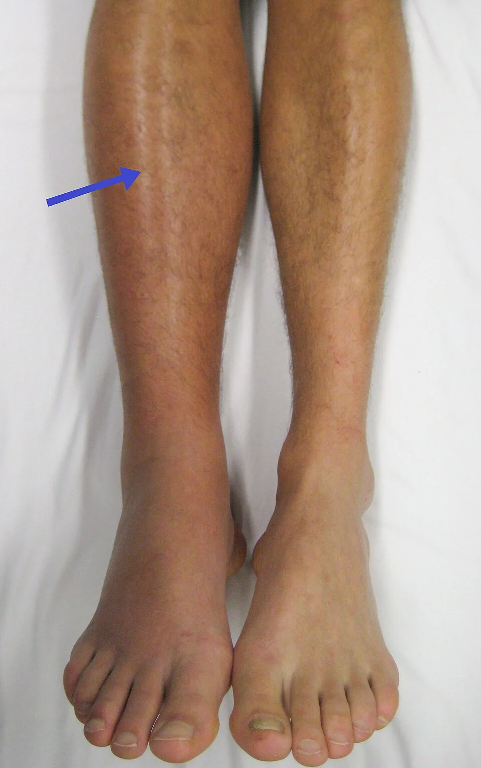
- Signs of DVT - a unilaterally swollen, warm, tender calf
- Signs of right heart strain - a raised JVP, a parasternal heave, a loud pulmonary second heart sound, and a right ventricular gallop
- Hypotension, cyanosis and cardiogenic shock in massive PE
Diagnosis
NICE recommends a two-step approach: assess clinical probability with the two-level Wells score, then investigate accordingly.1
| Feature | Points |
|---|---|
| Clinical signs and symptoms of DVT - leg swelling and pain on palpation of the deep veins | 3 |
| An alternative diagnosis is less likely than PE | 3 |
| Heart rate above 100 beats per minute | 1.5 |
| Immobilisation for more than 3 days, or surgery in the previous 4 weeks | 1.5 |
| Previous DVT or PE | 1.5 |
| Haemoptysis | 1 |
| Malignancy - treated within the last 6 months, or receiving palliative treatment | 1 |
| Score | Probability | Next step |
|---|---|---|
| More than 4 | PE likely | Immediate CTPA. If CTPA cannot be done at once, give interim therapeutic anticoagulation while waiting |
| 4 or less | PE unlikely | D-dimer. If positive, arrange a CTPA (with interim anticoagulation if delayed). If negative, PE is excluded - stop and look for another diagnosis |
D-dimer
- A fibrin degradation product, so it is highly sensitive but very poorly specific - it rises in infection, malignancy, pregnancy, trauma, surgery, inflammation, liver disease and simply with age
- Its value lies entirely in its negative predictive value: a negative result in a low-probability patient effectively excludes PE
- Never use it to rule PE in, and never send it in a patient in whom PE is likely - a positive result adds nothing and a negative one may mislead
- Age-adjusted D-dimer - in patients over 50, the threshold may be taken as age multiplied by 10 micrograms per litre, which reduces unnecessary imaging without missing significant events
Imaging
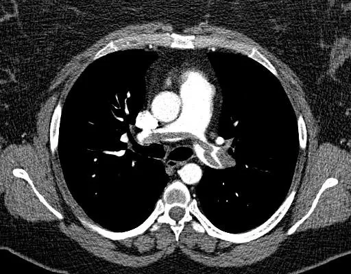
- CT pulmonary angiography (CTPA) - the investigation of choice. It is fast, widely available, and identifies alternative diagnoses such as pneumonia, dissection or malignancy when PE is absent.
- V/Q scan or V/Q SPECT - the alternative where CTPA is contraindicated: renal impairment, contrast allergy or a desire to limit breast radiation. It is less useful when the chest radiograph is abnormal or there is coexisting lung disease.
- Proximal leg vein ultrasound - if there are symptoms or signs of DVT. A positive scan in a patient with compatible symptoms allows treatment to begin without further imaging.
- Echocardiography - useful at the bedside in a haemodynamically unstable patient too unwell for CT. Right ventricular dilatation and strain support a presumptive diagnosis sufficient to justify thrombolysis.
Supporting investigations
- ECG - most commonly sinus tachycardia. Other findings include right axis deviation, right bundle branch block, and T wave inversion in V1 to V4 from right ventricular strain. The classical S1Q3T3 pattern - a deep S wave in lead I, a Q wave and inverted T wave in lead III - is well known but present in well under a fifth of cases.
- Chest X-ray - usually normal, which is itself a useful finding in a breathless hypoxic patient. Recognised but uncommon signs are a wedge-shaped peripheral opacity (Hampton's hump), regional oligaemia (Westermark sign) and a small pleural effusion.
- Arterial blood gas - typically type 1 respiratory failure with a low PaO2, low PaCO2 and respiratory alkalosis
- Troponin and BNP - raised levels indicate right ventricular strain and identify intermediate-risk patients needing closer monitoring
- FBC, U&Es, LFTs and clotting - baseline before anticoagulation, and renal function determines DOAC dosing
Management
Immediate assessment
Assess with an ABCDE approach, give oxygen to maintain saturations of 94-98%, gain intravenous access and obtain an ECG and blood tests. The first decision is whether the patient is haemodynamically stable.
Anticoagulation
Start anticoagulation as soon as PE is suspected, without waiting for imaging, unless there is a contraindication.1
| Situation | Anticoagulant |
|---|---|
| Most patients | A DOAC - apixaban or rivaroxaban - first line. Both begin with a higher loading dose for the first 7 or 21 days respectively. |
| DOAC unsuitable | LMWH for at least 5 days followed by dabigatran or edoxaban, or LMWH with a vitamin K antagonist for at least 5 days and until the INR is above 2 on two consecutive readings |
| Severe renal impairment (eGFR under 15) | LMWH, unfractionated heparin, or LMWH followed by a vitamin K antagonist |
| Antiphospholipid syndrome | LMWH with a vitamin K antagonist, target INR 2 to 3. DOACs are less effective in triple-positive antiphospholipid syndrome and should be avoided. |
| Active cancer | A DOAC, unless there is a gastrointestinal or genitourinary tumour or a high bleeding risk, when LMWH is preferred |
| Pregnancy | LMWH. DOACs and warfarin are contraindicated. |
| Haemodynamic instability | Unfractionated heparin infusion, because it is rapidly reversible if thrombolysis is needed |
Duration of anticoagulation
- Provoked PE - 3 months, provided the provoking factor has resolved
- Unprovoked PE - at least 3 months, then consider continuing indefinitely, weighing the recurrence risk against bleeding risk and patient preference. Many patients continue on a reduced-dose DOAC long term.
- Active cancer - 3 to 6 months, and continue while the cancer remains active or treatment continues
- Recurrent VTE, or antiphospholipid syndrome - long-term anticoagulation
- Review at 3 months in every patient, and document the decision and the discussion about bleeding risk
Other measures
- Inferior vena cava filter - only where anticoagulation is absolutely contraindicated, or where recurrent PE occurs despite adequate anticoagulation. Filters should be retrieved once anticoagulation can be started.
- Outpatient management - suitable for haemodynamically stable low-risk patients, identified with the PESI or simplified PESI score, provided there is adequate support and follow-up
- Investigating for underlying cancer - NICE no longer recommends extensive routine testing after an unprovoked VTE. Take a full history, examine the patient, and review the blood results and chest radiograph; investigate further only where something suggests a specific diagnosis.
- Thrombophilia testing - consider antiphospholipid antibody testing in unprovoked VTE if stopping anticoagulation is planned, and hereditary thrombophilia testing in unprovoked VTE with a first-degree relative who has had a VTE. Do not test while the patient is anticoagulated or in the acute phase, as results are unreliable.
- Stop the combined oral contraceptive or HRT and provide alternative contraception advice
- VTE prophylaxis for future admissions and surgery, and an alert on the record
Complications
- Death - untreated PE has a mortality of around 30%, falling to 2 to 8% with treatment
- Cardiac arrest - PE causes pulseless electrical activity and is one of the reversible causes in the ALS algorithm
- Right ventricular failure and obstructive shock
- Chronic thromboembolic pulmonary hypertension (CTEPH) - developing in around 2 to 4% of patients, presenting months to years later with progressive exertional breathlessness. It is potentially curable by pulmonary endarterectomy, so persistent breathlessness after a PE should always prompt investigation rather than reassurance.
- Recurrent VTE - highest in the first months and in unprovoked events
- Pulmonary infarction, with pleural effusion and infarct-related pain
- Bleeding from anticoagulation - including major gastrointestinal and intracranial haemorrhage
- Post-thrombotic syndrome - in those with an associated DVT, causing chronic leg pain, swelling and ulceration
- Heparin-induced thrombocytopenia - a fall in platelets 5 to 10 days after starting heparin, paradoxically causing thrombosis
Red flags
Prognosis
Prognosis depends principally on the haemodynamic consequences at presentation. Untreated PE carries a mortality of around 30%; treated, it falls to between 2 and 8%. Patients with massive PE and shock have a mortality above 25% even with treatment, while low-risk patients with a normal blood pressure, normal right ventricle and normal troponin have a mortality below 1% and can often be managed entirely as outpatients.
Most survivors recover fully, although breathlessness on exertion for weeks to months afterwards is common and expected. Persistent breathlessness at 3 to 6 months should never be dismissed - it may indicate chronic thromboembolic pulmonary hypertension, which is one of the few causes of pulmonary hypertension that can be cured surgically.
Perhaps the most important point is preventive. A large proportion of hospital-acquired VTE is avoidable, and risk assessment on admission with appropriate mechanical or pharmacological prophylaxis prevents more deaths than any advance in treatment. Assessing every admitted patient for VTE risk is a national requirement precisely because it works.
References
- NICE NG158. Venous thromboembolic diseases: diagnosis, management and thrombophilia testing. 2020, updated 2023. Available here
- NICE Clinical Knowledge Summaries. Pulmonary embolism. Available here
- Konstantinides SV, Meyer G, Becattini C et al. 2019 ESC Guidelines for the diagnosis and management of acute pulmonary embolism. European Heart Journal. 2020. Available here
- NICE NG89. Venous thromboembolism in over 16s: reducing the risk of hospital-acquired deep vein thrombosis or pulmonary embolism. 2018, updated 2019. Available here
- Royal College of Obstetricians and Gynaecologists. Green-top Guideline No. 37b: Thromboembolic disease in pregnancy and the puerperium. Available here
- Resuscitation Council UK. Adult advanced life support guidelines: reversible causes. Available here
- Wells PS, Anderson DR, Rodger M et al. Derivation of a simple clinical model to categorize patients probability of pulmonary embolism. Thrombosis and Haemostasis. 2000. Available here
- BNF. Apixaban, rivaroxaban and alteplase - indications and dosing. Available here
- Rvahudson, CC BY-SA 4.0, via Wikimedia Commons. Available here
- James Heilman MD, CC BY-SA 3.0, via Wikimedia Commons. Available here
This article is written for revision and education. It is not clinical guidance and must not be used to make decisions about the care of a patient. Always check current NICE guidance and local protocols.