
Cardiac Arrest: Advanced Life Support
Key points
- Cardiac arrest: the sudden cessation of effective cardiac output. Diagnosed clinically by unresponsiveness with absent or abnormal breathing - do not delay CPR to feel for a pulse.
- Shockable rhythms: ventricular fibrillation and pulseless ventricular tachycardia. Defibrillate immediately and resume compressions without a rhythm check.
- Non-shockable rhythms: pulseless electrical activity and asystole. Give adrenaline immediately and concentrate on finding a reversible cause.
- Adrenaline: 1 mg IV every 3-5 minutes. Given immediately in non-shockable rhythms, and after the third shock in shockable rhythms.
- Amiodarone: 300 mg IV after the third shock, with a further 150 mg after the fifth. It has no role in non-shockable arrest.
- High-quality CPR: compressions at 100-120 per minute to a depth of 5-6 cm, with full recoil, minimal interruption and a chest compression fraction above 60%.
- Reversible causes: the four Hs - hypoxia, hypovolaemia, hyper/hypokalaemia and metabolic, hypothermia - and the four Ts - thrombosis, tamponade, tension pneumothorax, toxins.
- Post-ROSC care: targeted oxygenation of 94-98%, normocapnia, a 12-lead ECG, urgent angiography if a cardiac cause is likely, temperature control and intensive care admission.
Introduction
Cardiac arrest is the abrupt loss of effective cardiac output, causing immediate cessation of cerebral and coronary perfusion. Without intervention, consciousness is lost within seconds and irreversible neurological injury begins within minutes.
Around 30,000 out-of-hospital cardiac arrests are attended by the emergency services in the UK each year, with overall survival to hospital discharge of roughly one in ten. In-hospital arrest is somewhat more survivable, in part because it is witnessed and treated earlier, though the underlying condition is often more advanced.
Three interventions determine outcome more than anything that happens later: early recognition, early high-quality CPR, and early defibrillation. Survival from a shockable rhythm falls by approximately 10% for every minute that defibrillation is delayed, which is why bystander CPR and public access defibrillators matter so much.
Recognition
Cardiac arrest is a clinical diagnosis. In an unresponsive patient who is not breathing normally, start CPR - do not delay for a prolonged pulse check.
- Unresponsive to voice and to a firm shake of the shoulders
- Absent or abnormal breathing - agonal gasping is present in up to 40% of arrests in the first minutes and is frequently mistaken for breathing by both bystanders and clinicians. Slow, laboured, noisy gasps are a sign of cardiac arrest, not of life.
- Absent central pulse - carotid or femoral, checked for no more than 10 seconds, and only by someone trained to do so. Pulse checks are unreliable even among experienced clinicians.
- Sudden collapse, sometimes preceded by seizure-like activity from cerebral hypoperfusion - another common source of misdiagnosis
In hospital, most arrests are preceded by hours of physiological deterioration. Recognising and acting on a rising early warning score prevents more deaths than any resuscitation skill, and this is the reasoning behind track-and-trigger systems and critical care outreach teams.
Basic life support
- Ensure safety for yourself, the patient and bystanders
- Check responsiveness - shake and shout
- Open the airway with a head tilt and chin lift, and look, listen and feel for normal breathing for up to 10 seconds
- Call for help - dial 2222 in hospital or 999 in the community, and send for a defibrillator
- Start chest compressions - 30 compressions, then 2 rescue breaths, continuing at 30:2
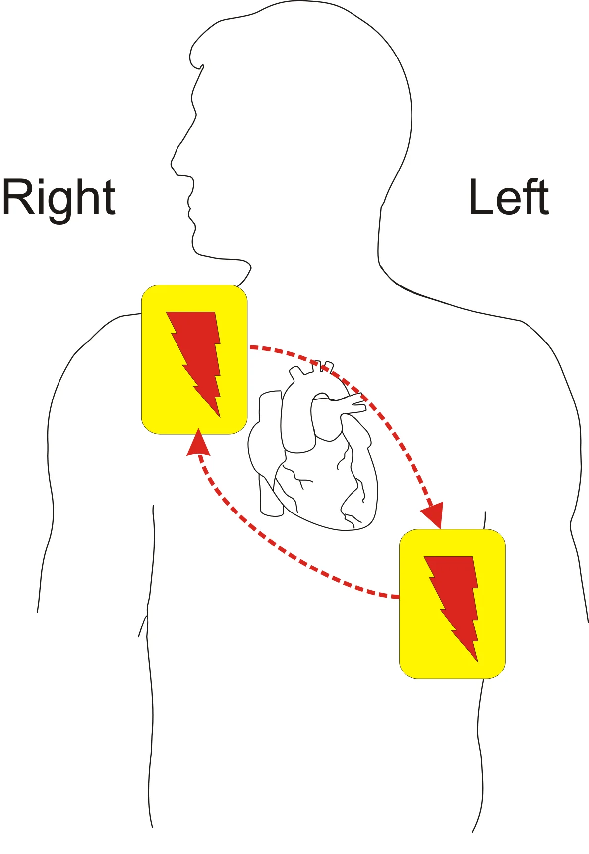
- Attach a defibrillator or AED as soon as it arrives and follow its prompts
The advanced life support algorithm
Once the rhythm is known, the algorithm divides into two branches. The critical difference is not only whether a shock is given, but when adrenaline is given.1
Shockable rhythms: VF and pulseless VT
- Defibrillate immediately - 150 J biphasic for the first shock (or per the manufacturer's guidance), escalating with subsequent shocks
- Resume chest compressions immediately for 2 minutes without checking the rhythm or the pulse. The heart rarely produces an effective output straight after a shock, and pausing wastes perfusion time.
- Reassess the rhythm after 2 minutes; if still shockable, deliver a second shock and resume compressions
- After the third shock, give adrenaline 1 mg IV and amiodarone 300 mg IV, then resume compressions
- Continue in 2-minute cycles, giving adrenaline 1 mg every 3-5 minutes (in practice, every other cycle)
- After the fifth shock, give a further amiodarone 150 mg IV
- Throughout, seek and treat reversible causes
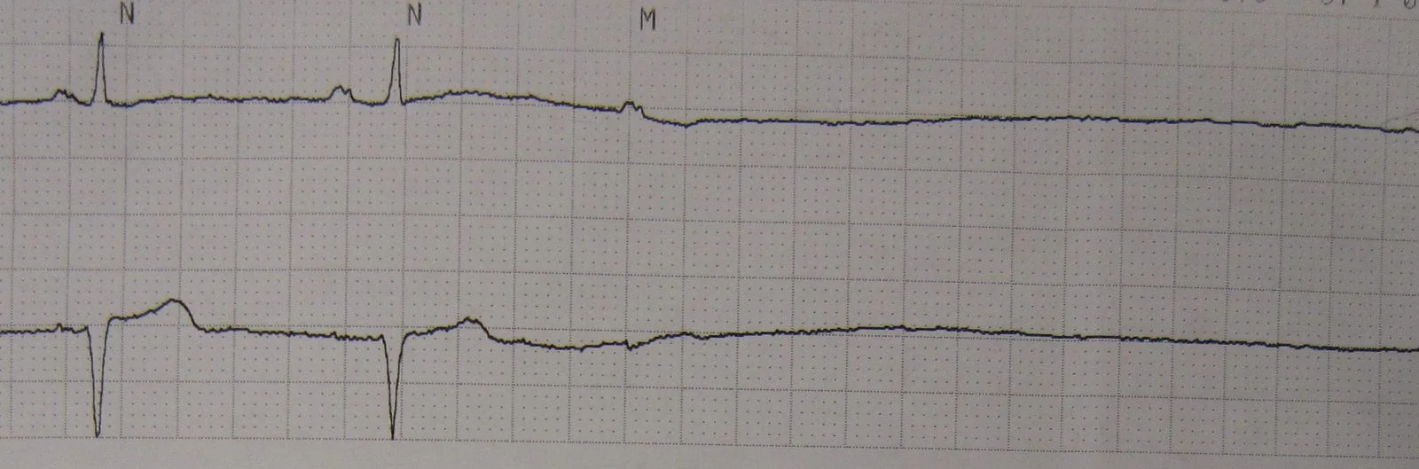
Non-shockable rhythms: PEA and asystole
- Give adrenaline 1 mg IV as soon as possible - unlike in shockable rhythms, it is not delayed
- Continue CPR in 2-minute cycles, reassessing the rhythm at the end of each
- Repeat adrenaline 1 mg every 3-5 minutes
- Amiodarone has no role in non-shockable arrest
- Concentrate on reversible causes - the yield here is far higher than in shockable rhythms, and identifying one is usually the only route to survival
- If the rhythm changes to a shockable one, switch to that branch of the algorithm
Reversible causes
Considering these is a mandatory part of every arrest, and should be spoken aloud during the resuscitation. They matter most in PEA and asystole, where treating the cause is often the only path to survival.
| Cause | Clues | Treatment |
|---|---|---|
| Hypoxia | Airway obstruction, respiratory arrest preceding collapse, cyanosis | Secure the airway, ventilate with high-concentration oxygen, confirm bilateral chest rise |
| Hypovolaemia | Trauma, gastrointestinal or obstetric haemorrhage, ruptured aortic aneurysm, sepsis, anaphylaxis | Rapid volume replacement, blood products, and surgical control of bleeding |
| Hyperkalaemia, hypokalaemia and metabolic | Renal failure, dialysis patient, crush injury, tall tented T waves before arrest | Calcium chloride 10 ml of 10% for hyperkalaemia, with insulin-dextrose; potassium and magnesium replacement for hypokalaemia |
| Hypothermia | Immersion, environmental exposure, older person found on the floor, J waves | Active rewarming; withhold drugs below 30 degrees and limit shocks to three until rewarmed. Do not stop until the patient is warm and dead. |
| Thrombosis | Coronary - known ischaemic heart disease, preceding chest pain. Pulmonary - recent surgery, immobility, malignancy, preceding breathlessness | Coronary: consider PCI during or immediately after resuscitation. Pulmonary: consider thrombolysis, and if given, continue CPR for 60-90 minutes |
| Tamponade | Recent cardiac surgery or intervention, penetrating chest trauma, malignancy, distended neck veins | Urgent pericardiocentesis or resuscitative thoracotomy in trauma |
| Tension pneumothorax | Trauma, central line insertion, asthma, mechanical ventilation, absent breath sounds, tracheal deviation | Immediate needle or finger thoracostomy, followed by a chest drain |
| Toxins | Overdose, occupational exposure, drug chart review | Specific antidotes - naloxone, digoxin-specific antibody fragments, intravenous lipid emulsion for local anaesthetic toxicity, glucagon for beta-blocker overdose |
Point-of-care ultrasound by a competent operator can identify tamponade, right heart strain suggesting massive pulmonary embolism, hypovolaemia and cardiac standstill. It must be used during the 10-second rhythm check without prolonging the pause in compressions.
During CPR
- Ensure high-quality compressions with minimal interruption - this outranks every other consideration
- Airway - a bag-valve-mask with an oropharyngeal airway is acceptable. A supraglottic airway or tracheal tube allows continuous compressions. Tracheal intubation should only be attempted by someone competent, and only with a pause of under 5 seconds.
- Oxygen - give the highest concentration available during the arrest itself
- Waveform capnography - continuous end-tidal CO2 monitoring confirms tracheal tube placement, gives real-time feedback on compression quality, and a sudden rise in ETCO2 is often the first indicator of return of spontaneous circulation. A persistently low value (under 1.3 kPa) after 20 minutes of ALS is a poor prognostic sign, though it must never be used alone to stop.
- Vascular access - intravenous first; if not obtained quickly, use the intraosseous route. The tracheal route is no longer recommended.
- Consider extracorporeal CPR (eCPR) in selected patients with a reversible cause in centres where it is available
Post-resuscitation care
Return of spontaneous circulation is the beginning of treatment, not the end of it. Post-cardiac arrest syndrome comprises hypoxic brain injury, myocardial dysfunction, a systemic ischaemia-reperfusion response resembling sepsis, and the persisting effects of the precipitating illness.
- Airway and breathing - titrate oxygen to saturations of 94-98%; hyperoxia after arrest worsens neurological injury. Aim for normocapnia (PaCO2 4.5-6.0 kPa), since hypocapnia causes cerebral vasoconstriction. Use lung-protective ventilation.
- Circulation - a 12-lead ECG immediately. Arrange urgent coronary angiography if there is ST elevation, and consider it in any arrest with a likely cardiac cause even without ST elevation. Target a mean arterial pressure sufficient for adequate urine output and lactate clearance.
- Disability - control seizures, maintain normoglycaemia, and avoid fever aggressively. Targeted temperature management with active prevention of fever (keeping temperature below 37.7 degrees) is recommended for at least 72 hours.
- Exposure and everything else - identify and treat the precipitating cause, correct electrolytes, insert a urinary catheter, and admit to intensive care
- Neuroprognostication - should be deferred for at least 72 hours after normothermia is restored and sedation has cleared, and must use a multimodal approach combining clinical examination, EEG, somatosensory evoked potentials, neuron-specific enolase and imaging. No single test is sufficient to withdraw treatment.
When not to start, and when to stop
These decisions are difficult, frequently examined in ethics stations, and best approached through a small number of clear principles.
Withholding resuscitation
- A valid DNACPR decision is in place. This records a clinical decision that CPR would not be successful or would not be in the patient's best interests. It applies only to CPR and must never restrict any other treatment.
- A valid and applicable advance decision to refuse treatment covering CPR
- Injuries incompatible with life - decapitation, hemicorporectomy, rigor mortis, hypostasis, or incineration
- CPR would be futile - where the patient is dying of an irreversible condition and cardiac arrest represents the expected end of that process
Stopping resuscitation
The decision rests with the team leader and is based on the overall clinical picture rather than any single factor. Considerations include the presenting rhythm and any change in it, the duration of the arrest, whether it was witnessed and whether bystander CPR was given, the response to treatment, the identification and correction of reversible causes, end-tidal CO2 values, and the patient's condition before the arrest.
Red flags
Prognosis
Overall survival to hospital discharge after out-of-hospital cardiac arrest in the UK is approximately 8-10%, though this conceals enormous variation. Survival exceeds 50% in a witnessed arrest with a shockable rhythm and immediate bystander CPR and defibrillation, and approaches zero in an unwitnessed asystolic arrest with no bystander intervention.
The determinants of outcome, in approximate order of importance, are: whether the arrest was witnessed, whether bystander CPR was given, the initial rhythm (shockable rhythms carry a far better prognosis), the time to defibrillation, and the presence of a reversible cause. Every one of the first four is influenced by public education and by the availability of defibrillators, which is why community resuscitation training is a genuine public health intervention.
In-hospital cardiac arrest has a survival to discharge of roughly 15-20%, better where arrest occurs in a monitored area and worse where it follows a prolonged deterioration on a general ward. Much of that gap is preventable through earlier recognition of the deteriorating patient.
Among survivors, neurological outcome dominates quality of life. The majority of those who survive to discharge have a good or moderate neurological recovery, but hypoxic brain injury is the commonest cause of death after successful initial resuscitation. This is why post-resuscitation care - oxygen targets, temperature control, avoidance of fever, and careful multimodal prognostication - is as much a part of resuscitation as the compressions themselves.
References
- Resuscitation Council UK. Adult advanced life support guidelines. 2021. Available here
- Resuscitation Council UK. Adult basic life support guidelines. 2021. Available here
- Resuscitation Council UK. Post-resuscitation care guidelines. 2021. Available here
- Resuscitation Council UK. Decisions relating to cardiopulmonary resuscitation (joint statement with the BMA and RCN). Available here
- Resuscitation Council UK. Special circumstances guidelines. 2021. Available here
- Nolan JP, Sandroni C, Bottiger BW et al. European Resuscitation Council and European Society of Intensive Care Medicine guidelines 2021: post-resuscitation care. Intensive Care Medicine. 2021. Available here
- British Heart Foundation. Out-of-hospital cardiac arrest statistics. Available here
- NICE CG50. Acutely ill adults in hospital: recognising and responding to deterioration. 2007. Available here
This article is written for revision and education. It is not clinical guidance and must not be used to make decisions about the care of a patient. Always check current NICE guidance and local protocols.