
Pacemakers and Implantable Cardioverter Defibrillators
Key points
- Pacemaker: a device that delivers an electrical impulse to maintain an adequate heart rate. Its principal indication is symptomatic or high-risk bradycardia.
- ICD: an implantable cardioverter defibrillator recognises and terminates ventricular arrhythmia by anti-tachycardia pacing or a shock. It prevents sudden arrhythmic death but does not treat heart failure.
- The pacing code: letter one is the chamber paced, letter two the chamber sensed, letter three the response to sensing, and letter four rate responsiveness. VVI, DDD and AAI are the common modes.
- Paced ECG: a pacing spike before the P wave indicates atrial pacing; a spike before a broad QRS indicates ventricular pacing, and right ventricular pacing normally produces a left bundle branch block pattern.
- CRT: cardiac resynchronisation therapy paces both ventricles to correct dyssynchrony. Consider it in heart failure with an ejection fraction of 35% or below and a QRS of 130 ms or more, particularly with LBBB.
- Malfunction: failure to capture (a spike with no complex), failure to output (no spike when one is due), failure to sense (inappropriate spikes) and oversensing (inappropriately withheld pacing).
- Complications: early - pneumothorax, haematoma, lead displacement, tamponade. Late - infection, lead fracture, venous obstruction and pacemaker syndrome.
- Practical advice: no MRI unless the system is MR-conditional, avoid diathermy where possible, keep mobile phones away from the device, and check DVLA rules, which differ between pacemakers and ICDs.
Introduction
Cardiac implantable electronic devices fall into three broad groups, and it is worth being clear about which problem each solves, because they are frequently confused.
- Pacemakers treat a rate that is too slow. They deliver an impulse when the heart fails to generate one.
- Implantable cardioverter defibrillators (ICDs) treat rhythms that are too fast and life-threatening. They detect ventricular tachycardia and fibrillation and terminate them. All ICDs can also pace for bradycardia.
- Cardiac resynchronisation therapy (CRT) treats mechanical dyssynchrony in heart failure. By pacing both ventricles it coordinates their contraction, improving symptoms and survival.
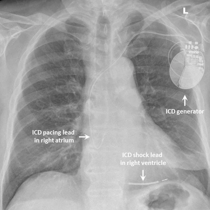
The categories combine: a CRT-D is a resynchronisation device with defibrillator capability, and a CRT-P provides resynchronisation pacing only. Over 50,000 devices are implanted annually in the UK, so students will meet these patients regularly, and being able to identify a device on a chest X-ray and interpret a paced ECG is a realistic examination expectation.
How a pacemaker works
A conventional transvenous system consists of a pulse generator - a titanium case containing a lithium battery and circuitry, implanted subcutaneously below the left clavicle - connected to one or more leads passed through the subclavian or cephalic vein into the right heart.
- Single-chamber - one lead, in either the right atrium or the right ventricle
- Dual-chamber - two leads, one in the right atrium and one in the right ventricle, allowing atrioventricular synchrony to be preserved
- Biventricular (CRT) - three leads, adding one passed through the coronary sinus to pace the lateral wall of the left ventricle
- Leadless pacemakers - a self-contained capsule implanted directly into the right ventricle via the femoral vein, avoiding leads and a subcutaneous pocket, though currently limited to single-chamber ventricular pacing
- Subcutaneous ICD (S-ICD) - electrodes placed outside the thorax with no intravascular components, useful where venous access is difficult or infection risk is high, but unable to provide anti-tachycardia or bradycardia pacing
The pacing code
Devices are described by a standard three- to five-letter code. The first three letters are the ones to know.
| Position | Meaning | Options |
|---|---|---|
| I | Chamber paced | A (atrium), V (ventricle), D (dual), O (none) |
| II | Chamber sensed | A, V, D, O |
| III | Response to a sensed event | I (inhibited), T (triggered), D (dual - atrial sensing triggers ventricular pacing, and ventricular sensing inhibits it), O (none) |
| IV | Rate modulation | R (rate responsive - the device increases rate with activity), O |
| V | Multisite pacing | A, V, D, O |
| Mode | Meaning | Typical indication |
|---|---|---|
| AAI | Paces and senses the atrium, inhibited by sensed atrial activity | Sinus node disease with intact AV conduction. Rarely used alone, as AV block may develop. |
| VVI | Paces and senses the ventricle, inhibited by sensed ventricular activity | Permanent atrial fibrillation with a slow ventricular response - there is no organised atrial activity worth sensing |
| DDD | Paces and senses both chambers, with dual response | AV block in a patient in sinus rhythm - preserves atrioventricular synchrony |
| DDDR | As DDD with rate response | AV block with chronotropic incompetence |
| VOO / DOO | Asynchronous pacing, no sensing | Temporary mode used during surgical diathermy, when a magnet is applied over the device |
Indications
Permanent pacing
- Complete (third degree) heart block - almost always, unless a reversible cause is identified
- Mobitz type II second degree AV block - even if asymptomatic, because progression is unpredictable
- Symptomatic sick sinus syndrome, including chronotropic incompetence
- Tachy-brady syndrome - to allow rate-controlling drugs to be given safely
- Symptomatic bradycardia from a cause that cannot be removed, for example essential beta-blockade after myocardial infarction
- Persistent AV block after anterior myocardial infarction
- Bifascicular or trifascicular block with syncope, or with documented intermittent complete heart block
- Recurrent syncope from carotid sinus hypersensitivity with a cardioinhibitory response
- After AV node ablation for refractory atrial fibrillation - the patient becomes pacemaker dependent
Implantable cardioverter defibrillators
| Category | Indication |
|---|---|
| Secondary prevention | Survived cardiac arrest due to VT or VF; sustained VT with syncope or haemodynamic compromise; sustained VT with an ejection fraction of 35% or below |
| Primary prevention | Ischaemic or non-ischaemic cardiomyopathy with an ejection fraction of 35% or below and NYHA class I-III symptoms, after at least 3 months of optimal medical therapy |
| Inherited conditions | High-risk hypertrophic cardiomyopathy, long QT syndrome, Brugada syndrome, ARVC and CPVT, guided by validated risk scores |
An ICD is not indicated where an arrhythmia had a fully reversible cause, such as ventricular fibrillation within 48 hours of an acute infarct that has been successfully revascularised, or torsades from a drug that has been withdrawn. Nor is it appropriate where life expectancy from other causes is under a year, since it would prevent a sudden death only to permit a slower one.1
Cardiac resynchronisation therapy
In heart failure with left bundle branch block, the left ventricular free wall contracts substantially after the septum. This dyssynchrony wastes contractile work and worsens mitral regurgitation. Biventricular pacing restores coordination.
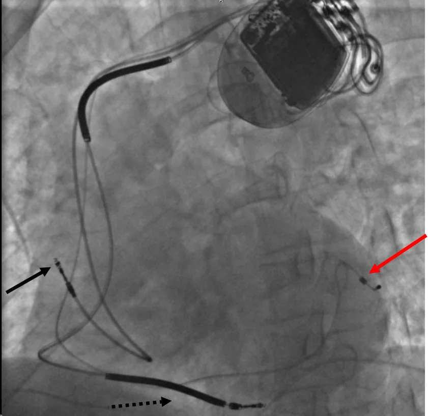
Consider CRT in patients with symptomatic heart failure, an ejection fraction of 35% or below, and a QRS duration of 130 ms or more despite optimal medical therapy. The benefit is greatest with left bundle branch block morphology and a QRS above 150 ms, and is uncertain or absent where the QRS is narrow. Whether a defibrillator is added (CRT-D rather than CRT-P) depends on the arrhythmic risk and the aetiology.1,2
The paced ECG
Pacing spikes are narrow, sharp vertical deflections, often only a millimetre or two tall and easily missed on a poor-quality trace.
| Finding | Interpretation |
|---|---|
| Spike before a P wave | Atrial pacing |
| Spike before a broad QRS | Ventricular pacing |
| Spikes before both P wave and QRS | Dual-chamber pacing (AV sequential) |
| Spike followed by a QRS with LBBB morphology | Normal right ventricular apical pacing - the right ventricle is activated first |
| Spike followed by a QRS with RBBB morphology | Left ventricular pacing, as in CRT, or a lead in an unexpected position |
| Intermittent spikes with intrinsic beats in between | Normal demand pacing - the device is inhibited when the heart's own rate is adequate |
| No spikes at all | Normal if the intrinsic rate is above the programmed lower rate; abnormal if the patient is bradycardic |
Device malfunction
| Problem | ECG appearance | Common causes |
|---|---|---|
| Failure to capture | A pacing spike is present but is not followed by a P wave or QRS | Lead displacement or fracture, myocardial fibrosis at the lead tip, electrolyte disturbance (especially hyperkalaemia), antiarrhythmic drugs raising the capture threshold, battery depletion |
| Failure to output | No pacing spike when one should be delivered, with resulting bradycardia or asystole | Battery depletion, lead fracture, generator failure, oversensing causing inappropriate inhibition |
| Undersensing | Pacing spikes appear inappropriately, including within or after intrinsic complexes | Lead displacement, poor lead position, an intrinsic signal too small to detect, programming error |
| Oversensing | Pacing is inappropriately withheld | Detection of myopotentials (diaphragmatic or pectoral muscle activity), T wave oversensing, electromagnetic interference, lead insulation failure |
Any suspected malfunction requires device interrogation by the cardiac physiology service. In a patient who is pacemaker dependent, failure to output is a medical emergency requiring external pacing while the device is assessed.
Complications
Early (procedural)
- Pneumothorax - from subclavian puncture; a chest X-ray is performed routinely after implantation
- Pocket haematoma - commoner in anticoagulated patients
- Lead displacement - most likely in the first six weeks, before fibrosis anchors the lead. Patients are advised to avoid raising the arm above shoulder height on the implant side during this period.
- Cardiac perforation and tamponade - rare but life-threatening
- Arrhythmia during lead placement
- Diaphragmatic or pectoral stimulation - causing hiccups or twitching, corrected by reprogramming
Late
- Infection - of the pocket or, more seriously, of the leads causing device-related infective endocarditis. This almost always requires complete extraction of the entire system, since antibiotics alone rarely clear infection on a foreign body. Fever in a patient with a device should always prompt consideration of this.
- Erosion of the generator through the skin
- Lead fracture or insulation failure
- Venous obstruction or thrombosis of the subclavian vein
- Tricuspid regurgitation - from a lead crossing the valve
- Pacemaker syndrome - loss of AV synchrony in single-chamber ventricular pacing
- Pacing-induced cardiomyopathy - a high burden of right ventricular pacing creates dyssynchrony resembling LBBB and can reduce ejection fraction over years. This is why modern devices are programmed to minimise unnecessary ventricular pacing, and why conduction system pacing is increasingly used.
- Battery depletion - generators typically last 6-15 years and are replaced in a straightforward procedure that leaves the leads in place
- Psychological effects - anxiety and depression are common, particularly after ICD shocks
Practical advice for patients
| Topic | Advice |
|---|---|
| MRI scanning | Only if the system is MR-conditional and the scan is done under a specific protocol with device reprogramming. Always check the device card before requesting an MRI. |
| Surgery and diathermy | Inform the surgical team. Use bipolar diathermy where possible, position the return plate away from the device, and arrange perioperative device management. A magnet may be used to switch a pacemaker to asynchronous mode or to suspend ICD therapies. |
| Airport security | Walk through steadily rather than lingering; carry the device identification card and request a hand search if concerned |
| Mobile phones | Keep at least 15 cm from the device and use the opposite ear. Do not carry the phone in a breast pocket over the generator. |
| Induction hobs and electrical equipment | Keep at least 60 cm away from induction hobs. Domestic appliances and microwaves are safe. |
| Arc welding and industrial equipment | Strong electromagnetic fields should be avoided; occupational assessment may be needed |
| Sport and activity | Normal activity is encouraged after the initial healing period. Avoid contact sports that risk a direct blow to the generator. |
| Follow-up | Regular device checks, in person or by remote monitoring, to assess battery life, lead integrity and stored arrhythmia data |
| End of life | Deactivating the tachyarrhythmia function of an ICD should be discussed proactively in advancing terminal illness, so that a dying patient is not shocked repeatedly. Pacing function is usually left on, as switching it off does not shorten life in most patients but may cause symptoms. |
Red flags
Prognosis
Permanent pacing for complete heart block restores life expectancy close to that of the general population and produces a marked improvement in symptoms and quality of life. It is among the most reliably effective interventions in cardiology.
ICDs reduce mortality substantially in appropriately selected patients, both for secondary prevention after a survived arrhythmic arrest and for primary prevention in those with significantly impaired ventricular function. The benefit depends heavily on selection: patients who are likely to die of progressive heart failure or non-cardiac disease derive little advantage, since the device converts a sudden death into a slower one.
Cardiac resynchronisation therapy improves symptoms, ejection fraction, hospitalisation rates and survival in patients with heart failure and a broad QRS, with the greatest benefit in those with left bundle branch block and a QRS above 150 ms. Around a third of patients are non-responders, which is why patient selection and careful device optimisation matter.
Generators require replacement every 6-15 years depending on the device type and pacing burden, and each replacement carries a small additional infection risk. Long-term outcomes are otherwise excellent, and the great majority of patients live entirely normal lives with their device.
References
- NICE TA314. Implantable cardioverter defibrillators and cardiac resynchronisation therapy for arrhythmias and heart failure. 2014. Available here
- Glikson M, Nielsen JC, Kronborg MB et al. 2021 ESC Guidelines on cardiac pacing and cardiac resynchronization therapy. European Heart Journal. 2021. Available here
- DVLA. Assessing fitness to drive: a guide for medical professionals. Available here
- NICE NG106. Chronic heart failure in adults: diagnosis and management. 2018. Available here
- Resuscitation Council UK. Adult advanced life support guidelines. Available here
- Zeppenfeld K, Tfelt-Hansen J, de Riva M et al. 2022 ESC Guidelines for the management of patients with ventricular arrhythmias and the prevention of sudden cardiac death. European Heart Journal. 2022. Available here
- British Heart Foundation. Pacemakers. Available here
- MHRA. Guidance on the safe use of cardiac implantable electronic devices. Available here
This article is written for revision and education. It is not clinical guidance and must not be used to make decisions about the care of a patient. Always check current NICE guidance and local protocols.