
Acute Coronary Syndrome: Recognition and Emergency Management
Key points
- Acute coronary syndrome: a spectrum of unstable angina, NSTEMI and STEMI, all caused by acute plaque rupture or erosion with thrombus formation.
- The distinction: STEMI has ST elevation on the ECG; NSTEMI does not but has a raised troponin; unstable angina has neither.
- Presentation: central crushing chest pain lasting more than 15 minutes, at rest, with sweating, nausea and breathlessness. It can be silent in diabetes, older people and women.
- First action: a 12-lead ECG within 10 minutes of first medical contact.
- STEMI: primary PCI if it can be delivered within 120 minutes of the time fibrinolysis could have been given, and within 12 hours of symptom onset.
- NSTEMI: risk stratify with the GRACE score; angiography within 72 hours for anyone at intermediate or higher risk.
- Oxygen: only if saturations are below 94%. Routine oxygen in a normoxic patient causes harm.
- Secondary prevention: dual antiplatelet therapy, a beta-blocker, an ACE inhibitor, a high-intensity statin and cardiac rehabilitation.
Introduction
Acute coronary syndrome (ACS) is the umbrella term for the acute presentations of coronary artery disease. It covers three diagnoses that share one mechanism: unstable angina, non-ST-elevation myocardial infarction (NSTEMI) and ST-elevation myocardial infarction (STEMI).1
They are separated by two pieces of information available within the first hour: the ECG, and the troponin. That separation matters because it determines how quickly the patient needs to reach a catheter laboratory.
| ECG | Troponin | Pathology | |
|---|---|---|---|
| Unstable angina | No ST elevation | Normal | Partial occlusion, no myocardial necrosis |
| NSTEMI | No ST elevation (may show ST depression or T inversion) | Raised | Partial occlusion with subendocardial necrosis |
| STEMI | ST elevation or new left bundle branch block | Raised | Complete occlusion with transmural necrosis |
ACS remains one of the commonest causes of death in the UK, though mortality has fallen substantially with primary PCI and modern secondary prevention.2 It is also the presentation where a student's actions in the first ten minutes matter most, which is why it is examined so heavily.
Aetiology
The overwhelming majority of ACS results from atherosclerotic plaque rupture or erosion. The exposed lipid core and subendothelial collagen are highly thrombogenic, triggering platelet adhesion, activation and aggregation, and then the coagulation cascade.
What happens next depends on whether the resulting thrombus occludes the vessel completely:
- Complete occlusion causes full-thickness ischaemia of the territory supplied. This produces ST elevation and, untreated, transmural infarction - a STEMI.
- Partial occlusion, or complete occlusion with good collateral supply, causes subendocardial ischaemia. The subendocardium is the most vulnerable layer because it is furthest from the epicardial vessels and subject to the highest wall stress. This produces an NSTEMI, or unstable angina if no myocyte necrosis occurs.
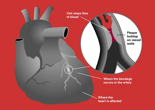
The plaque responsible is frequently not the most stenotic one. Plaques with a large lipid core, a thin fibrous cap and dense macrophage infiltration are the most likely to rupture, and these may cause only modest narrowing beforehand. This is why a patient can have a normal exercise tolerance the week before a large infarct.
Less common causes
- Coronary artery dissection - spontaneous coronary artery dissection (SCAD) is an important cause in younger women and in the peripartum period, and is easily missed
- Coronary embolism - from infective endocarditis, a prosthetic valve, or atrial fibrillation
- Coronary spasm - including cocaine-induced, where beta-blockers are relatively contraindicated
- Coronary vasculitis - Kawasaki disease, Takayasu arteritis
- Type 2 myocardial infarction - supply-demand mismatch without an acute plaque event, for example in severe sepsis, anaemia or tachyarrhythmia. Managing the precipitant matters more than antiplatelet therapy here.
Risk factors
The risk factors are those for atherosclerosis: smoking, hypertension, diabetes mellitus, dyslipidaemia, obesity, physical inactivity, increasing age, male sex, South Asian ethnicity, family history of premature coronary disease and chronic kidney disease.
Cocaine and amphetamine use deserves a specific question in any young patient with chest pain. Both cause coronary vasospasm and accelerate atherosclerosis, and cocaine-associated chest pain is managed differently: benzodiazepines and nitrates first, with beta-blockers avoided because of the risk of unopposed alpha-adrenergic vasoconstriction.
Clinical features
The classical presentation is severe central chest pain, described as crushing, heavy or like a band, lasting more than 15 minutes, occurring at rest, and not relieved by GTN. It may radiate to the jaw, neck, shoulders or either arm.
Associated autonomic features are important and often more discriminating than the pain itself:
- Sweating and clamminess
- Nausea and vomiting
- Breathlessness
- Palpitations or syncope
- A sense of impending doom (angor animi)
A useful discriminator against stable angina is the pattern: ACS pain occurs at rest, lasts longer, is more severe, and does not settle with GTN. A crescendo pattern of angina - more frequent, less exertion needed, longer episodes - is unstable angina even before any troponin is back.
Clinical examination
Examination is frequently unremarkable and a normal examination never excludes ACS. Its purpose is to identify haemodynamic compromise, complications and alternative diagnoses.
- General: pallor, sweating, distress, vomiting
- Haemodynamics: heart rate, blood pressure in both arms, and signs of shock. Hypotension in an inferior infarct suggests right ventricular involvement.
- Cardiovascular: JVP, a third or fourth heart sound, and a new murmur - a pansystolic murmur may mean acute mitral regurgitation from papillary muscle rupture or a ventricular septal defect
- Respiratory: basal crackles indicating pulmonary oedema; use the Killip class to grade it
- Peripheral: pulses in all four limbs, and any radio-radial or radio-femoral delay suggesting dissection
Differential diagnosis
- Aortic dissection - tearing pain to the back, pulse or blood pressure differential
- Pulmonary embolism - pleuritic pain, breathlessness, hypoxia, VTE risk factors
- Tension pneumothorax - sudden breathlessness, tracheal deviation, absent breath sounds
- Pericarditis - sharp, positional pain relieved by sitting forward, widespread saddle-shaped ST elevation with PR depression
- Myocarditis - often young, preceding viral illness, raised troponin without occlusive disease
- Oesophageal rupture - vomiting then severe pain, surgical emphysema
- Gastro-oesophageal reflux and oesophageal spasm - burning, food-related, may respond to GTN
- Musculoskeletal pain - reproducible on palpation
- Takotsubo cardiomyopathy - emotional or physical trigger, apical ballooning, unobstructed coronaries
Investigations
ECG
A 12-lead ECG within 10 minutes of first medical contact is the single most important investigation, because it determines whether the patient needs immediate reperfusion. A normal ECG does not exclude ACS; repeat it if pain recurs or the patient deteriorates.1
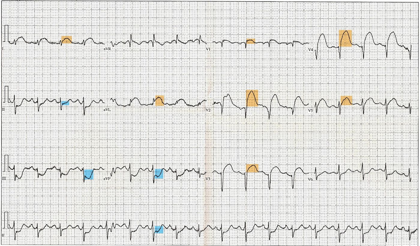
| Leads | Territory | Artery |
|---|---|---|
| II, III, aVF | Inferior | Right coronary artery (usually) |
| V1-V2 | Septal | Left anterior descending |
| V3-V4 | Anterior | Left anterior descending |
| V5-V6, I, aVL | Lateral | Left circumflex |
| V7-V9 | Posterior | Right coronary or circumflex |
| V1-V6, I, aVL | Extensive anterior | Proximal left anterior descending |
In an inferior STEMI, record right-sided leads (V4R in particular). Right ventricular infarction occurs in up to a third of inferior infarcts and changes management: these patients are preload-dependent, so nitrates and opioids can cause profound hypotension and fluid resuscitation is often needed instead.
Troponin
Cardiac troponin I or T is the biomarker of myocardial necrosis. High-sensitivity assays allow a rule-out or rule-in decision with paired samples, typically at presentation and 3 hours later, though many units now use 0- and 1-hour protocols.3
Troponin begins to rise within 3-4 hours, peaks at 24-48 hours, and can remain detectable for up to 10-14 days. Its main limitation is specificity: it marks myocardial injury of any cause, not coronary occlusion.
Other investigations
- FBC - anaemia both precipitates ischaemia and affects antiplatelet decisions
- U&Es - renal function before contrast, and potassium, since hypokalaemia is arrhythmogenic
- Glucose and HbA1c, and a lipid profile within 24 hours
- Coagulation screen before antithrombotic therapy
- Chest X-ray - pulmonary oedema, and to look for a widened mediastinum, though it must not delay reperfusion
- Echocardiogram - regional wall motion abnormalities, left ventricular function, and mechanical complications
Management
Immediate measures for all suspected ACS
- 12-lead ECG within 10 minutes, and continuous cardiac monitoring in a resuscitation area
- Aspirin 300 mg, chewed or dispersed
- Sublingual or intravenous GTN for pain, provided the patient is not hypotensive and there is no suspicion of right ventricular infarction
- Intravenous morphine with an antiemetic if pain is severe and not relieved by nitrates
- Oxygen only if oxygen saturations are below 94% (88-92% if at risk of hypercapnic respiratory failure)
- IV access, bloods including troponin, and analgesia
STEMI: reperfusion
Reperfusion is time-critical - myocardium infarcts progressively from the moment of occlusion. Offer reperfusion to patients presenting within 12 hours of symptom onset.1
Primary PCI is the preferred strategy if it can be delivered within 120 minutes of the time when fibrinolysis could have been given. If it cannot, give fibrinolysis and transfer for angiography: perform rescue PCI if reperfusion fails, and angiography within 6-24 hours if it succeeds.
Antithrombotic therapy alongside primary PCI is aspirin plus prasugrel in patients not already taking an oral anticoagulant, with unfractionated heparin where radial access is used. Patients on an oral anticoagulant receive clopidogrel instead.1
NSTEMI and unstable angina: risk stratification
Here the priority is not immediate reperfusion but working out who benefits from early angiography. Use the GRACE score, which estimates 6-month mortality from age, heart rate, blood pressure, creatinine, Killip class, cardiac arrest at admission, ST deviation and raised biomarkers.
| 6-month mortality | Risk | Strategy |
|---|---|---|
| 1.5% or below | Low | Conservative management; consider angiography if ischaemia recurs |
| Above 3% | Intermediate to high | Coronary angiography within 72 hours, with PCI if indicated |
| Any, if unstable | Very high | Immediate angiography - haemodynamic instability, ongoing pain despite treatment, dynamic ECG changes or life-threatening arrhythmia |
Give aspirin 300 mg to everyone. Add fondaparinux unless immediate angiography is planned or bleeding risk is high, and a second antiplatelet such as ticagrelor.
Secondary prevention
Started before discharge in every patient, and worth memorising as a list because it is a reliable exam question:1
- Dual antiplatelet therapy - aspirin 75 mg indefinitely, plus a second agent (ticagrelor, prasugrel or clopidogrel) usually for 12 months
- Beta-blocker - continue for at least 12 months, and indefinitely if left ventricular function is reduced
- ACE inhibitor - started early and titrated; an ARB if not tolerated
- High-intensity statin - atorvastatin 80 mg
- Aldosterone antagonist - if there is heart failure with reduced ejection fraction after MI
- Cardiac rehabilitation - offered to all, and one of the few interventions that improves both mortality and quality of life
- Lifestyle - smoking cessation, Mediterranean-style diet, activity, and alcohol within limits
Complications
Complications are best organised by when they occur after the infarct, which is also how they tend to be examined.
| Timing | Complication | Features |
|---|---|---|
| Minutes to hours | Ventricular fibrillation or tachycardia | The commonest cause of death before reaching hospital |
| Hours to days | Cardiogenic shock | Hypotension, oliguria, cool peripheries; mortality remains high |
| Hours to days | Acute heart failure and pulmonary oedema | Graded by Killip class |
| Days 1-5 | Bradyarrhythmia and heart block | Especially after inferior MI, since the RCA usually supplies the AV node |
| Days 3-5 | Papillary muscle rupture | Acute severe mitral regurgitation, pansystolic murmur, flash pulmonary oedema |
| Days 3-5 | Ventricular septal defect | New harsh pansystolic murmur with a step-up in oxygen saturation on right heart catheterisation |
| Days 3-5 | Free wall rupture | Tamponade, electromechanical dissociation, usually fatal |
| Days to weeks | Left ventricular aneurysm | Persistent ST elevation, heart failure, mural thrombus with embolic risk |
| 2-6 weeks | Dressler's syndrome | Autoimmune pericarditis with fever, pleuritic pain, raised inflammatory markers and a pericardial effusion |
Mechanical complications cluster at days 3-5 because that is when the infarcted myocardium is at its weakest, before fibrous scar has formed. A new murmur with sudden deterioration in that window should trigger an urgent echocardiogram.
Red flags
Prognosis
Outcomes have improved markedly with primary PCI networks, early risk stratification and modern secondary prevention. In-hospital mortality after STEMI treated with primary PCI is now well below the rates seen in the fibrinolysis era.2
The strongest determinants of long-term outcome are the extent of myocardial damage, particularly left ventricular ejection fraction, the presence of heart failure, age, renal function and diabetes. This is the reasoning behind the emphasis on door-to-balloon time: myocardium salvaged in the first hours translates directly into ejection fraction, and ejection fraction translates into survival.
Adherence to secondary prevention is the other major factor, and the one most amenable to influence in clinic. Cardiac rehabilitation attendance is associated with reduced cardiovascular mortality and rehospitalisation, yet uptake remains well short of the eligible population.
References
- NICE NG185. Acute coronary syndromes. 2020, updated 2025. Available here
- British Heart Foundation. UK Factsheet: heart and circulatory disease statistics. Available here
- NICE DG40. High-sensitivity troponin tests for the early rule out of NSTEMI. 2020. Available here
- Cabello JB, Burls A, Emparanza JI et al. Oxygen therapy for acute myocardial infarction. Cochrane Database of Systematic Reviews. 2016. Available here
- DVLA. Assessing fitness to drive: a guide for medical professionals. Available here
- NICE Clinical Knowledge Summaries. Acute coronary syndrome. Available here
- Thygesen K, Alpert JS, Jaffe AS et al. Fourth universal definition of myocardial infarction. European Heart Journal. 2018. Available here
- Resuscitation Council UK. Adult advanced life support guidelines. Available here
- BNF. Ticagrelor - indications and dosing. Available here
This article is written for revision and education. It is not clinical guidance and must not be used to make decisions about the care of a patient. Always check current NICE guidance and local protocols.