
Stable Angina: Diagnosis and Management
Key points
- Stable angina: chest discomfort caused by reversible myocardial ischaemia, brought on by exertion and relieved by rest or GTN.
- Mechanism: a fixed atherosclerotic stenosis limits coronary flow, so supply cannot meet the rise in myocardial oxygen demand on exertion.
- Classification: three features define it - constricting chest discomfort, precipitated by exertion, relieved by rest or GTN within about 5 minutes. All three is typical, two is atypical, one or none is non-anginal.
- First-line investigation: CT coronary angiography for anyone with typical or atypical anginal pain.
- Symptom relief: sublingual GTN, with clear instructions on when to call 999.
- First-line antianginal: a beta-blocker or a rate-limiting calcium channel blocker.
- Secondary prevention: aspirin 75 mg and a high-intensity statin in everyone, plus risk factor modification.
- Revascularisation: PCI or CABG when symptoms are not controlled on two antianginal drugs, or where the anatomy carries a prognostic benefit.
Introduction
Stable angina is chest discomfort caused by reversible myocardial ischaemia. It occurs when a fixed coronary stenosis prevents blood flow from rising to meet an increase in myocardial oxygen demand, most often during exertion.1
The word stable is doing important work. It means the pattern of symptoms has been unchanged over recent weeks: the same amount of exertion brings on the same discomfort, relieved in the same way. A pattern that is worsening, occurring at rest, or lasting longer is by definition unstable and is managed as an acute coronary syndrome.
It is common, affecting an estimated 3-4% of UK adults, and it is a frequent presentation in both primary care and rapid access chest pain clinics.2 It is also a favourite exam topic because the diagnosis rests almost entirely on the history.
Aetiology
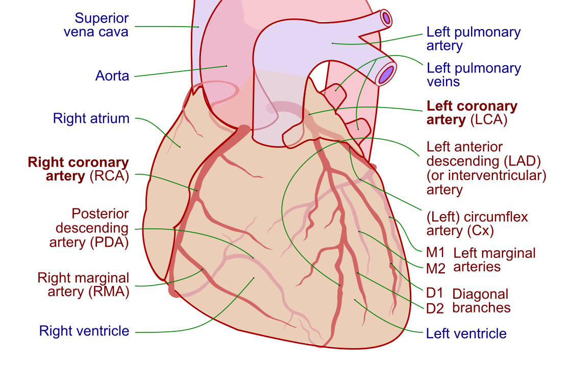
Myocardial oxygen supply depends chiefly on coronary blood flow, which occurs during diastole, and on the oxygen-carrying capacity of blood. Demand is determined by heart rate, contractility and ventricular wall stress. Angina is the symptom produced when demand outstrips supply.
Understanding this balance explains both the causes and the treatments: every antianginal drug works by reducing demand, improving supply, or both.
Reduced supply
- Coronary atherosclerosis - by far the commonest cause. A stenosis generally has to occlude around 70% of the lumen before flow is limited on exertion.
- Aortic stenosis - reduces coronary perfusion pressure while the hypertrophied ventricle demands more oxygen
- Hypertrophic cardiomyopathy - the same combination of increased muscle mass and impaired perfusion
- Anaemia - reduces oxygen-carrying capacity, and can unmask angina at a stenosis that was previously asymptomatic
- Coronary artery spasm (vasospastic or Prinzmetal angina) - typically occurs at rest, often at night
- Coronary microvascular dysfunction - angina with unobstructed epicardial arteries, more common in women
Increased demand
- Tachyarrhythmia
- Thyrotoxicosis
- Left ventricular hypertrophy, usually secondary to hypertension
- Fever or sepsis
Risk factors
The risk factors are those for atherosclerosis generally, since that is the underlying process.
- Smoking
- Hypertension
- Diabetes mellitus
- Dyslipidaemia
- Obesity and physical inactivity
- Increasing age and male sex
- Family history of premature coronary artery disease (first-degree relative affected before 55 in men or 65 in women)
- Chronic kidney disease
Clinical features
NICE defines anginal pain by three features, and the number present determines how the patient is classified and investigated:3
- Constricting discomfort in the front of the chest, or in the neck, shoulders, jaw or arms
- Precipitated by physical exertion
- Relieved by rest or by GTN within about 5 minutes
| Features present | Classification | Implication |
|---|---|---|
| All three | Typical angina | Investigate with CT coronary angiography |
| Two of three | Atypical angina | Investigate with CT coronary angiography |
| One or none | Non-anginal chest pain | Investigate only if the resting ECG is abnormal; otherwise consider other causes |
Patients frequently describe the sensation as heaviness, tightness or a band around the chest rather than pain, and may deny 'pain' if asked directly. Associated breathlessness is common. Nausea, sweating and pain at rest point away from stable angina and towards an acute coronary syndrome.
Anginal symptoms are more likely to be atypical in women, in older people and in those with diabetes, where autonomic neuropathy may blunt the sensation. Breathlessness on exertion may be the only symptom, sometimes called an anginal equivalent.
Grading severity
The Canadian Cardiovascular Society classification grades functional limitation and is useful for documenting change over time.4
| Grade | Description |
|---|---|
| I | Angina only with strenuous or prolonged exertion; ordinary activity is unaffected |
| II | Slight limitation of ordinary activity, for example walking uphill or climbing more than one flight of stairs quickly |
| III | Marked limitation - angina on walking one or two blocks on the level, or one flight of stairs at normal pace |
| IV | Inability to carry out any physical activity without discomfort; angina may be present at rest |
Points to cover in the history
- Site, character, radiation, and what brings it on and relieves it
- Exercise tolerance in concrete terms - how far on the flat, how many stairs - and whether that has changed
- Duration of a typical episode, and whether any episode has occurred at rest
- Whether GTN is already being used, how quickly it works, and how many doses are needed
- Full cardiovascular risk factor history including smoking pack-years and family history
- Occupation and driving, particularly vocational licence holders
- Symptoms suggesting an alternative cause: reflux, pleuritic pain, tenderness, anxiety
Clinical examination
Examination is frequently normal in stable angina, which does not exclude the diagnosis. Its value lies in identifying risk factors, alternative causes and complications.
- General: BMI, tar staining, corneal arcus, xanthelasma, tendon xanthomata, and signs of anaemia
- Cardiovascular: pulse rate and rhythm, blood pressure in both arms, apex beat, and auscultation for murmurs - an ejection systolic murmur radiating to the carotids suggests aortic stenosis as the cause
- Heart failure: raised JVP, displaced apex, third heart sound, basal crackles, peripheral oedema
- Peripheral vessels: carotid, femoral and pedal pulses, plus carotid and abdominal bruits, since coexistent peripheral disease is common
- Chest wall: reproducible tenderness suggests a musculoskeletal cause, though it does not exclude ischaemia
Differential diagnosis
The differential for exertional chest pain is broad, and several alternatives are immediately life-threatening:
- Acute coronary syndrome: pain at rest, prolonged, or a crescendo pattern - this is the diagnosis to exclude first
- Aortic dissection: sudden, severe, tearing pain radiating to the back, with a blood pressure differential between arms
- Pulmonary embolism: pleuritic pain, breathlessness, tachycardia, risk factors for venous thromboembolism
- Pericarditis: sharp pain, worse lying flat and relieved by sitting forward, with a friction rub
- Aortic stenosis: may itself cause angina, and coexists with coronary disease
- Gastro-oesophageal reflux: burning pain, related to meals and posture, may also respond to GTN, which is a well-known diagnostic trap
- Musculoskeletal chest pain: localised, reproducible on palpation or movement
- Anxiety or panic disorder: associated with paraesthesiae and a sense of impending doom, but a diagnosis of exclusion
Investigations
Bedside and laboratory
- 12-lead ECG - often normal, which does not exclude angina. Look for pathological Q waves indicating prior infarction, left bundle branch block, ST or T wave changes, and left ventricular hypertrophy.
- FBC - anaemia can precipitate or worsen angina
- HbA1c and lipid profile - both to identify risk factors and to guide secondary prevention
- U&Es, TFTs and LFTs - renal function before contrast imaging, thyrotoxicosis as a precipitant, and a baseline before starting a statin
- Troponin - only if an acute coronary syndrome is suspected. It has no role in the assessment of stable symptoms.
Imaging
CT coronary angiography (CTCA) is the first-line investigation in the UK for people with typical or atypical anginal pain, and for those with non-anginal pain whose resting ECG shows ST or T wave changes or pathological Q waves.3 It is non-invasive, and its strength is its very high negative predictive value: a normal CTCA effectively excludes obstructive coronary disease.
If CTCA shows disease of uncertain functional significance, the next step is non-invasive functional imaging to demonstrate reversible ischaemia. Options include stress echocardiography, myocardial perfusion scintigraphy with SPECT, and stress cardiac MRI.
Invasive coronary angiography is reserved for those in whom functional imaging is inconclusive, or where revascularisation is being actively considered. It remains the definitive assessment of coronary anatomy and allows measurement of fractional flow reserve to determine whether a stenosis is functionally significant.
Management
Management has three strands that run in parallel: relieving the acute episode, preventing further episodes, and reducing the risk of myocardial infarction and death. Students often focus on the antianginals and forget the third, which is the part that changes prognosis.
Immediate symptom relief
Sublingual GTN spray is given to all patients, both to treat an episode and to take before predictably strenuous activity. Counselling on how to use it is a common OSCE task.
Antianginal therapy
First-line is either a beta-blocker (for example bisoprolol) or a rate-limiting calcium channel blocker (diltiazem or verapamil), titrated to symptoms and heart rate.5
If symptoms are not controlled on one agent at the maximum tolerated dose, switch to the other, or combine them.
If symptoms persist on two drugs, or a second drug is not tolerated, add or substitute one of: a long-acting nitrate such as isosorbide mononitrate, ivabradine, nicorandil, or ranolazine. NICE advises adding a third antianginal only while the patient is awaiting revascularisation or when revascularisation is not appropriate.
Secondary prevention
This is the component that alters prognosis and should be addressed at the same consultation:
- Aspirin 75 mg once daily - clopidogrel if aspirin is not tolerated
- High-intensity statin - atorvastatin 80 mg, targeting LDL cholesterol of 2.0 mmol/L or below
- ACE inhibitor - if the patient also has diabetes, hypertension, heart failure or chronic kidney disease
- Optimise risk factors - smoking cessation, blood pressure control, glycaemic control, weight and activity
- Cardiac rehabilitation and a structured exercise programme
- Annual influenza vaccination
Revascularisation
Revascularisation is considered when symptoms are not controlled by optimal medical therapy, or where the coronary anatomy carries a prognostic benefit from intervention.
| Percutaneous coronary intervention | Coronary artery bypass grafting | |
|---|---|---|
| Best suited to | Single or two-vessel disease; anatomically straightforward lesions | Left main stem disease; three-vessel disease, especially with diabetes or impaired LV function |
| Recovery | Day case or overnight stay | Sternotomy, several days in hospital, weeks of recovery |
| Durability | Higher rate of repeat revascularisation | More durable, with better long-term survival in complex disease |
| Afterwards | Dual antiplatelet therapy for a defined period | Ongoing single antiplatelet therapy |
Neither procedure removes the need for secondary prevention or antianginal therapy, and both are decided by a multidisciplinary heart team where the anatomy is complex.
Complications
The principal concern is progression to an acute coronary syndrome through plaque rupture. Other consequences include ischaemic cardiomyopathy and heart failure, ventricular arrhythmias, and a substantial impact on quality of life and independence where symptoms limit activity.
Treatment carries its own risks. Beta-blockers cause fatigue, bradycardia, cold peripheries and bronchospasm in asthma. Dihydropyridine calcium channel blockers cause ankle oedema and flushing. Nicorandil can cause troublesome mucosal and gastrointestinal ulceration, which is easily missed if the drug history is not reviewed.
Red flags
Prognosis
Prognosis in stable angina is generally good with optimal medical therapy, and depends chiefly on left ventricular function, the extent of coronary disease, and how well risk factors are controlled. Annual mortality in contemporary cohorts on modern therapy is low.
An important and frequently examined point is that revascularisation improves symptoms but does not reliably improve survival in stable disease, outside specific anatomical subgroups such as left main stem or extensive three-vessel disease. The ORBITA trial found that PCI produced little symptomatic benefit over a placebo procedure in single-vessel disease, and ISCHEMIA found no reduction in cardiovascular events from routine invasive management compared with optimal medical therapy alone.8,9
The practical consequence is that optimal medical therapy is not a holding measure while waiting for a stent. It is the treatment, and revascularisation is added for symptoms that it fails to control or for anatomy that carries a prognostic benefit.
References
- Knuuti J, Wijns W, Saraste A et al. ESC Guidelines for the diagnosis and management of chronic coronary syndromes. European Heart Journal. 2019. Available here
- NICE Clinical Knowledge Summaries. Angina. Available here
- NICE CG95. Recent-onset chest pain of suspected cardiac origin: assessment and diagnosis. 2010, updated 2016. Available here
- Campeau L. Grading of angina pectoris. Circulation. 1976. Available here
- NICE CG126. Stable angina: management. 2011, updated 2016. Available here
- BNF. Verapamil hydrochloride - interactions and cautions. Available here
- DVLA. Assessing fitness to drive: a guide for medical professionals. Available here
- Al-Lamee R, Thompson D, Dehbi HM et al. Percutaneous coronary intervention in stable angina (ORBITA): a double-blind, randomised controlled trial. The Lancet. 2018. Available here
- Maron DJ, Hochman JS, Reynolds HR et al. Initial invasive or conservative strategy for stable coronary disease (ISCHEMIA). New England Journal of Medicine. 2020. Available here
- NICE NG238. Cardiovascular disease: risk assessment and reduction, including lipid modification. 2023. Available here
This article is written for revision and education. It is not clinical guidance and must not be used to make decisions about the care of a patient. Always check current NICE guidance and local protocols.