
Hypokalaemia
Key points
- Hypokalaemia: serum potassium below 3.5 mmol/L. Mild 3.0-3.5, moderate 2.5-2.9, severe below 2.5 or symptomatic at any level.
- Commonest causes: GI losses (vomiting, diarrhoea) and drugs - above all loop and thiazide diuretics, plus beta-2 agonists, insulin, corticosteroids and laxative misuse.
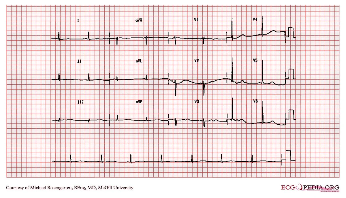
- ECG changes: small or inverted T waves, prominent U waves, ST depression and a prolonged QT/QU interval - risking torsades de pointes and ventricular arrhythmia.
- Always check magnesium: hypomagnesaemia causes refractory hypokalaemia by increasing renal potassium wasting. Potassium cannot be corrected until magnesium is replaced - a classic clinical trap.
- Use blood pressure to find the cause: hypertension suggests hyperaldosteronism, Cushing's, renal artery stenosis or liquorice; normotension suggests GI losses, diuretics, or Gitelman/Bartter syndromes.
- Oral replacement: preferred where possible - Sando-K or equivalent, with dietary advice. Recheck within a few days.
- Intravenous replacement: maximum 10 mmol/hour via a peripheral line, and never more than 20 mmol/hour even centrally, with cardiac monitoring. Concentrated potassium is a never event if given undiluted.
- Digoxin interaction: hypokalaemia potentiates digoxin toxicity, because both compete at the Na⁺/K⁺-ATPase - a low potassium can precipitate toxicity at therapeutic digoxin levels.
Introduction
Hypokalaemia is defined as a serum potassium below 3.5 mmol/L and is one of the most frequently encountered electrolyte abnormalities in hospital practice, found in up to a fifth of inpatients. It is usually mild, but it becomes clinically important because of its effects on cardiac excitability, skeletal muscle and the kidney.1
As with hyperkalaemia, it helps to remember that 98% of body potassium is intracellular. A low serum potassium may therefore reflect true total body depletion (from renal or gastrointestinal loss, or poor intake) or simply a shift into cells with normal total body stores. Distinguishing the two matters, because a shift-related hypokalaemia will correct itself and over-replacement risks rebound hyperkalaemia.
Causes
Gastrointestinal losses
- Diarrhoea - the commonest GI cause; loses potassium and bicarbonate, producing a normal anion gap metabolic acidosis with hypokalaemia
- Vomiting and nasogastric aspiration - note the mechanism is largely renal rather than direct loss: volume depletion drives secondary hyperaldosteronism, and the metabolic alkalosis shifts potassium into cells. This produces the classic hypokalaemic, hypochloraemic metabolic alkalosis seen in pyloric stenosis
- Laxative misuse - consider in unexplained hypokalaemia, particularly with eating disorders
- Villous adenoma of the rectum - secretes large volumes of potassium-rich mucus
- Fistulae, ileostomy losses and short bowel syndrome
Renal losses
- Loop and thiazide diuretics - by a clear margin the commonest drug cause; they increase distal sodium delivery and therefore potassium excretion
- Hyperaldosteronism - primary (Conn syndrome) or secondary; causes hypokalaemia with hypertension and metabolic alkalosis
- Cushing syndrome and high-dose corticosteroids - cortisol has mineralocorticoid activity at high concentrations; ectopic ACTH classically causes profound hypokalaemia
- Renal artery stenosis and renin-secreting tumours - secondary hyperaldosteronism
- Liquorice excess and carbenoxolone - inhibit 11-beta-hydroxysteroid dehydrogenase, allowing cortisol to act on mineralocorticoid receptors (apparent mineralocorticoid excess)
- Renal tubular acidosis types 1 and 2 - hypokalaemia with a normal anion gap metabolic acidosis, in contrast to most other renal causes which cause alkalosis
- Bartter and Gitelman syndromes - inherited tubulopathies causing hypokalaemic alkalosis with normal or low blood pressure; Bartter mimics a loop diuretic, Gitelman a thiazide (and Gitelman characteristically also causes hypomagnesaemia and hypocalciuria)
- Hypomagnesaemia - see below; a critically important and often missed cause
- Drugs - amphotericin B, aminoglycosides, cisplatin, high-dose penicillins
- Osmotic diuresis - as in diabetic ketoacidosis and hyperglycaemia
- Recovery phase of AKI and post-obstructive diuresis
Shift into cells
- Insulin - including treatment of DKA, where potassium falls precipitously; and insulin-glucose given for hyperkalaemia
- Beta-2 agonists - salbutamol, especially nebulised and repeated in acute asthma
- Metabolic alkalosis - potassium moves into cells in exchange for hydrogen ions
- Refeeding syndrome - insulin release after starvation drives potassium, phosphate and magnesium intracellularly
- Catecholamines - stress, sepsis, myocardial infarction, and inotropes
- Hypokalaemic periodic paralysis, including the thyrotoxic form
Inadequate intake
Rarely a sole cause in a patient with normal kidneys, since renal conservation is efficient - but it contributes in malnutrition, alcohol dependence, anorexia nervosa and prolonged inadequate parenteral nutrition, and it compounds losses from other sources.
Clinical features
Mild hypokalaemia is usually asymptomatic and detected incidentally. Symptoms generally appear below 3.0 mmol/L and reflect the effect of potassium on excitable membranes - cardiac, skeletal and smooth muscle.
- Generalised muscle weakness, fatigue and myalgia - typically proximal and ascending; severe cases progress to flaccid paralysis and, rarely, respiratory muscle failure
- Muscle cramps and tetany
- Palpitations and arrhythmias - ectopics, atrial fibrillation, ventricular tachycardia and torsades de pointes
- Constipation, ileus and abdominal distension - from smooth muscle involvement
- Polyuria and polydipsia - hypokalaemia causes a nephrogenic diabetes insipidus-like state by impairing renal concentrating ability
- Rhabdomyolysis - severe hypokalaemia impairs muscle blood flow and can cause muscle breakdown, which is worth knowing because it then releases potassium and confuses the picture
- Paraesthesiae and reduced reflexes
- Worsening of glycaemic control - potassium is required for insulin secretion
- Exacerbation of hepatic encephalopathy - hypokalaemia increases renal ammonia production
- Features of the underlying cause - hypertension in Conn's, cushingoid appearance, dental erosion or parotid swelling in bulimia, diarrhoea or vomiting
Investigations
- 12-lead ECG - essential in moderate or severe hypokalaemia, and in anyone on digoxin or with cardiac disease
- Serum magnesium - check in every case; hypomagnesaemia is a common cause of refractory hypokalaemia
- U&Es, creatinine and eGFR - renal function and associated abnormalities
- Venous blood gas or bicarbonate - to establish whether there is a metabolic alkalosis or acidosis, which discriminates between causes
- Glucose - DKA and refeeding risk
- Calcium and phosphate - often disturbed together, especially in refeeding syndrome
- Urinary potassium and creatinine (spot sample or 24-hour) - low suggests GI or dietary cause, high indicates renal wasting
- Urinary chloride - helps separate vomiting (low urinary chloride) from diuretic use (high)
- Aldosterone and renin (aldosterone-to-renin ratio) - if hypokalaemia is associated with hypertension, to screen for primary hyperaldosteronism
- Cortisol testing - overnight dexamethasone suppression or 24-hour urinary free cortisol if Cushing syndrome is suspected
- Thyroid function - thyrotoxic periodic paralysis
- Digoxin level - if the patient takes digoxin, since hypokalaemia potentiates toxicity
- Creatine kinase - if there is significant weakness, to detect rhabdomyolysis
- A careful drug and laxative history - including over-the-counter laxatives, salbutamol use and liquorice consumption
ECG changes
- Small, flattened or inverted T waves
- Prominent U waves - an additional deflection after the T wave, best seen in the precordial leads; the classic finding
- ST segment depression
- Prolonged PR interval and increased P wave amplitude
- Apparent QT prolongation - in reality often a fused QU interval, but functionally it carries the same risk of torsades de pointes
- Ventricular and atrial ectopics, progressing to VT, VF and torsades
Management
Management has three components: replace potassium safely, correct magnesium, and treat the underlying cause. The second of these is the one most often forgotten.
Oral replacement
- Preferred where the patient is stable, able to swallow, and potassium is above about 2.5 mmol/L without ECG changes
- Potassium chloride preparations - e.g. Sando-K (12 mmol K⁺ per tablet), typically 2 tablets three times daily, adjusted to response
- Take with food - the main adverse effects are nausea, gastric irritation and, rarely, ulceration
- Dietary advice - bananas, oranges and orange juice, tomatoes, potatoes, avocado, dried fruit, nuts and beans
- Recheck potassium within a few days, and continue until the cause is corrected
Intravenous replacement
| Parameter | Guidance |
|---|---|
| When to use IV | Severe hypokalaemia (<2.5 mmol/L), ECG changes, arrhythmia, significant weakness, or the patient cannot take oral |
| Peripheral line - maximum rate | 10 mmol/hour, at a concentration no greater than 40 mmol/L (e.g. 40 mmol in 1 litre of 0.9% sodium chloride over 4+ hours) |
| Central line / high dependency | Up to 20 mmol/hour with continuous cardiac monitoring and frequent rechecks - only in a monitored setting |
| Diluent | Sodium chloride 0.9% is generally preferred. Avoid glucose-containing fluids initially - glucose stimulates insulin and can drive potassium further into cells, worsening the hypokalaemia |
| Monitoring | Recheck potassium every 1-4 hours during active replacement; cardiac monitoring for severe hypokalaemia or rapid infusion |
| Peripheral vein irritation | Potassium is irritant and painful; use a large vein, dilute adequately, and consider central access if repeated infusions are needed |
Treating the underlying cause
- Review and adjust diuretics - consider switching to or adding a potassium-sparing diuretic (spironolactone, amiloride) where ongoing diuresis is needed
- Treat diarrhoea and vomiting, and replace ongoing GI losses
- Address laxative misuse and eating disorders with appropriate specialist input
- Investigate and treat hyperaldosteronism - spironolactone or adrenalectomy in Conn syndrome
- Stop liquorice, and review corticosteroid dosing
- In DKA, anticipate the fall in potassium: withhold insulin if potassium is below 3.5 mmol/L until replacement has begun, and add potassium to fluids once it is below 5.5
- In refeeding syndrome, replace potassium, phosphate and magnesium, give thiamine, and reintroduce feeding slowly
- Review salbutamol use in asthma, where repeated nebulisers commonly cause transient hypokalaemia
- Check and manage digoxin - hypokalaemia potentiates toxicity
Complications
- Ventricular arrhythmias - ventricular tachycardia, ventricular fibrillation and torsades de pointes; the principal danger
- Digoxin toxicity - precipitated at otherwise therapeutic levels because potassium and digoxin compete at the Na⁺/K⁺-ATPase
- Atrial fibrillation and other supraventricular arrhythmias
- Cardiac arrest
- Flaccid paralysis and respiratory muscle weakness in severe depletion
- Rhabdomyolysis - from impaired muscle perfusion
- Ileus and constipation
- Nephrogenic diabetes insipidus with polyuria and polydipsia; chronic hypokalaemia can cause hypokalaemic nephropathy with interstitial fibrosis
- Impaired glucose tolerance, since potassium is needed for insulin release
- Precipitation or worsening of hepatic encephalopathy
- Complications of treatment - rebound hyperkalaemia from over-rapid replacement, phlebitis and pain from peripheral infusion, and the catastrophic consequences of a potassium bolus
Red flags
Prognosis
Mild hypokalaemia is generally benign and corrects readily once the underlying cause is addressed. Most cases in practice arise from diuretics or a self-limiting episode of vomiting or diarrhoea, respond to oral replacement, and cause no lasting harm. The important qualification is that even mild hypokalaemia is not harmless in the wrong context - in patients with ischaemic heart disease, heart failure, left ventricular hypertrophy or digoxin therapy, potassium levels in the 3.0-3.5 range are independently associated with increased arrhythmic risk and mortality. Many cardiologists therefore aim to keep potassium above 4.0 mmol/L in such patients rather than merely within the reference range.
Severe hypokalaemia is potentially fatal but fully reversible with prompt treatment. Arrhythmias and muscle weakness resolve as potassium is restored, and there is usually no residual deficit. The risks in this group come as much from treatment as from the abnormality itself - over-rapid replacement causing rebound hyperkalaemia, and the well-documented catastrophe of concentrated potassium given as a bolus.
Long-term outcome depends on the underlying cause. Diuretic-induced hypokalaemia is easily managed with dose adjustment, potassium-sparing agents or supplementation. Primary hyperaldosteronism is potentially curable by adrenalectomy, and identifying it converts a lifetime of refractory hypertension and potassium supplements into a treatable diagnosis - which is why hypokalaemia with hypertension should always be investigated rather than simply corrected. Conversely, chronic untreated hypokalaemia can cause hypokalaemic nephropathy, with tubulointerstitial fibrosis and progressive renal impairment, and hypokalaemia arising from eating disorders or laxative misuse carries the prognosis of the underlying psychiatric condition, which is considerably more serious than the electrolyte result alone suggests.
References
- NICE Clinical Knowledge Summaries. Hypokalaemia. Available here
- UK Kidney Association. Clinical practice guidelines - electrolyte disorders. Available here
- NHS England. Never Events list - maladministration of potassium-containing solutions. Available here
- Joint British Diabetes Societies. The Management of Diabetic Ketoacidosis in Adults (potassium replacement). Available here
- NICE NG32 / CG32. Nutrition support in adults - refeeding syndrome. Available here
- BNF. Potassium chloride - oral and intravenous replacement. Available here
- Michael Rosengarten BEng MD, McGill (CardioNetworks ECGpedia), CC BY-SA 3.0, via Wikimedia Commons. Available here
This article is written for revision and education. It is not clinical guidance and must not be used to make decisions about the care of a patient. Always check current NICE guidance and local protocols.