
Infantile Hypertrophic Pyloric Stenosis
Key points
- Definition: hypertrophy of the circular smooth muscle of the pylorus causing progressive gastric outlet obstruction in infancy.
- Who gets it: 2-4 per 1,000 live births, males four to five times more often than females, classically the firstborn son, presenting between 2 and 8 weeks with a peak at 3-6 weeks.
- The history: non-bilious projectile vomiting immediately after feeds, in a baby who is then immediately hungry again - the hungry vomiter.
- Biochemistry: hypochloraemic, hypokalaemic metabolic alkalosis with paradoxical aciduria - the single most examined fact in paediatrics.
- Examination: a test feed showing visible gastric peristalsis moving left to right, and a palpable olive-sized mass in the right upper quadrant.
- Investigation: blood gas and U&Es first, then abdominal ultrasound - muscle thickness above 3 mm and canal length above 15-17 mm.
- This is a medical emergency, not a surgical one: the alkalosis and dehydration must be corrected before anaesthesia, or the infant risks postoperative apnoea.
- Treatment: Ramstedt pyloromyotomy - splitting the hypertrophied muscle down to, but not through, the mucosa. Curative, with no long-term sequelae.
Introduction
Infantile hypertrophic pyloric stenosis is progressive hypertrophy and hyperplasia of the circular muscle of the pylorus, narrowing the pyloric canal until the stomach can no longer empty. It affects 2-4 per 1,000 UK live births and is one of the commonest conditions requiring surgery in the first months of life.
It is a favourite exam topic for two reasons. The clinical picture is highly stereotyped, so it can be described in three lines of a question stem; and it produces a metabolic disturbance that can be reasoned out from first principles rather than memorised - which is exactly what examiners like to test.
The one clinical point that repeatedly catches students out is the sequencing. Pyloric stenosis is not treated with urgent surgery. The infant is dehydrated and profoundly alkalotic, and taking them to theatre in that state risks postoperative apnoea. Resuscitate first, operate later - often 24 to 48 hours later - and be able to say why.
Aetiology
The cause is not fully understood. The muscle is normal at birth and hypertrophies over the first weeks, which is why the condition presents at 3-6 weeks rather than on day one. Reduced expression of neuronal nitric oxide synthase in the pyloric muscle, with consequently impaired smooth muscle relaxation, is the best-supported mechanism, superimposed on a clear genetic predisposition.
- Male sex - a four to fivefold excess, and it is disproportionately the firstborn
- Family history - present in around 15%. If the mother was affected, up to 20% of her sons will be; the risk from an affected father is lower.
- Macrolide exposure in the first 2 weeks of life - erythromycin carries the strongest association, with a relative risk of roughly eightfold, and azithromycin carries a weaker but real signal3
- Maternal macrolide use in late pregnancy or while breastfeeding
- Bottle feeding rather than breastfeeding
- Maternal smoking
- Associations with Turner syndrome, trisomy 18, Cornelia de Lange syndrome and oesophageal atresia
Pathophysiology and the metabolic derangement
The obstruction is at the gastric outlet, proximal to the ampulla of Vater. The infant therefore vomits stomach contents only - milk, hydrogen ions, chloride and some sodium and potassium - and never bile. Everything else follows from what is being lost and how the kidney responds.
- Loss of hydrochloric acid in the vomit produces a hypochloraemic metabolic alkalosis. Chloride falls furthest and is the most useful single marker of severity.
- Loss of fluid causes volume depletion, activating the renin-angiotensin-aldosterone system.
- Aldosterone drives sodium reabsorption in the distal nephron in exchange for potassium and hydrogen ions, deepening both the hypokalaemia and the alkalosis.
- Early on the kidney tries to compensate by excreting bicarbonate, and the urine is alkaline.
- As volume depletion worsens, the kidney prioritises sodium and water retention over acid-base balance. With chloride unavailable as the accompanying anion, sodium is reabsorbed in exchange for hydrogen ions, so acid is excreted into the urine despite a systemic alkalosis - the classic paradoxical aciduria.
- Hypokalaemia compounds this: potassium shifts out of cells in exchange for hydrogen ions moving in, and the kidney excretes more hydrogen to conserve potassium.
The end result is a dehydrated, hypochloraemic, hypokalaemic, alkalotic infant passing paradoxically acidic urine. Severe alkalosis depresses central respiratory drive and shifts the oxyhaemoglobin dissociation curve to the left, which is the physiological reason for the postoperative apnoea risk if the infant is anaesthetised before it is corrected.
Clinical features
- Projectile, non-bilious vomiting, occurring immediately or shortly after a feed and becoming more forceful and more frequent over days to weeks
- Immediate hunger after vomiting - the infant feeds avidly and then vomits again. This distinguishes pyloric stenosis from sepsis or a systemic illness, in which the baby is anorexic and unwell.
- Weight loss or failure to gain weight, and reduced wet nappies
- Constipation, with small infrequent stools, because so little reaches the intestine
- Dehydration - sunken fontanelle, dry mucous membranes, reduced skin turgor, lethargy, and in severe cases shock
- Jaundice in around 2-5% - unconjugated, and attributed to reduced glucuronosyltransferase activity in the starved infant. It resolves after surgery.
Assessing severity
Severity is judged by the degree of dehydration and the extent of the biochemical derangement rather than by the frequency of vomiting, and both determine how long resuscitation will take.
- Weight against birth weight and the growth chart - a fall across centiles quantifies how long this has been going on
- Perfusion and hydration: capillary refill, mucous membranes, fontanelle, skin turgor, urine output in wet nappies, and conscious level
- Chloride - a chloride below about 90 mmol/L indicates severe depletion and predicts a longer period of preoperative correction
- Bicarbonate and base excess - a bicarbonate above 30 mmol/L is a marker of severe alkalosis
- Potassium, which is often deceptively normal at first and falls further once volume is restored and acidosis corrected
- Lethargy or apnoea - late features indicating profound alkalosis and dehydration, and a reason for urgent senior involvement
The test feed
This is a classic examination station. The infant is given a feed - by bottle or breast - while the examiner sits on the baby's left with a warm hand on the abdomen.
- Visible gastric peristalsis - waves passing from the left upper quadrant towards the right, best seen with tangential lighting
- A palpable pyloric mass - firm, smooth and olive-sized, in the right upper quadrant or epigastrium, lateral to the rectus muscle and often easiest to feel just after a vomit when the stomach is empty
- The mass is palpable in a majority of cases in experienced hands, and a confidently palpated olive is sufficient to make the diagnosis without imaging
- Assess hydration and weight at the same time, and plot the weight against the birth weight
Differential diagnosis
| Diagnosis | Distinguishing features |
|---|---|
| Pyloric stenosis | Projectile, non-bilious, hungry after vomiting, weight loss, hypochloraemic hypokalaemic alkalosis |
| Gastro-oesophageal reflux | Effortless posseting rather than projectile, thriving, no metabolic disturbance, often positional2 |
| Overfeeding | Large volumes, excessive weight gain, settles with feed volume advice |
| Gastroenteritis | Diarrhoea, contact history, may be bilious, no palpable mass |
| Cow's milk protein allergy | Eczema, blood or mucus in the stool, symptoms with feeds, family history of atopy |
| Sepsis or urinary tract infection | Unwell, anorexic rather than hungry, fever or temperature instability, raised inflammatory markers |
| Congenital adrenal hyperplasia | Hyponatraemia, hyperkalaemia and metabolic acidosis, hypoglycaemia, ambiguous genitalia in girls, circulatory collapse |
| Malrotation with volvulus | Bilious vomiting - a surgical emergency, and a completely different pathway |
| Raised intracranial pressure | Bulging fontanelle, rapidly increasing head circumference, sunsetting eyes, abnormal neurology |
| Inborn errors of metabolism | Acidosis, hypoglycaemia, hyperammonaemia, encephalopathy, deterioration after feeds |
Investigations
Biochemistry first
- Capillary or venous blood gas - metabolic alkalosis with a raised bicarbonate and base excess
- U&Es and chloride - low chloride, low potassium, and often a low sodium. Chloride is the best single measure of severity and of the adequacy of resuscitation.
- Glucose, since prolonged poor intake causes hypoglycaemia
- Urine dipstick may demonstrate the paradoxical aciduria, though it is rarely needed clinically
Imaging
Abdominal ultrasound is the investigation of choice, and is diagnostic where the pyloric mass has not been palpated. Note what is not needed: an abdominal radiograph adds nothing beyond a distended stomach, and contrast studies carry an aspiration risk in a vomiting infant. Bloods beyond a gas, electrolytes and glucose are not routinely required.
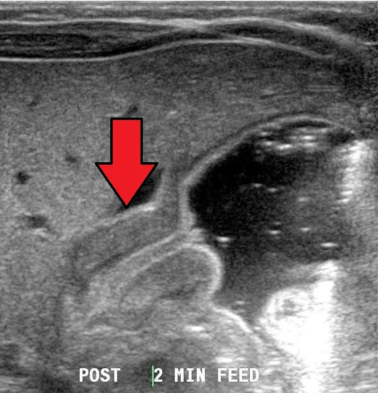
| Measurement | Threshold |
|---|---|
| Pyloric muscle thickness | Greater than 3 mm |
| Pyloric canal length | Greater than 15-17 mm |
| Pyloric transverse diameter | Greater than 13 mm |
| Dynamic findings | Failure of the channel to open, with vigorous gastric peristalsis ending at a closed pylorus |
Management
Step 1: resuscitation and correction
- Nil by mouth, with a nasogastric tube on free drainage aspirated 4-hourly
- A 10-20 mL/kg bolus of 0.9% sodium chloride if the infant is shocked
- Maintenance and deficit replacement with a sodium chloride and glucose solution containing potassium chloride, typically at around 1.5 times maintenance. Providing chloride is the key to correcting the alkalosis, and potassium cannot be replaced properly until urine output is established.6
- Replace nasogastric losses millilitre for millilitre with 0.9% sodium chloride with added potassium
- Repeat the gas and U&Es every 6-12 hours
- Book theatre only when the biochemistry has normalised - conventionally chloride above 100 mmol/L, bicarbonate below 26-28 mmol/L, potassium normal, and the infant clinically rehydrated with good urine output
Step 2: surgery
- Ramstedt pyloromyotomy - a longitudinal incision through the serosa and hypertrophied circular muscle of the pylorus, spreading the muscle fibres apart down to but not through the mucosa, which then bulges into the defect and relieves the obstruction
- Performed open, through a right upper quadrant or circumumbilical incision, or laparoscopically, with comparable outcomes
- The stomach is decompressed and the mucosa checked for perforation before closure, often by insufflating air down the nasogastric tube
- Feeding is restarted within 6-24 hours and increased over the next day or so
- Most infants go home within 1-2 days
Complications
- Preoperative: dehydration, hypovolaemic shock, severe electrolyte disturbance, hypoglycaemia and aspiration of vomit
- Postoperative apnoea where the alkalosis was not fully corrected before anaesthesia
- Mucosal perforation during myotomy - usually recognised and repaired at the time, most often at the duodenal end where the mucosa is thinnest
- Incomplete myotomy, causing persistent vomiting and requiring reoperation
- Wound infection, dehiscence and incisional hernia, all uncommon
- Duodenal injury and, rarely, bleeding
Prognosis
Pyloromyotomy is curative. Once feeding is established the infant catches up in weight rapidly, the jaundice resolves, and there are no long-term gastrointestinal consequences - no increased risk of reflux, ulceration or malabsorption in later life. Mortality in the UK is well under 0.5%, and the deaths that do occur are almost entirely attributable to delayed presentation with severe dehydration rather than to the operation.5
Parents should be warned before discharge that a small amount of vomiting for a day or two is expected, that feeds should be built back up gradually as advised by the surgical team, and that they should seek review for persistent vomiting, a fever, or redness or discharge from the wound. Weight is checked in the community over the following weeks, and virtually all infants have regained their expected centile within a month or two.
The two things to take from this topic are therefore practical rather than surgical. First, think of pyloric stenosis in any infant of 2-8 weeks with worsening vomiting and poor weight gain, and check a gas and electrolytes rather than labelling it reflux - the biochemistry makes the diagnosis obvious. Second, once the diagnosis is made, resist the instinct to escalate straight to theatre; the emergency is metabolic, and correcting it properly is the intervention that determines how safely the child gets through the anaesthetic.1
References
- Advanced Life Support Group. Advanced Paediatric Life Support: A Practical Approach to Emergencies. Available here
- NICE NG1. Gastro-oesophageal reflux disease in children and young people: diagnosis and management. 2015, updated 2019. Available here
- Eberly MD, Eide MB, Thompson JL, Nylund CM. Azithromycin in early infancy and pyloric stenosis. Pediatrics. 2015;135:483-8. Available here
- BNF for Children. Erythromycin. Available here
- British Association of Paediatric Surgeons. Congenital Anomalies Surveillance System (BAPS-CASS). Available here
- NICE NG29. Intravenous fluid therapy in children and young people in hospital. 2015, updated 2020. Available here
This article is written for revision and education. It is not clinical guidance and must not be used to make decisions about the care of a patient. Always check current NICE guidance and local protocols.