
Biliary Atresia
Key points
- Definition: progressive fibro-obliterative destruction of the extrahepatic bile ducts, causing obstructive cholestasis and, untreated, biliary cirrhosis and death by 2 years.
- Incidence: around 1 in 15,000-19,000 live births in the UK. Rare, but the commonest indication for liver transplantation in children.
- The classic triad: prolonged jaundice, pale chalky stools and dark urine in a baby who otherwise looks well and is gaining weight.
- The essential test: a split (conjugated) bilirubin in any baby jaundiced beyond 14 days at term or 21 days if preterm. Conjugated bilirubin above 25 micromol/L is always pathological.
- Ultrasound: an absent or small gallbladder and the triangular cord sign at the porta hepatis support the diagnosis, and exclude a choledochal cyst.
- Definitive diagnosis: intraoperative cholangiogram, usually preceded by a percutaneous liver biopsy showing bile ductular proliferation and bile plugs.
- Treatment: Kasai portoenterostomy, performed at one of three UK supraregional centres, ideally before 60 days of age.
- Long term: around half clear their jaundice after a Kasai, but most eventually need a liver transplant. Ten-year survival with transplantation exceeds 90%.
Introduction
Biliary atresia is an inflammatory, progressively obliterative disease of the extrahepatic bile ducts, beginning in the perinatal period. Bile cannot leave the liver, so it accumulates and destroys the hepatic parenchyma, producing biliary cirrhosis and portal hypertension within months.4
It is rare - a UK GP might see one case in a career - but it appears repeatedly in exams and in national screening policy for one reason: outcome depends almost entirely on how quickly it is diagnosed. Surgery performed before 60 days of age gives a substantially better chance of clearing jaundice and preserving the native liver than the same surgery performed a month later.
The obstacle to early diagnosis is that these babies look well. A 5-week-old who is breastfeeding happily, gaining weight and simply a bit yellow is the most reassuring thing on a paediatric ward round, and prolonged jaundice in a thriving breastfed infant is overwhelmingly likely to be breast milk jaundice. The whole point of the prolonged jaundice screen is that you cannot tell the difference by looking - only by measuring a conjugated bilirubin and inspecting a nappy.
Aetiology and classification
The cause is unknown. The leading hypotheses are a perinatal viral infection - cytomegalovirus, reovirus and rotavirus have all been implicated - triggering an autoimmune or immune-mediated destruction of bile duct epithelium in a genetically susceptible infant; a toxin exposure, following the identification of the plant toxin biliatresone as a cause of an identical picture in animal models; and a primary defect of biliary morphogenesis in the subgroup with other congenital anomalies. It is not inherited, and recurrence in a subsequent pregnancy is very unusual.
Clinical subtypes
- Isolated (perinatal) biliary atresia - around 80% of cases. The baby is normal at birth with normally pigmented stools, and cholestasis develops over the following weeks, blending seamlessly into physiological jaundice.
- Biliary atresia splenic malformation (BASM) syndrome - around 10%. An embryonic form associated with polysplenia, situs inversus, a preduodenal portal vein, an absent inferior vena cava, intestinal malrotation and cardiac defects. It presents earlier and carries a worse prognosis.
- Cystic biliary atresia - around 5%, in which a cyst forms within the obliterated ducts. It may be detected antenatally and must be distinguished from a choledochal cyst, which has a far better prognosis and entirely different surgery.
- Cytomegalovirus-associated biliary atresia - a subgroup with evidence of CMV infection, older at presentation and with poorer outcomes after the Kasai
Anatomical classification
| Type | Level of atresia | Frequency |
|---|---|---|
| I | Common bile duct, with patent proximal ducts | Around 5% |
| II | Common hepatic duct | Uncommon |
| III | Porta hepatis, with no patent duct at the hilum | Over 90% - the so-called non-correctable type, which is what the Kasai procedure was designed for |
Clinical features
The presentation is defined by obstructive cholestasis, and every feature follows from bile failing to reach the gut.
- Prolonged jaundice - persisting beyond 14 days in a term baby or 21 days in a preterm baby, and characteristically with a slightly greenish tinge rather than the lemon-yellow of unconjugated jaundice
- Pale, chalky, putty-coloured (acholic) stools - the single most important sign. Bile pigment is what makes stool brown, and without it stools become clay-coloured. Look at a nappy yourself rather than relying on a parental description.
- Dark urine that stains the nappy - conjugated bilirubin is water-soluble and is excreted renally, which never happens with unconjugated jaundice
- Hepatomegaly, often with a firm liver edge, and later splenomegaly as portal hypertension develops
- A well, thriving baby in the early weeks - the feature that most often delays diagnosis
- Later features: faltering growth, pruritus, abdominal distension with ascites, and the consequences of fat-soluble vitamin malabsorption
Differential diagnosis
The differential is that of conjugated neonatal hyperbilirubinaemia, and the whole point of the workup is to identify the treatable and the time-critical causes.
| Category | Causes | Distinguishing features |
|---|---|---|
| Obstructive | Biliary atresia, choledochal cyst, inspissated bile syndrome, gallstones, bile duct stenosis | Pale stools, dark urine, markedly raised GGT; a choledochal cyst is visible as a cystic structure on ultrasound |
| Genetic and metabolic | Alpha-1 antitrypsin deficiency, cystic fibrosis, galactosaemia, tyrosinaemia, bile acid synthesis defects, progressive familial intrahepatic cholestasis | Family history; galactosaemia presents with vomiting, E. coli sepsis and cataracts; PFIC types 1 and 2 have a characteristically low or normal GGT |
| Syndromic | Alagille syndrome (JAG1) | Bile duct paucity with a triangular facies, posterior embryotoxon, butterfly vertebrae and peripheral pulmonary stenosis |
| Infective | Congenital CMV, toxoplasmosis, rubella, syphilis, herpes simplex; urinary tract infection; neonatal sepsis | Growth restriction, hepatosplenomegaly, thrombocytopenia, chorioretinitis, intracranial calcification |
| Endocrine | Hypopituitarism, hypothyroidism | Hypoglycaemia, micropenis, midline defects; check the newborn blood spot |
| Acquired | Parenteral nutrition-associated cholestasis | History of prolonged parenteral nutrition, usually in a preterm infant |
| Idiopathic | Neonatal hepatitis | A diagnosis of exclusion - giant cell transformation on biopsy without ductular proliferation |
Investigations
First line
- Split bilirubin - the test that opens the pathway. A conjugated fraction above 25 micromol/L, or more than 20% of the total, is always abnormal.1
- LFTs - a markedly raised gamma-glutamyl transferase is typical of biliary obstruction, alongside a raised alkaline phosphatase and modestly raised transaminases. A normal or low GGT points instead towards progressive familial intrahepatic cholestasis or a bile acid synthesis disorder.
- Coagulation screen - prolonged INR from vitamin K deficiency, which must be corrected with intravenous vitamin K before any biopsy
- FBC, U&Es, albumin and glucose
- Direct inspection of the stool by a clinician, and use of a stool colour chart with the family3
Excluding other causes
- Alpha-1 antitrypsin level and phenotype
- Thyroid function, and confirmation of the newborn blood spot result
- Urine for reducing substances and urine culture
- Infection screen - CMV urine PCR, TORCH serology
- Sweat test or CF genetics, and the newborn screening result
- Metabolic screen including plasma amino acids, urine organic acids and urine succinylacetone for tyrosinaemia
Imaging and tissue diagnosis
- Abdominal ultrasound, performed fasted by an experienced paediatric radiologist: a small, absent or non-contractile gallbladder, an absent common bile duct, and the triangular cord sign - a triangle of echogenic fibrous tissue at the porta hepatis. Crucially, it also identifies a choledochal cyst, and looks for the polysplenia and vascular anomalies of BASM syndrome. A normal ultrasound does not exclude biliary atresia.
- Hepatobiliary scintigraphy (HIDA or TEBIDA, with phenobarbital priming) - shows good hepatic uptake but no excretion into the bowel. Sensitive but not specific, since any severe cholestasis can produce the same picture.
- Percutaneous liver biopsy - the most accurate preoperative test, showing bile ductular proliferation, bile plugs, portal tract oedema and expanding portal fibrosis
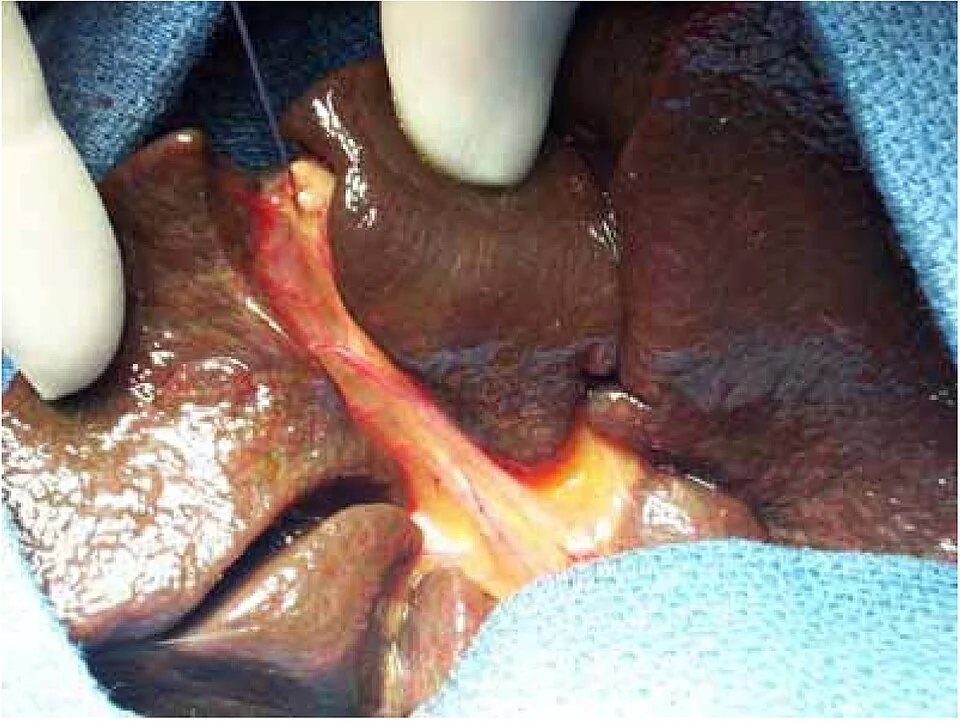
- Intraoperative cholangiogram - the definitive diagnostic test, performed at laparotomy, with the surgeon proceeding directly to a Kasai procedure if the ducts are shown to be obliterated
Management
The Kasai portoenterostomy
Since more than 90% of cases have no patent duct at the hilum, there is nothing to anastomose to in the conventional sense. The Kasai operation instead excises the fibrous biliary remnant back to the surface of the porta hepatis, where microscopic residual ductules still communicate with the intrahepatic biliary tree, and brings up a Roux-en-Y loop of jejunum to drain directly from that transected surface.
- Timing is the dominant prognostic factor. Performed before 60 days, around 60-70% of infants clear their jaundice; beyond 90 days the figure falls substantially, because portal fibrosis has already advanced.
- Success is defined as clearance of jaundice, conventionally a total bilirubin below 20-25 micromol/L within 6 months of surgery
- The Kasai is not a cure. It buys native liver function and growth, and defers or occasionally avoids transplantation.
- A failed Kasai - persistent jaundice at 3-6 months - is an indication to list for transplantation rather than to reoperate
Postoperative and supportive care
- Prophylactic antibiotics for the first months, since the Roux loop allows ascending bacterial cholangitis
- Ursodeoxycholic acid to improve bile flow and reduce the toxicity of retained bile acids
- Fat-soluble vitamin supplementation (A, D, E and K) with monitoring of levels, since deficiency continues while cholestasis persists
- Nutritional support with a feed enriched in medium-chain triglycerides, which are absorbed without bile salts, plus a high calorie intake - growth failure both worsens outcome and complicates transplantation
- Corticosteroids after the Kasai remain contentious; the START randomised trial did not show a significant improvement in bile drainage, and practice varies between centres
- Surveillance for portal hypertension with regular ultrasound, endoscopy where varices are suspected, and monitoring of growth, synthetic liver function and bilirubin
Liver transplantation
Biliary atresia is the single commonest indication for liver transplantation in children, accounting for roughly half of paediatric transplants. Indications are a failed Kasai, decompensated cirrhosis, recurrent cholangitis, intractable pruritus, portal hypertension with variceal bleeding, hepatopulmonary syndrome, and growth failure unresponsive to nutritional support. Outcomes are excellent, with 10-year survival well above 90%.
Complications
- Ascending cholangitis - most frequent in the first two years after surgery
- Portal hypertension with oesophageal and gastric varices, splenomegaly, hypersplenism and variceal haemorrhage
- Ascites and spontaneous bacterial peritonitis
- Fat-soluble vitamin deficiency - rickets from vitamin D deficiency, coagulopathy from vitamin K deficiency, neurological and haematological effects of vitamin E deficiency, and visual impairment from vitamin A deficiency
- Growth failure and malnutrition, which are among the strongest predictors of poor transplant outcome
- Hepatopulmonary syndrome and portopulmonary hypertension
- Hepatocellular carcinoma, uncommon but described in the cirrhotic native liver
- Complications of transplantation - rejection, hepatic artery thrombosis, biliary strictures, and the long-term effects of immunosuppression including post-transplant lymphoproliferative disorder
Prognosis and screening
Untreated, biliary atresia is uniformly fatal, with death from liver failure and its complications typically before the second birthday. With modern management the picture is very different: overall survival, combining native liver survival and transplantation, exceeds 90% at 10 years in UK series.4,5
Native liver survival is a more sober figure. Roughly half of infants clear their jaundice after a Kasai, and of those, many still develop progressive fibrosis over years, so that around 40-50% survive to 10 years with their own liver and the majority will need a transplant at some point in childhood or early adult life. Age at surgery, the anatomical type, the presence of BASM syndrome and the degree of fibrosis at operation all influence this.
Because outcome depends so heavily on age at surgery, several countries have introduced stool colour card screening, in which parents compare their baby's stool against printed reference colours in the first weeks. Taiwan's national programme substantially increased the proportion of infants operated on before 60 days. In the UK the equivalent is the Children's Liver Disease Foundation Yellow Alert campaign, together with the routine prolonged jaundice check at 14 days.3
The practical implication for anyone seeing babies is small and specific: for every infant still jaundiced at 2 weeks, send a split bilirubin and look at a nappy. Doing that consistently is what turns an untreatable disease into a treatable one.2
References
- NICE CG98. Jaundice in newborn babies under 28 days. 2010, updated 2023. Available here
- NICE Clinical Knowledge Summaries. Jaundice in the newborn. Available here
- Children's Liver Disease Foundation. Yellow Alert: jaundice in newborn babies and the stool colour chart. Available here
- Hartley JL, Davenport M, Kelly DA. Biliary atresia. The Lancet. 2009;374:1704-13. Available here
- Chardot C. Biliary atresia. Orphanet Journal of Rare Diseases. 2006;1:28. Available here
- British Society of Paediatric Gastroenterology, Hepatology and Nutrition (BSPGHAN). Available here
This article is written for revision and education. It is not clinical guidance and must not be used to make decisions about the care of a patient. Always check current NICE guidance and local protocols.