
Hepatocellular Carcinoma: Surveillance, Diagnosis and Management
Key points
- Hepatocellular carcinoma (HCC): the commonest primary liver malignancy, arising from hepatocytes and developing in a cirrhotic liver in around 80-90% of cases.
- Main risk factors: cirrhosis of any cause, chronic hepatitis B (which causes HCC even without cirrhosis), hepatitis C, alcohol, MASLD and aflatoxin exposure.
- Surveillance: six-monthly liver ultrasound, with or without alpha-fetoprotein, in all patients with cirrhosis and in selected non-cirrhotic hepatitis B carriers.
- Presentation: often silent until advanced; suspect it when a patient with stable cirrhosis unexpectedly decompensates.
- Diagnosis: uniquely among solid tumours, HCC can be diagnosed radiologically without biopsy in a cirrhotic liver, using characteristic contrast enhancement.
- Radiological hallmark: arterial phase hyperenhancement with washout in the portal venous or delayed phase, reflecting the tumour's arterial blood supply.
- Staging: the Barcelona Clinic Liver Cancer (BCLC) system uniquely combines tumour burden, liver function and performance status to guide treatment.
- Curative options: resection, liver transplantation (within the Milan criteria) and ablation; TACE and systemic therapy for intermediate and advanced disease.
Introduction
Hepatocellular carcinoma (HCC) is a malignant tumour of hepatocytes and the commonest primary liver cancer, accounting for around 75-85% of cases. Globally it is the third leading cause of cancer death, and its incidence in the UK has more than tripled over the last three decades, driven by chronic viral hepatitis, alcohol and the rising prevalence of MASLD.1
The defining feature that shapes its management is that HCC arises on a background of cirrhosis in 80-90% of cases. This means the clinician must always contend with two diseases simultaneously - the tumour and the underlying liver failure - and treatment that would be straightforward in a healthy liver may be impossible in a decompensated one. It is also what makes surveillance viable, since the at-risk population is clearly identifiable.
Risk factors
| Risk factor | Notes |
|---|---|
| Cirrhosis of any cause | The dominant risk factor - annual HCC incidence of around 2-4% in established cirrhosis |
| Chronic hepatitis B | Causes HCC with or without cirrhosis through viral genome integration. The leading global cause |
| Chronic hepatitis C | Causes HCC almost exclusively once cirrhosis has developed. Risk persists after viral cure, so surveillance continues |
| Alcohol-related liver disease | A major UK cause, acting synergistically with viral hepatitis and obesity |
| MASLD/MASH | The fastest-rising cause; may occasionally cause HCC without cirrhosis |
| Haemochromatosis | High risk once cirrhotic; risk persists despite venesection |
| Aflatoxin B1 | A mycotoxin from Aspergillus contaminating stored grains and nuts. Causes a characteristic p53 tumour suppressor gene mutation; a major cause in sub-Saharan Africa and East Asia |
| Other | Alpha-1 antitrypsin deficiency, autoimmune hepatitis, primary biliary cholangitis, Wilson disease, type 2 diabetes, obesity, smoking, anabolic steroids, and the oral contraceptive pill (weakly) |
Additional determinants of risk within a cirrhotic population include male sex (2-4 times the risk), older age, ongoing alcohol use, high viral load in hepatitis B, and coexisting diabetes.
Surveillance
Because HCC is asymptomatic until advanced but curable when small, surveillance of at-risk populations is central to improving outcomes and is one of the few genuinely effective cancer surveillance programmes in hepatology.2
- Six-monthly abdominal ultrasound, with or without serum alpha-fetoprotein (AFP). The six-month interval reflects the typical tumour doubling time
- Offer to: all patients with cirrhosis (of any cause) who would be candidates for treatment; non-cirrhotic patients with chronic hepatitis B according to age, ethnicity, viral load and family history; and patients with hepatitis C who have achieved viral cure but had established cirrhosis beforehand
- Do not offer surveillance to patients whose liver disease or comorbidity is so advanced that they could not tolerate any treatment, as it cannot benefit them
- A new focal lesion on surveillance ultrasound, or a rising AFP, triggers four-phase contrast CT or contrast-enhanced MRI
AFP alone is not adequate for surveillance: it is neither sensitive (normal in around a third of HCCs, including many small ones) nor specific (raised in chronic hepatitis, cirrhotic regeneration, pregnancy and germ cell tumours). Its value lies in trend monitoring, in prognostication - a very high AFP suggests aggressive biology - and in assessing treatment response.
Clinical features
Early HCC is asymptomatic, which is precisely why surveillance matters. When symptoms do appear, they are frequently those of the underlying cirrhosis rather than the tumour itself.
- Unexpected decompensation of previously stable cirrhosis - new or worsening ascites, jaundice, encephalopathy or variceal bleeding. This is often the first clue and should always prompt imaging
- Right upper quadrant pain or a dragging discomfort
- Weight loss, anorexia, fatigue and cachexia
- Hepatomegaly, sometimes with a palpable irregular mass, or an audible arterial bruit over the liver
- Jaundice, from tumour bulk or biliary obstruction
- Acute abdomen with haemoperitoneum from tumour rupture - a rare but dramatic and life-threatening presentation
- Fever of unknown origin
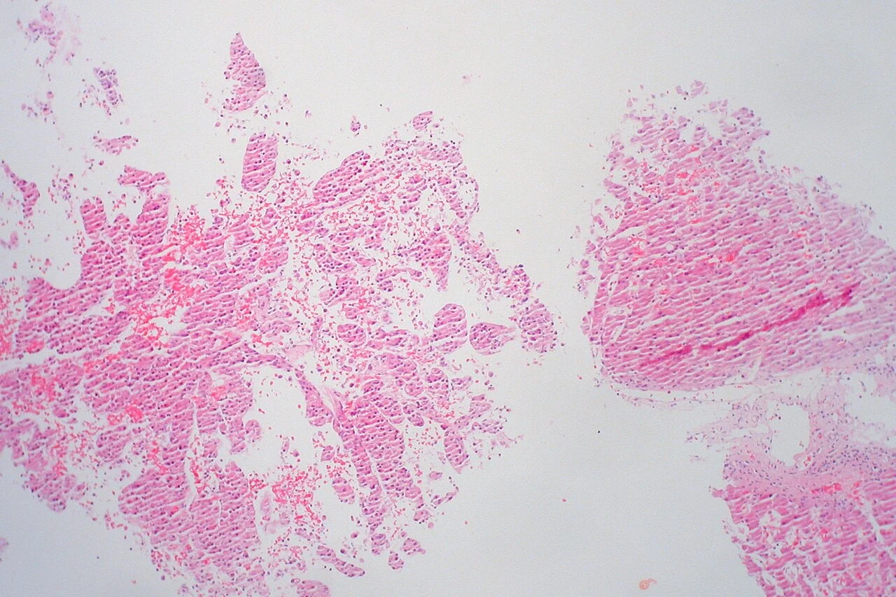
Paraneoplastic syndromes
HCC produces several recognised paraneoplastic phenomena, which are worth knowing because they can be the presenting feature:
- Hypoglycaemia - from tumour production of insulin-like growth factor II, or from massive tumour glucose consumption
- Hypercalcaemia - from parathyroid hormone-related peptide
- Erythrocytosis - from ectopic erythropoietin production, which is counterintuitive in a patient who might be expected to be anaemic
- Hypercholesterolaemia - from unregulated tumour cholesterol synthesis
- Watery diarrhoea, and cutaneous features such as porphyria cutanea tarda and pityriasis rotunda
Investigations
Radiological diagnosis
HCC is unusual among solid tumours in that a confident diagnosis can be made on imaging alone, without histology, in a patient with cirrhosis. This is possible because HCC has a distinctive vascular signature.2
A normal liver receives around 75% of its blood from the portal vein, but HCC develops a neovascular arterial supply as it grows. On four-phase contrast CT or contrast-enhanced MRI this produces the characteristic pattern:
- Arterial phase hyperenhancement - the lesion enhances brightly and earlier than the surrounding liver, because contrast reaches it through the hepatic artery first
- Washout in the portal venous or delayed phase - the lesion becomes relatively hypodense compared with the surrounding parenchyma, which is still enhancing from portal venous inflow
- A capsule or pseudocapsule may be seen with delayed enhancement
In a cirrhotic liver, a lesion over 1 cm showing this arterial hyperenhancement with washout is diagnostic of HCC and requires no biopsy. This is reported using the LI-RADS classification. Biopsy is generally avoided where imaging is diagnostic, both because it is unnecessary and because of a small risk of tumour seeding along the needle track and of bleeding in a coagulopathic patient. Biopsy is reserved for indeterminate lesions, or where the liver is non-cirrhotic and the diagnosis is uncertain.
Other investigations
- Alpha-fetoprotein (AFP) - for prognostication and monitoring rather than diagnosis
- Liver function tests, albumin, INR and bilirubin - to calculate the Child-Pugh score, which is essential for treatment planning
- Full blood count - thrombocytopenia indicates portal hypertension; erythrocytosis may be paraneoplastic
- Urea and electrolytes, calcium and glucose - for paraneoplastic hypercalcaemia and hypoglycaemia
- Hepatitis B and C serology, and a full aetiological screen if not already established
- CT chest for staging, as lung is the commonest site of extrahepatic spread
- Doppler ultrasound or cross-sectional imaging of the portal vein - portal vein invasion is common and profoundly alters staging and treatment options
- Upper GI endoscopy to assess varices, and performance status assessment
Staging
Conventional TNM staging is inadequate for HCC because it ignores liver function, which frequently determines survival more than the tumour does. The Barcelona Clinic Liver Cancer (BCLC) system is therefore used, uniquely integrating tumour burden, liver function (Child-Pugh) and performance status, and linking each stage directly to a treatment recommendation.3
| Stage | Features | Treatment |
|---|---|---|
| 0 - Very early | Single lesion under 2 cm, Child-Pugh A, performance status 0 | Resection or ablation |
| A - Early | Single lesion, or up to 3 nodules each under 3 cm; Child-Pugh A-B; performance status 0 | Curative intent: resection, liver transplantation, or ablation |
| B - Intermediate | Large or multinodular disease, Child-Pugh A-B, performance status 0 | Transarterial chemoembolisation (TACE) |
| C - Advanced | Portal vein invasion, extrahepatic spread, or performance status 1-2 | Systemic therapy - immunotherapy or tyrosine kinase inhibitors |
| D - Terminal | Child-Pugh C, or performance status 3-4 | Best supportive and palliative care |
The Milan criteria
These define which patients with HCC will do well after liver transplantation, and are widely used for listing: a single tumour of 5 cm or less, or up to three tumours each 3 cm or less, with no vascular invasion and no extrahepatic spread. Patients within these criteria achieve 5-year survival after transplantation of around 70%, comparable to transplantation for non-malignant indications. Transplantation is particularly attractive because it treats both the tumour and the underlying cirrhosis simultaneously.
Management
Management is directed by a specialist hepatobiliary multidisciplinary team, balancing tumour control against the patient's residual liver function.3
Curative treatments
- Surgical resection: the treatment of choice for a solitary tumour in a patient with preserved liver function and no significant portal hypertension. Cirrhosis limits how much liver can safely be removed, so careful assessment of future liver remnant is essential. Recurrence in the remaining diseased liver is common, at around 70% at 5 years
- Liver transplantation: the ideal treatment where criteria are met, since it removes both the tumour and the cirrhotic liver. Limited by organ availability and waiting-list dropout; patients may receive bridging TACE or ablation while waiting
- Ablation: radiofrequency or microwave ablation for small tumours (generally under 3 cm), particularly in patients unfit for resection. Comparable to resection for very small tumours and far less invasive
Non-curative and palliative treatments
- Transarterial chemoembolisation (TACE): chemotherapy delivered directly into the hepatic artery supplying the tumour, followed by embolisation. It exploits the tumour's arterial dependence while the normal liver survives on portal supply. Standard for BCLC stage B. Contraindicated in portal vein thrombosis and decompensated disease
- Selective internal radiotherapy (SIRT) with yttrium-90 microspheres, and stereotactic body radiotherapy, in selected patients
- Systemic therapy for advanced disease: atezolizumab plus bevacizumab (immunotherapy with an anti-VEGF agent) is now first-line, having superseded sorafenib, a multikinase inhibitor, which remains an option. Durvalumab with tremelimumab and lenvatinib are alternatives
- Best supportive care for BCLC stage D, with early palliative care involvement for pain, ascites and nutritional support
Prevention
Prevention is the most effective strategy of all: universal hepatitis B vaccination (which has measurably reduced childhood HCC in Taiwan and elsewhere), antiviral suppression of hepatitis B, curative direct-acting antivirals for hepatitis C, alcohol abstinence, weight management in MASLD, and reduction of aflatoxin exposure through improved grain storage. Coffee consumption is consistently associated with reduced HCC risk in cirrhosis.
Complications
- Decompensation of the underlying cirrhosis - ascites, encephalopathy, variceal bleeding and jaundice
- Portal vein invasion and thrombosis, which precludes resection, transplantation and TACE
- Tumour rupture with haemoperitoneum - a surgical emergency with high mortality
- Extrahepatic metastasis - most commonly lung, then bone (often osteolytic and painful), adrenal glands and lymph nodes
- Biliary obstruction and obstructive jaundice
- Paraneoplastic syndromes - hypoglycaemia, hypercalcaemia and erythrocytosis
- Cachexia and malnutrition
- Recurrence after treatment, which is the rule rather than the exception given the field change in the remaining cirrhotic liver
- Treatment complications: post-embolisation syndrome after TACE, post-hepatectomy liver failure, and the immune-related adverse effects of checkpoint inhibitors
Red flags
Prognosis
Prognosis depends on both the tumour stage and the underlying liver function, which is exactly why the BCLC system incorporates the two. Overall 5-year survival in the UK remains poor at around 10-15%, chiefly because many patients present with advanced disease.1
The outlook for early-stage disease detected through surveillance is far better: 5-year survival after resection or ablation for BCLC stage 0-A is around 50-70%, and after transplantation within the Milan criteria approaches 70%. This gulf between screen-detected and symptomatic disease is the entire justification for the six-monthly surveillance programme, and the strongest argument for ensuring patients with cirrhosis are actually enrolled in it.
Intermediate disease treated with TACE has a median survival of roughly 2-2.5 years, while advanced disease on modern systemic therapy achieves a median survival of around 19 months with atezolizumab plus bevacizumab - a meaningful improvement over the sorafenib era. Recurrence remains the central problem even after curative treatment, since the whole cirrhotic liver is at risk, so lifelong surveillance continues after successful treatment.
References
- Cancer Research UK. Liver cancer statistics. Available here
- European Association for the Study of the Liver. EASL Clinical Practice Guidelines: management of hepatocellular carcinoma. J Hepatol. 2018. Available here
- Reig M et al. BCLC strategy for prognosis prediction and treatment recommendation: the 2022 update. J Hepatol. 2022. Available here
- NICE NG50. Cirrhosis in over 16s: assessment and management. 2016. Available here
- Ed Uthman, CC BY 2.0, via Wikimedia Commons. Available here
- Mazzaferro V et al. Liver transplantation for the treatment of small hepatocellular carcinomas (the Milan criteria). N Engl J Med. 1996. Available here
- NICE TA666. Atezolizumab with bevacizumab for treating advanced or unresectable hepatocellular carcinoma. 2020. Available here
This article is written for revision and education. It is not clinical guidance and must not be used to make decisions about the care of a patient. Always check current NICE guidance and local protocols.