
Variceal Bleeding: Emergency Management and Prophylaxis
Key points
- Varices: dilated portosystemic collateral veins that form when portal pressure rises, most importantly at the gastro-oesophageal junction.
- Cause: portal hypertension, most often from liver cirrhosis; bleeding risk rises once the hepatic venous pressure gradient exceeds 12 mmHg.
- Presentation: large-volume haematemesis and melaena, often with haemodynamic compromise and signs of chronic liver disease.
- Immediate drugs: terlipressin and prophylactic broad-spectrum antibiotics, both started as soon as variceal bleeding is suspected - before endoscopy.
- Endoscopy: within 12 hours of presentation. Band ligation is first-line for oesophageal varices; injection of a tissue adhesive for gastric varices.
- Rescue therapy: balloon tamponade as a temporising bridge, and TIPS for bleeding refractory to endoscopic control.
- Transfusion: use a restrictive threshold (target haemoglobin around 70-80 g/L); over-transfusion raises portal pressure and worsens rebleeding.
- Prophylaxis: non-selective beta-blockers or band ligation for primary prophylaxis; both beta-blockade and banding after a bleed.
Introduction
Varices are dilated collateral veins that develop as a consequence of portal hypertension, providing an alternative route for portal blood to reach the systemic circulation. Those at the gastro-oesophageal junction are the most clinically important, because they are thin-walled, superficial, and prone to catastrophic rupture.1
Variceal haemorrhage is one of the most feared complications of cirrhosis. It accounts for a minority of upper GI bleeds overall but a disproportionate share of the mortality, with a six-week mortality of roughly 15-20% even with modern management.2 Around half of patients with cirrhosis have varices at diagnosis, and roughly a third of those with varices will bleed.
Pathophysiology
In cirrhosis, fibrosis and regenerative nodules increase resistance to portal blood flow, while splanchnic vasodilatation increases portal inflow. Together these raise portal venous pressure. Portal hypertension is quantified by the hepatic venous pressure gradient (HVPG), normally 1-5 mmHg.
| HVPG | Significance |
|---|---|
| 1-5 mmHg | Normal |
| Above 5 mmHg | Portal hypertension present |
| Above 10 mmHg | Clinically significant portal hypertension - varices begin to form |
| Above 12 mmHg | Threshold above which varices may bleed, and ascites develops |
As pressure rises, portosystemic collaterals open at sites where the portal and systemic circulations meet: the gastro-oesophageal junction (oesophageal and gastric varices), the umbilicus (caput medusae), the rectum (rectal varices, distinct from haemorrhoids), and the retroperitoneum. Variceal rupture is governed by wall tension, so it is driven by large variceal size, thin wall (seen endoscopically as red wale marks) and high portal pressure.1
Causes of portal hypertension
- Pre-hepatic: portal vein thrombosis, splenic vein thrombosis
- Hepatic (commonest): cirrhosis of any cause - alcohol-related liver disease, viral hepatitis, MASLD; also schistosomiasis worldwide
- Post-hepatic: Budd-Chiari syndrome, right heart failure, constrictive pericarditis
Clinical features
Variceal bleeding classically presents with large-volume haematemesis and melaena, frequently with haemodynamic compromise. Because bleeding can be torrential, patients may present collapsed or in shock, and a brisk bleed can present as fresh blood per rectum.
Look for evidence of chronic liver disease and portal hypertension, which raises the pre-test probability considerably:
- Stigmata of chronic liver disease: jaundice, spider naevi, palmar erythema, gynaecomastia, Dupuytren contracture, leuconychia
- Signs of portal hypertension: ascites, splenomegaly, caput medusae
- Signs of decompensation: hepatic encephalopathy with asterixis and confusion, which GI bleeding itself precipitates
- A history of alcohol excess, viral hepatitis, or known cirrhosis with previous variceal banding
Emergency management
This is a medical emergency requiring simultaneous resuscitation, drug therapy and arrangement of urgent endoscopy.3
Resuscitation
- Airway: a high risk of aspiration with massive haematemesis or encephalopathy - involve anaesthetics early and consider intubation before endoscopy
- Circulation: two large-bore cannulae, urgent bloods including full blood count, urea and electrolytes, liver function, clotting and crossmatch, and activation of the major haemorrhage protocol if needed
- Restrictive transfusion: target a haemoglobin of around 70-80 g/L. Over-transfusion increases portal pressure and paradoxically worsens rebleeding and mortality
- Correct coagulopathy with vitamin K and blood products as indicated, though routine correction of a prolonged INR in cirrhosis is not recommended, as it does not reflect true bleeding risk
Drug therapy - start before endoscopy
Terlipressin, a vasopressin analogue, causes splanchnic vasoconstriction and reduces portal inflow and pressure. It is started as soon as variceal bleeding is suspected and continued for up to 5 days. Caution is needed in ischaemic heart disease and peripheral vascular disease, and it can cause hyponatraemia.3
Prophylactic broad-spectrum antibiotics (e.g. a quinolone or third-generation cephalosporin) are given to every patient with cirrhosis and upper GI bleeding, irrespective of whether infection is evident. Bacterial infection is common in this setting and antibiotic prophylaxis reduces rebleeding, infection and mortality - this is one of the highest-yield interventions in the whole pathway.
Endoscopic and rescue therapy
Upper GI endoscopy should be performed within 12 hours of presentation, once the patient has been resuscitated.3
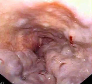
Endoscopic therapy
- Oesophageal varices: endoscopic variceal band ligation is first-line. Bands are applied to suck up and strangulate the varix, causing thrombosis and eventual obliteration. It is superior to sclerotherapy, which is now reserved for cases where banding is technically impossible
- Gastric varices: injection of a tissue adhesive (N-butyl-2-cyanoacrylate glue) is first-line, as banding is less effective at this site
Rescue therapy for uncontrolled bleeding
Balloon tamponade with a Sengstaken-Blakemore or Minnesota tube mechanically compresses the varices. It is highly effective but only a temporising bridge for a maximum of around 24 hours, because of the risk of pressure necrosis and oesophageal rupture; the airway should be protected first.
Transjugular intrahepatic portosystemic shunt (TIPS) creates a channel between the hepatic and portal veins within the liver, decompressing the portal system. It is used for bleeding refractory to endoscopic therapy, and is increasingly used early in high-risk patients. Its major complication is hepatic encephalopathy, since blood bypasses the liver's detoxifying function; it can also worsen liver function. Self-expanding covered metal oesophageal stents are an alternative bridge to TIPS.
Prophylaxis
Patients with cirrhosis undergo screening endoscopy to identify varices before they bleed, with the interval determined by whether varices are present and their size.
Primary prophylaxis (preventing a first bleed)
For medium or large varices, either a non-selective beta-blocker (propranolol or carvedilol) or endoscopic band ligation is offered.4 Non-selective beta-blockers reduce portal pressure by reducing cardiac output and, through unopposed alpha-adrenergic activity, causing splanchnic vasoconstriction - selective beta-blockers do not achieve this and are ineffective.
Secondary prophylaxis (preventing rebleeding)
After an index bleed, the risk of rebleeding is high, so both a non-selective beta-blocker and a programme of repeated band ligation are used, with banding sessions typically every 2-4 weeks until the varices are obliterated. TIPS is considered where bleeding recurs despite this combination. Patients should also be assessed for liver transplantation and receive treatment of the underlying liver disease, including alcohol cessation support.
Complications
- Hypovolaemic shock, multi-organ failure and death from exsanguination
- Rebleeding, which is common and carries a high mortality; the risk is greatest in the first 5 days
- Hepatic encephalopathy, precipitated by the protein load of blood in the gut - treat with lactulose and rifaximin
- Aspiration pneumonia, a major cause of death in this group
- Spontaneous bacterial peritonitis and other infections, hence routine antibiotic prophylaxis
- Hepatorenal syndrome and acute kidney injury following the hypotensive insult
- Complications of therapy: post-banding ulceration and stricture, oesophageal rupture from balloon tamponade, encephalopathy after TIPS
Red flags
Prognosis
Variceal bleeding carries a six-week mortality of around 15-20%, having improved substantially with the combination of vasoactive drugs, antibiotic prophylaxis, early endoscopic banding and early TIPS.2 Prognosis is determined less by the bleed itself than by the severity of the underlying liver disease, assessed with the Child-Pugh or MELD score: a patient with well-compensated cirrhosis has a far better outlook than one with decompensated disease. Rebleeding risk is highest in the first five days and remains substantial for six weeks, which is why secondary prophylaxis is started before discharge.
References
- Tripathi D et al. UK guidelines on the management of variceal haemorrhage in cirrhotic patients. Gut. 2015. Available here
- European Association for the Study of the Liver. EASL Clinical Practice Guidelines for decompensated cirrhosis. J Hepatol. 2018. Available here
- NICE CG141. Acute upper gastrointestinal bleeding in over 16s: management. 2012 (updated 2016). Available here
- NICE Clinical Knowledge Summaries (CKS). Cirrhosis. 2023. Available here
- Samir, Public domain, via Wikimedia Commons. Available here
- NICE NG50. Cirrhosis in over 16s: assessment and management. 2016. Available here
- British Society of Gastroenterology. Management of acute upper gastrointestinal bleeding. Gut. 2019. Available here
This article is written for revision and education. It is not clinical guidance and must not be used to make decisions about the care of a patient. Always check current NICE guidance and local protocols.