
Upper Gastrointestinal Bleed: Assessment and Management
Key points
- Upper GI bleed: bleeding from a source proximal to the ligament of Treitz, presenting with haematemesis and/or melaena.
- Common causes: peptic ulcer disease is the most common cause overall; other causes include oesophageal varices, Mallory-Weiss tears, oesophagitis, gastritis and malignancy.
- Initial assessment: ABCDE approach with immediate resuscitation; assess severity using the shock index and clinical signs of haemodynamic compromise.
- Risk scoring: Glasgow-Blatchford score before endoscopy to guide admission and timing; Rockall score after endoscopy to estimate mortality and rebleed risk.
- Resuscitation: IV access, crystalloid, and blood transfusion for major bleeding, guided by a restrictive transfusion threshold (haemoglobin around 70-80 g/L).
- Endoscopy: definitive diagnosis and treatment; within 24 hours for most patients, immediately after resuscitation if unstable or variceal bleeding is suspected.
- Variceal bleeding: terlipressin and prophylactic antibiotics before endoscopy, with band ligation as first-line endoscopic therapy.
- Non-variceal bleeding: endoscopic haemostasis (adrenaline injection, thermal coagulation or clips), followed by high-dose PPI therapy.
Introduction
Upper gastrointestinal (GI) bleeding is bleeding arising proximal to the ligament of Treitz, encompassing the oesophagus, stomach and duodenum.1 It is one of the most common gastrointestinal emergencies, with an annual UK incidence of roughly 100-150 per 100,000 adults, and carries an overall mortality of around 10%, higher in elderly patients and those with major comorbidity.2
Rapid recognition, risk stratification and resuscitation, run in parallel with arranging endoscopy, are the priorities in management.
Aetiology
Peptic ulcer disease is the single most common cause, accounting for roughly a third to a half of cases.1 Other important causes include:
- Oesophageal or gastric varices: secondary to portal hypertension, usually from cirrhosis; less common overall but a major cause of massive, life-threatening bleeding
- Mallory-Weiss tear: a mucosal tear at the gastro-oesophageal junction, classically after repeated retching or vomiting
- Erosive oesophagitis or gastritis: from acid reflux, NSAIDs, alcohol, or critical illness
- Malignancy: gastric or oesophageal cancer
- Angiodysplasia and vascular lesions
- Dieulafoy lesion: a large, tortuous submucosal artery that erodes through the mucosa, causing sudden severe bleeding from a small mucosal defect
Clinical features
The hallmark presentations are haematemesis (vomiting blood, which may be fresh red or altered "coffee-ground" in appearance) and melaena (black, tarry, offensive-smelling stool from digested blood). Bleeding severe enough to cause rapid transit can present with haematochezia (fresh red blood per rectum), which usually indicates a large-volume upper GI bleed rather than a lower GI source.
Features of haemodynamic compromise include tachycardia, hypotension, postural drop, pallor, cool peripheries, reduced urine output and, in severe cases, syncope or collapse. Important points in the history include:
- Prior peptic ulcer disease, dyspepsia or known H. pylori status
- NSAID, aspirin or anticoagulant use
- Alcohol intake and known or suspected liver disease (varices)
- Retching or vomiting preceding the bleed (Mallory-Weiss tear)
- Weight loss, dysphagia or anorexia (malignancy)
- Previous GI bleeding or endoscopic findings
Initial assessment and resuscitation
Upper GI bleeding is managed with an ABCDE approach, with resuscitation starting immediately and in parallel with, not after, further assessment.2
- Airway and breathing: protect the airway if there is reduced consciousness or massive haematemesis; give high-flow oxygen if needed
- Circulation: insert two large-bore IV cannulae, send bloods (full blood count, urea and electrolytes, liver function tests, coagulation screen, group and save/crossmatch, venous lactate), and give IV crystalloid
- Transfusion: give blood for major bleeding, using a restrictive threshold in stable patients (transfuse to a haemoglobin target of around 70-80 g/L rather than higher, since over-transfusion in variceal bleeding can worsen portal pressure and rebleeding)
- Correct coagulopathy: reverse anticoagulation where appropriate (e.g. vitamin K and prothrombin complex concentrate for warfarin), and correct significant thrombocytopenia or coagulopathy before endoscopy
- Disability and exposure: assess conscious level and look for stigmata of chronic liver disease
The shock index (heart rate divided by systolic blood pressure) is a simple bedside marker of severity; a value above 1 suggests significant haemodynamic compromise and correlates with the need for intervention and higher mortality.
Risk stratification
Validated scoring systems guide urgency of endoscopy, admission decisions, and prognosis.
Glasgow-Blatchford score
Calculated on initial assessment, before endoscopy, using urea, haemoglobin, systolic blood pressure, heart rate, and the presence of melaena, syncope, hepatic disease or cardiac failure.3 A score of 0 identifies very low-risk patients who may be safe for outpatient management; higher scores indicate a need for admission, transfusion or endoscopic intervention.
Rockall score
Calculated in two stages: an initial clinical score (age, shock, comorbidity) before endoscopy, and a full score incorporating the endoscopic diagnosis and stigmata of recent haemorrhage afterwards. It estimates the risk of rebleeding and mortality, and is used after the acute event rather than to decide the timing of endoscopy.3
Investigations
Upper GI endoscopy (OGD) is both diagnostic and therapeutic, and is the key investigation.2 Timing depends on severity: endoscopy is performed immediately after resuscitation in unstable patients or suspected variceal bleeding, and within 24 hours of admission for other patients.
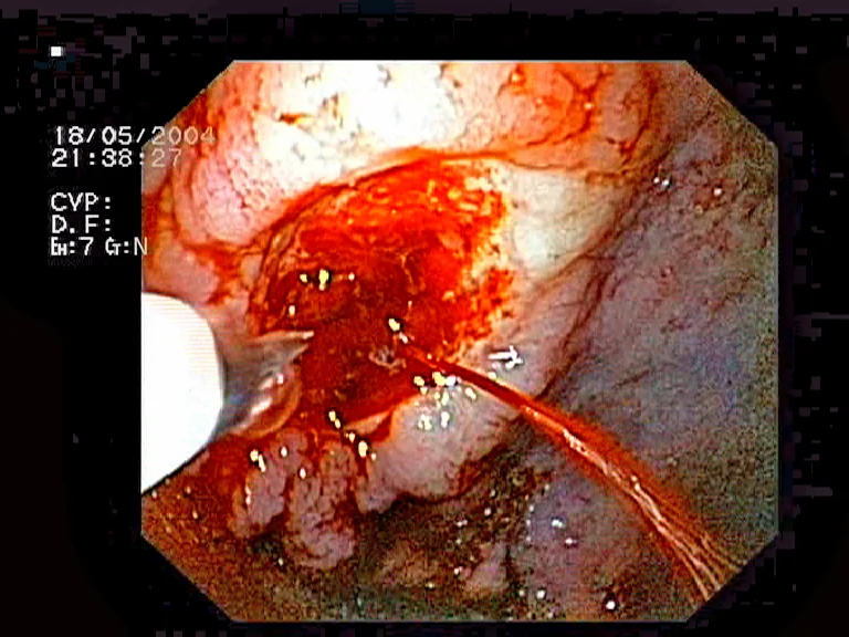
Forrest classification
Endoscopic appearance of a bleeding ulcer is described using the Forrest classification, which predicts the risk of rebleeding and guides the decision to intervene endoscopically.4
| Class | Appearance | Rebleed risk |
|---|---|---|
| Ia | Active spurting haemorrhage | High |
| Ib | Active oozing haemorrhage | High |
| IIa | Visible non-bleeding vessel | High |
| IIb | Adherent clot | Intermediate |
| IIc | Flat pigmented spot | Low |
| III | Clean ulcer base | Very low |
Forrest Ia-IIb lesions generally require endoscopic haemostasis; IIc and III lesions have a low rebleed risk and are usually managed medically alone.
Management
Suspected variceal bleeding
In patients with known or suspected liver disease, start terlipressin (a vasopressin analogue that reduces portal pressure) and prophylactic broad-spectrum antibiotics as soon as variceal bleeding is suspected, before endoscopy confirms the diagnosis.5 At endoscopy, band ligation is first-line for oesophageal varices; balloon tamponade (e.g. a Sengstaken-Blakemore tube) is used as a temporising measure for uncontrolled bleeding, and a transjugular intrahepatic portosystemic shunt (TIPS) is considered for bleeding refractory to endoscopic therapy.
Non-variceal bleeding
PPIs are not given routinely before endoscopy in suspected non-variceal bleeding, as this does not improve outcomes, though local practice varies.2 At endoscopy, haemostasis is achieved using injection (adrenaline), thermal coagulation, mechanical clips, or a combination. High-dose IV PPI therapy is started after endoscopic treatment of a bleeding peptic ulcer, which reduces rebleeding risk by maintaining a higher intragastric pH that stabilises clot formation.
Ongoing or recurrent bleeding
Rebleeding after initial endoscopic control is managed with repeat endoscopy in the first instance. Interventional radiology (angiographic embolisation) or surgery is considered if endoscopic haemostasis fails or is not feasible.
Complications
- Hypovolaemic shock and its sequelae, including acute kidney injury and myocardial ischaemia in patients with coronary disease
- Rebleeding, which is the strongest predictor of mortality
- Aspiration pneumonia, particularly with massive haematemesis or reduced consciousness
- Hepatic encephalopathy, precipitated by the protein load of GI bleeding in patients with cirrhosis
- Death, particularly in elderly patients, those with major comorbidity, and variceal bleeding
Red flags
Prognosis
Overall mortality from upper GI bleeding is around 10%, but this varies enormously with cause and patient factors: low-risk peptic ulcer bleeds managed promptly have an excellent prognosis, while variceal bleeding in decompensated cirrhosis carries a much higher mortality.2 Rebleeding is the single most important predictor of death, which is why risk stratification, prompt endoscopy, and post-endoscopic PPI or vasoactive therapy are central to management.
References
- NICE CG141. Acute upper gastrointestinal bleeding in over 16s: management. 2012 (updated 2016). Available here
- British Society of Gastroenterology. Management of acute upper gastrointestinal bleeding. 2019. Available here
- Blatchford O et al. A risk score to predict need for treatment for upper-gastrointestinal haemorrhage. Lancet. 2000. Available here
- Forrest JA, Finlayson ND, Shearman DJ. Endoscopy in gastrointestinal bleeding. Lancet. 1974. Available here
- NICE CG141. Variceal haemorrhage - recommendations. Available here
- Jeremias, CC BY-SA 3.0, via Wikimedia Commons. Available here
- NHS. Internal bleeding. 2022. Available here
This article is written for revision and education. It is not clinical guidance and must not be used to make decisions about the care of a patient. Always check current NICE guidance and local protocols.