
Lower Gastrointestinal Bleed: Assessment and Management
Key points
- Lower GI bleed: bleeding from a source distal to the ligament of Treitz, typically presenting with fresh or dark red blood per rectum.
- Common causes: diverticular disease is the most common cause of major lower GI bleeding; haemorrhoids are the commonest cause of minor bleeding.
- Key distinction: a brisk upper GI bleed can present as fresh rectal bleeding with haemodynamic compromise, and must be excluded first.
- Assessment: ABCDE approach with resuscitation, then risk stratification (e.g. the Oakland score) to decide on admission or outpatient management.
- Investigation: CT angiography for unstable patients with active bleeding; colonoscopy for stable patients, usually after bowel preparation.
- Management: most bleeds stop spontaneously; options include endoscopic haemostasis, angiographic embolisation, and surgery as a last resort.
- Red flags: haemodynamic instability, ongoing bleeding, or associated weight loss and change in bowel habit suggesting malignancy.
- Prognosis: roughly 80% of lower GI bleeds settle without intervention, but mortality rises in older patients with comorbidity.
Introduction
Lower gastrointestinal (GI) bleeding is bleeding that originates distal to the ligament of Treitz, that is, from the small bowel beyond the duodenojejunal flexure, the colon, the rectum or the anus.1 It usually presents as haematochezia: the passage of fresh or dark red blood per rectum, with or without clots.
It is around a fifth as common as upper GI bleeding but becomes markedly more frequent with age, reflecting the age distribution of diverticular disease and angiodysplasia. Most episodes are self-limiting, but a minority represent major haemorrhage requiring urgent intervention.
Aetiology
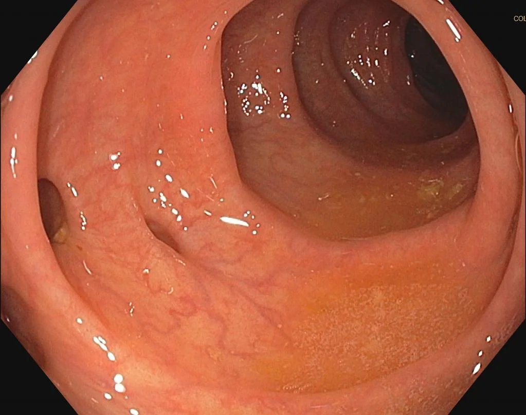
The likely cause depends heavily on the patient's age and the volume of bleeding.1,2
| Cause | Typical features |
|---|---|
| Diverticular disease | Commonest cause of major lower GI bleeding; painless, large-volume, often sudden and self-limiting |
| Haemorrhoids and anal fissure | Commonest cause of minor bleeding; fresh blood on the paper or coating the stool, fissure gives severe pain on defecation |
| Colorectal cancer or polyps | Bleeding with change in bowel habit, weight loss, or iron-deficiency anaemia |
| Angiodysplasia | Painless, recurrent bleeding, more common in older patients and those with aortic stenosis or renal failure |
| Inflammatory bowel disease | Bloody diarrhoea with urgency, mucus, and systemic upset |
| Infective colitis | Bloody diarrhoea with fever and recent travel, antibiotics or contact history |
| Ischaemic colitis | Crampy abdominal pain then bloody diarrhoea, typically at the splenic flexure in an arteriopath |
| Post-polypectomy bleeding | Within 2 weeks of a recent colonoscopic procedure |
Clinical features
The character of the bleeding gives useful clues to the site. Bright red blood suggests a distal (anorectal or left-sided colonic) source, while darker, maroon-coloured blood mixed with stool suggests a more proximal colonic or small bowel source. Melaena points strongly to an upper GI source rather than a lower one.
Important features to establish in the history:
- Colour, volume and duration of bleeding, and whether blood is mixed with stool or separate
- Associated pain: painless bleeding suggests diverticular disease or angiodysplasia, whereas pain suggests ischaemic colitis, IBD or an anal fissure
- Change in bowel habit, tenesmus or weight loss (malignancy)
- Recent colonoscopy or polypectomy
- Anticoagulant, antiplatelet or NSAID use
- Known diverticular disease, IBD, or previous bleeding episodes
- Vascular risk factors and recent hypotension (ischaemic colitis)
Examination should include abdominal examination, digital rectal examination and, where appropriate, proctoscopy or rigid sigmoidoscopy to identify an obvious anorectal source and to assess for a rectal mass.
Initial assessment and resuscitation
As with upper GI bleeding, the priority is an ABCDE approach with resuscitation running alongside assessment.2 Large-bore IV access, bloods (full blood count, urea and electrolytes, liver function, clotting, group and save or crossmatch), fluid resuscitation and transfusion where needed all take precedence over identifying the exact bleeding point.
Anticoagulation is reviewed and reversed where the bleeding is major, balancing the thrombotic risk of stopping it against the bleeding risk of continuing.
Risk stratification
The Oakland score is used to identify patients with lower GI bleeding who are at low risk and can safely be managed as outpatients.2 It incorporates age, sex, previous admission with lower GI bleeding, findings on digital rectal examination, heart rate, systolic blood pressure and haemoglobin. A score of 8 or less identifies patients suitable for discharge with outpatient follow-up, while higher scores indicate a need for admission and inpatient investigation.
Regardless of score, any patient with ongoing bleeding or haemodynamic instability requires admission and urgent investigation.
Investigations
In unstable patients
CT angiography (CTA) is the first-line investigation in haemodynamically unstable patients with suspected ongoing bleeding. It can localise active bleeding rapidly and allows immediate progression to catheter angiography and embolisation if a bleeding point is identified.2
In stable patients
Colonoscopy is the investigation of choice for stable patients, usually performed after bowel preparation during the admission. It is both diagnostic and therapeutic, allowing haemostasis with clips, thermal coagulation or adrenaline injection.
Obscure bleeding
If both upper GI endoscopy and colonoscopy are negative, a small bowel source is considered. Capsule endoscopy is the usual next step, with CT enterography or double-balloon enteroscopy as alternatives. A Meckel's diverticulum should be considered in younger patients, and can be identified with a technetium-99m pertechnetate (Meckel's) scan.
Management
The majority of lower GI bleeds (around 80%) stop spontaneously with supportive care alone, so management is often conservative with observation, fluid resuscitation and transfusion as required.1
Interventional options
- Endoscopic haemostasis: clips, thermal coagulation, or adrenaline injection at colonoscopy
- Angiographic embolisation: superselective embolisation of the bleeding vessel at catheter angiography, usually following a positive CTA; carries a small risk of bowel ischaemia
- Surgery: segmental or, rarely, subtotal colectomy, reserved for uncontrolled bleeding where the source has been localised and other measures have failed
Treating the underlying cause
Definitive management addresses the specific diagnosis: haemorrhoid banding or surgery, treatment of IBD, resection for colorectal cancer, or argon plasma coagulation for angiodysplasia. Anticoagulation is restarted once bleeding has settled, with the timing individualised to the thrombotic risk.
Complications
- Hypovolaemic shock, acute kidney injury and myocardial ischaemia from major blood loss
- Rebleeding, which is common in diverticular and angiodysplastic bleeding
- Bowel ischaemia or infarction following angiographic embolisation
- Complications of blood transfusion and of emergency surgery in frail patients
- Delayed diagnosis of colorectal cancer if bleeding is attributed to haemorrhoids without further investigation
Red flags
Prognosis
Most lower GI bleeding settles spontaneously and has a good prognosis, with overall mortality lower than for upper GI bleeding at roughly 3-5%.2 Outcomes are worse in older patients, those bleeding while already an inpatient for another condition, those on anticoagulants, and those with significant comorbidity. Diverticular bleeding recurs in around a quarter of patients, and angiodysplasia has a particular tendency to rebleed, sometimes requiring repeated endoscopic treatment.
References
- British Society of Gastroenterology. Guidelines on the management of acute lower gastrointestinal bleeding. Gut. 2019. Available here
- Oakland K et al. Derivation and validation of a novel risk score for safe discharge after acute lower gastrointestinal bleeding. Lancet Gastroenterol Hepatol. 2017. Available here
- NICE NG12. Suspected cancer: recognition and referral. 2015 (updated 2023). Available here
- NICE Clinical Knowledge Summaries (CKS). Diverticular disease. 2024. Available here
- Jmarchn, CC BY-SA 3.0, via Wikimedia Commons. Available here
- NHS. Blood in your poo. 2024. Available here
- Strate LL, Gralnek IM. ACG clinical guideline: management of patients with acute lower gastrointestinal bleeding. Am J Gastroenterol. 2016. Available here
This article is written for revision and education. It is not clinical guidance and must not be used to make decisions about the care of a patient. Always check current NICE guidance and local protocols.