
Diverticular Disease and Diverticulitis: Diagnosis and Management
Key points
- Diverticulosis: the presence of colonic diverticula without symptoms - extremely common, affecting most people over 80, and usually an incidental finding.
- Diverticular disease: diverticula causing symptoms such as intermittent lower abdominal pain and altered bowel habit, without acute inflammation.
- Diverticulitis: acute inflammation or infection of one or more diverticula, causing left iliac fossa pain, fever and raised inflammatory markers.
- Pathophysiology: raised intraluminal pressure in a low-fibre colon forces mucosa through weak points where vasa recta penetrate the muscle wall - these are false (pulsion) diverticula.
- Site: the sigmoid colon in over 90% of Western patients, as it has the narrowest lumen and highest pressures; right-sided disease predominates in Asia.
- Investigation: CT abdomen and pelvis with contrast is the investigation of choice in acute diverticulitis; avoid colonoscopy acutely because of perforation risk.
- Uncomplicated diverticulitis: many patients can be managed at home without antibiotics; use analgesia (not NSAIDs or opioids), fluids and safety-netting.
- Complications: perforation, abscess, fistula (commonly colovesical), obstruction from stricture, and diverticular bleeding.
Introduction and terminology
Colonic diverticula are small outpouchings of the bowel wall. They are extremely common in Western populations, present in around 50% of people over 50 and the majority of those over 80, and in most cases cause no symptoms at all.1
The terminology is frequently confused, and getting it right matters because management differs substantially at each stage:
| Term | Definition |
|---|---|
| Diverticulum | An outpouching of the bowel wall |
| Diverticulosis | The presence of diverticula without symptoms - usually an incidental finding at colonoscopy or on CT |
| Diverticular disease | Diverticula causing symptoms (intermittent abdominal pain, bloating, altered bowel habit) without acute inflammation |
| Diverticulitis | Acute inflammation or infection of one or more diverticula |
| Complicated diverticulitis | Diverticulitis with abscess, perforation, fistula, obstruction or significant bleeding |
Pathophysiology and risk factors
Colonic diverticula are false (pulsion) diverticula: they contain only mucosa and submucosa herniating through the muscular wall, not all layers. They form at the points of anatomical weakness where the vasa recta penetrate the circular muscle layer to supply the mucosa - which is precisely why diverticular bleeding can be brisk, as the herniating mucosa sits directly against an artery.2
A low-fibre diet produces smaller, harder stools requiring higher intraluminal pressures to propel. These pressures are highest in the sigmoid colon, which has the narrowest lumen (by the law of Laplace, pressure is inversely related to radius), explaining why over 90% of Western diverticular disease is sigmoid. In Asian populations, right-sided diverticula predominate and are more often true diverticula.
Diverticulitis arises when a diverticulum becomes obstructed, classically by inspissated faecal material, leading to bacterial overgrowth, local ischaemia of the thin wall, inflammation and micro- or macroperforation. Contemporary understanding places more emphasis on chronic low-grade inflammation and altered microbiota than on simple faecalith obstruction alone.
Risk factors
- Increasing age - the dominant risk factor
- Low dietary fibre intake
- Obesity and physical inactivity
- Smoking
- NSAIDs, aspirin, opioids and corticosteroids - associated with perforation and bleeding
- A diet high in red meat
- Connective tissue disorders such as Ehlers-Danlos and Marfan syndromes, which cause diverticula at a younger age
- Family history and genetic predisposition
Clinical features
Diverticulosis and diverticular disease
Diverticulosis is asymptomatic by definition. Diverticular disease causes intermittent, crampy left iliac fossa pain, often relieved by defecation or passing flatus, with bloating and altered bowel habit. Systemic features are absent, and inflammatory markers are normal - a picture that overlaps considerably with irritable bowel syndrome.
Acute diverticulitis
The classic presentation is constant left iliac fossa pain with fever and localised tenderness, sometimes described as "left-sided appendicitis". Features include:
- Constant lower abdominal pain, usually left iliac fossa, sometimes suprapubic
- Localised tenderness, with guarding or rebound if there is local peritonism; a tender mass may be palpable if an abscess or phlegmon has formed
- Fever and malaise
- Altered bowel habit - constipation more often than diarrhoea
- Nausea and vomiting
- Urinary symptoms (frequency, dysuria) if the inflamed sigmoid lies against the bladder - and pneumaturia or faecaluria if a colovesical fistula has developed
- Per rectum bleeding is uncommon in acute diverticulitis; diverticular bleeding is typically painless and separate from an inflammatory episode
- Signs of generalised peritonitis, sepsis or shock in perforation
Differential diagnosis
- Colorectal cancer: the single most important differential, as it can present with identical symptoms and a similar CT appearance - which is why interval investigation of the colon is considered after an episode
- Irritable bowel syndrome: overlapping symptoms, but no fever or raised inflammatory markers
- Inflammatory bowel disease: bloody diarrhoea, systemic upset, raised calprotectin
- Appendicitis: right-sided, but a long or pelvic appendix, or right-sided diverticulitis, can mimic
- Ischaemic colitis: pain out of proportion, bloody diarrhoea, vascular risk factors
- Gynaecological: ectopic pregnancy, ovarian cyst torsion or rupture, pelvic inflammatory disease - always perform a pregnancy test in women of childbearing age
- Urological: renal or ureteric colic, urinary tract infection
- Infective colitis, and incarcerated hernia
Investigations
- Bloods: full blood count (leucocytosis), CRP (a key marker of severity and of complicated disease), urea and electrolytes, and group and save if surgery is possible
- Urine dipstick and culture: to exclude urinary tract infection and to detect the sterile pyuria or organisms suggesting a colovesical fistula
- Pregnancy test in all women of childbearing age
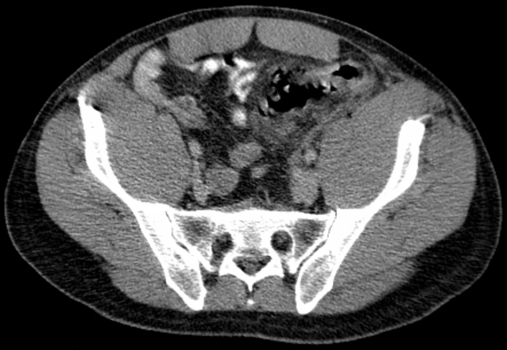
- CT abdomen and pelvis with contrast: the investigation of choice in suspected acute diverticulitis. It confirms the diagnosis, assesses severity, and identifies complications such as abscess, perforation, free gas and fistula. Typical findings are colonic wall thickening, pericolic fat stranding and the presence of diverticula1
- Erect chest X-ray: may show free air under the diaphragm in perforation, though CT is far more sensitive
- Ultrasound: an alternative in young or pregnant patients to avoid radiation, though it is operator-dependent
Hinchey classification
Perforated diverticulitis is graded on CT using the Hinchey classification, which guides management:
| Stage | Findings | Typical management |
|---|---|---|
| I | Pericolic phlegmon or small confined abscess | Antibiotics; percutaneous drainage if the abscess exceeds about 3-4 cm |
| II | Pelvic, retroperitoneal or distant abscess | Antibiotics with radiologically guided percutaneous drainage |
| III | Purulent peritonitis (generalised) | Emergency surgery - laparoscopic lavage in selected cases, or resection |
| IV | Faeculent peritonitis (generalised) | Emergency surgery - Hartmann procedure, with high mortality |
Management
Diverticulosis and diverticular disease
Asymptomatic diverticulosis needs no treatment, only reassurance and dietary advice. For symptomatic diverticular disease, advise a high-fibre diet introduced gradually with adequate fluid intake, bulk-forming laxatives if constipated, weight loss, smoking cessation and exercise.1 Simple analgesia with paracetamol is preferred; avoid NSAIDs and opioids, both of which are associated with perforation. The historical advice to avoid nuts, seeds and popcorn has been disproven and should no longer be given.
Acute uncomplicated diverticulitis
Practice has shifted markedly away from routine antibiotics. NICE advises that for uncomplicated diverticulitis in a systemically well patient with no significant comorbidity, it is reasonable to manage without antibiotics, using oral fluids, paracetamol, and clear safety-netting advice to return if symptoms worsen or do not improve within 48 hours.1
Oral antibiotics (typically co-amoxiclav, or a cephalosporin with metronidazole in penicillin allergy) are offered where the patient is systemically unwell, immunosuppressed, frail, or has significant comorbidity. Admission for IV antibiotics, IV fluids and analgesia is indicated for patients unable to tolerate oral intake, with uncontrolled pain, who are frail or immunosuppressed, or who fail to improve on oral treatment.
Complicated diverticulitis
- Abscess: IV antibiotics, with radiologically guided percutaneous drainage for collections above roughly 3-4 cm
- Perforation with peritonitis (Hinchey III-IV): resuscitation, IV antibiotics and emergency surgery
- Hartmann procedure - sigmoid resection with an end colostomy and closure of the rectal stump - is the standard emergency operation, with reversal considered months later; some patients are suitable for primary anastomosis with or without a defunctioning stoma
- Laparoscopic lavage may be used in selected Hinchey III cases
- Fistula: most commonly colovesical (pneumaturia, faecaluria, recurrent UTIs), also colovaginal; usually requires elective resection
- Stricture and obstruction: elective resection, or stenting as a bridge in selected cases
- Diverticular bleeding: usually stops spontaneously; managed as a lower GI bleed with resuscitation, CT angiography if unstable, and colonoscopic or angiographic haemostasis if needed
Elective surgery
Elective sigmoid resection is no longer recommended purely on the basis of a set number of previous episodes. It is considered on an individualised basis for patients with recurrent, disabling symptoms significantly affecting quality of life, complications such as fistula or stricture, or immunosuppression, weighing the benefits against the significant operative morbidity.
Complications
- Perforation with purulent or faeculent peritonitis, sepsis and shock
- Abscess formation (pericolic, pelvic or intra-abdominal)
- Fistula: colovesical (commonest, causing pneumaturia, faecaluria and recurrent urinary infection), colovaginal, colocutaneous and coloenteric
- Stricture from repeated inflammation and fibrosis, causing large bowel obstruction
- Diverticular bleeding: typically painless, brisk and self-limiting, and the commonest cause of major lower GI bleeding
- Recurrent diverticulitis
- Portal pyaemia and liver abscess (rare)
- Post-surgical complications, including stoma-related problems and anastomotic leak
Red flags
Prognosis
Most patients with diverticulosis remain asymptomatic for life, and only around 4-5% ever develop diverticulitis.1 The majority of acute uncomplicated episodes settle with conservative management, and increasingly without antibiotics at all, with a good long-term outlook.
Around a fifth of patients experience a recurrence, and recurrent episodes are, reassuringly, not usually more severe than the first - it is the initial presentation that most often involves perforation. Complicated diverticulitis carries considerably greater morbidity, and emergency surgery for faeculent peritonitis (Hinchey IV) has a high mortality, particularly in older and comorbid patients. Long-term dietary fibre, weight management, smoking cessation and avoidance of NSAIDs reduce the risk of further episodes.
References
- NICE NG147. Diverticular disease: diagnosis and management. 2019. Available here
- Tursi A et al. Colonic diverticular disease. Nat Rev Dis Primers. 2020. Available here
- NICE Clinical Knowledge Summaries (CKS). Diverticular disease. 2024. Available here
- Hinchey EJ, Schaal PG, Richards GK. Treatment of perforated diverticular disease of the colon. Adv Surg. 1978. Available here
- Hellerhoff, CC BY-SA 3.0, via Wikimedia Commons. Available here
- NHS. Diverticular disease and diverticulitis. 2023. Available here
- NICE NG12. Suspected cancer: recognition and referral. 2015 (updated 2023). Available here
This article is written for revision and education. It is not clinical guidance and must not be used to make decisions about the care of a patient. Always check current NICE guidance and local protocols.