
Ectopic Pregnancy
Key points
- Definition: implantation of a pregnancy outside the endometrial cavity, most often in the fallopian tube.
- Incidence: approximately 1 in 90 pregnancies in the UK; it remains a leading cause of first-trimester maternal death.
- Site: the ampulla is the commonest site; the isthmus and the interstitial portion are the most dangerous because they rupture early and bleed heavily.
- Presentation: 6-8 weeks of amenorrhoea with unilateral lower abdominal pain and dark vaginal bleeding; shoulder-tip pain and collapse indicate rupture.
- Diagnosis: transvaginal ultrasound showing an adnexal mass separate from the ovary, with an empty uterus and a positive pregnancy test.
- hCG: a rise of less than 63% or a fall of less than 50% over 48 hours in a pregnancy of unknown location is suspicious for an ectopic.
- Management: expectant, single-dose intramuscular methotrexate, or laparoscopic salpingectomy, decided by symptoms, hCG level and mass size.
- Emergency: haemodynamic instability means rupture until proven otherwise; resuscitate and go straight to theatre without waiting for imaging.
Introduction
An ectopic pregnancy is one that implants anywhere other than the endometrial cavity. It affects around 1 in 90 pregnancies in the UK, approximately 11 per 1,000, and although mortality has fallen substantially it remains one of the leading causes of death in early pregnancy. Deaths continue to occur, and the recurring theme in confidential enquiries is not failure of surgery but failure of recognition: the woman sent home with a diagnosis of gastroenteritis or urinary infection because a pregnancy test was never done.1,2
The operative rule is therefore simple. Any woman of reproductive age presenting with abdominal pain, vaginal bleeding, collapse, or unexplained gastrointestinal or urinary symptoms should have a pregnancy test. If it is positive and the pregnancy has not been localised, an ectopic pregnancy is the working diagnosis until ultrasound proves otherwise.
The clinical challenge is that ectopic pregnancy is now often diagnosed before it causes any dramatic symptoms, in a stable woman attending an early pregnancy assessment unit for a routine scan. Much of the management is therefore about matching the intensity of treatment to the actual risk, and preserving future fertility where it is safe to do so.
Sites and pathophysiology
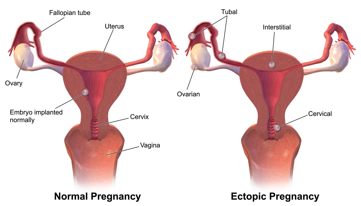
Over 95% of ectopic pregnancies are tubal. The distribution within the tube matters because it determines how a rupture behaves.
| Site | Approximate frequency | Significance |
|---|---|---|
| Ampulla | About 70% | The widest part of the tube, so it distends before rupturing; often presents later |
| Isthmus | About 12% | Narrow and muscular, so ruptures earlier and more violently |
| Fimbrial end | About 11% | May undergo tubal abortion, expelling the pregnancy into the peritoneal cavity |
| Interstitial (cornual) | 2-3% | Implanted in the myometrial segment of the tube; ruptures late, around 8-16 weeks, with catastrophic bleeding from the uterine and ovarian arteries |
| Ovarian, cervical, caesarean scar, abdominal | Under 3% combined | Rare, difficult to manage, and associated with major haemorrhage |
The underlying mechanism is impaired tubal transport of the blastocyst. Anything that damages the ciliated epithelium or distorts the tubal lumen delays passage, so the blastocyst reaches implantation competence while still in the tube. The trophoblast then invades the tubal wall, which lacks a decidual layer and cannot accommodate an expanding pregnancy. As the trophoblast erodes into the muscularis and its vessels, the tube either bleeds slowly into the peritoneal cavity or ruptures.
Risk factors
Risk factors reflect tubal damage or altered tubal transport, but around a third of women have none at all, which is precisely why their absence must never be used to exclude the diagnosis.
- Previous ectopic pregnancy - the strongest single risk factor, with a recurrence rate of around 10%
- Pelvic inflammatory disease, particularly chlamydial salpingitis, which scars the tubal epithelium
- Previous tubal surgery, including sterilisation and reversal of sterilisation
- Assisted reproductive technology - IVF carries an increased risk, including of heterotopic pregnancy
- Intrauterine contraception in situ - the coil is highly effective, but if pregnancy does occur it is more likely to be ectopic
- Progestogen-only contraception, which reduces tubal motility
- Endometriosis and pelvic adhesions
- Smoking, which impairs ciliary function, and increasing maternal age
Clinical features
The classic presentation is a woman with 6-8 weeks of amenorrhoea, unilateral lower abdominal pain and vaginal bleeding. The bleeding is characteristically darker and less heavy than a period, sometimes described as looking like prune juice, because it is decidua being shed from the uterus rather than fresh bleeding from the implantation site. Pain typically precedes the bleeding, whereas in a miscarriage the bleeding usually comes first.
Blood tracking into the peritoneal cavity produces symptoms that mislead. Diaphragmatic irritation refers pain to the shoulder tip. Blood in the pouch of Douglas irritates the rectum, producing tenesmus, an urge to defaecate, or diarrhoea. Some women present with dizziness or syncope alone. The commonest misdiagnoses given to women who later prove to have an ectopic are gastroenteritis, urinary tract infection and constipation.1
Examination
- Observations, looking for tachycardia, hypotension and a narrowed pulse pressure
- Abdominal examination for tenderness, guarding, rebound and generalised peritonism
- Speculum examination to assess bleeding and the cervical os, which is closed in an ectopic
- Bimanual examination for cervical excitation, adnexal tenderness and any adnexal mass
- A uterus that is smaller than expected for the stated gestation
Investigations
Pregnancy test
A urinary hCG test is the first and most important investigation. It is the step that is most often omitted and the omission that causes the deaths.
Transvaginal ultrasound
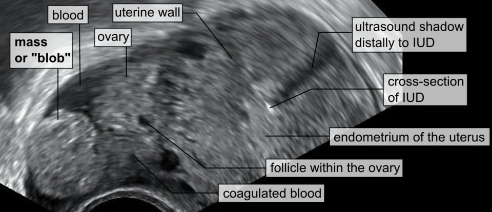
Transvaginal ultrasound is the diagnostic investigation of choice. The findings that establish the diagnosis are an adnexal mass that moves separately from the ovary, together with an empty uterus.
- Blob sign - an inhomogeneous adnexal mass adjacent to but distinct from the ovary, the commonest appearance
- Bagel or tubal ring sign - an empty gestational sac with a thick hyperechoic rim
- Live ectopic - a gestational sac containing a yolk sac or fetal pole, sometimes with cardiac activity
- Free fluid in the pouch of Douglas; echogenic free fluid suggests haemoperitoneum
- A pseudosac in the uterus - a centrally placed collection of fluid within the cavity that can be mistaken for an early intrauterine sac, in contrast to the eccentrically placed true sac with a double decidual ring
Pregnancy of unknown location
In around 10% of early pregnancy scans no pregnancy is seen anywhere despite a positive test. This is a pregnancy of unknown location, not a diagnosis but a state of uncertainty which may resolve into an intrauterine pregnancy, a failing pregnancy, or an ectopic. Serial serum hCG measured exactly 48 hours apart is used to discriminate.1
| 48-hour change | Interpretation | Action |
|---|---|---|
| Rise of more than 63% | Likely developing intrauterine pregnancy | Repeat transvaginal scan when hCG exceeds 1,500 IU/L |
| Fall of more than 50% | Likely failing pregnancy of any location | Urine pregnancy test in 14 days; if still positive, review |
| Change between these values | Suspicious for ectopic pregnancy | Review in the early pregnancy unit within 24 hours |
A single hCG value does not diagnose an ectopic. The concept of a discriminatory zone, usually taken as 1,500 IU/L, refers to the level above which an intrauterine pregnancy should be visible transvaginally; failure to see one above that threshold raises suspicion but is not proof, and in multiple pregnancy the threshold does not apply. Check the full blood count and group and save in every case, and crossmatch if there is any suspicion of rupture.
Differential diagnosis
- Miscarriage - bleeding usually precedes pain, the pain is central and crampy, and the os may be open
- Ovarian cyst accident: rupture, haemorrhage or torsion
- Pelvic inflammatory disease - bilateral pain, fever, discharge, cervical excitation
- Appendicitis - migratory pain, anorexia, fever; a pregnancy test is still mandatory
- Urinary tract infection and renal colic
- Gastroenteritis and constipation - the two diagnoses most often given in error
Management
The unstable patient
Expectant management
Some ectopic pregnancies resolve without intervention. NICE supports expectant management where the woman is clinically stable and pain-free, the ectopic is unruptured with an adnexal mass under 35 mm and no visible heartbeat, the serum hCG is below 1,000 IU/L, and she is able and willing to attend for follow-up. hCG is repeated on days 2, 4 and 7, and if it falls by at least 15% between measurements it is then checked weekly until under 20 IU/L.1
Methotrexate
Methotrexate is a folate antagonist that halts trophoblastic proliferation. It is given as a single intramuscular dose of 50 mg/m2 body surface area. The criteria are similar to those for expectant management but permit a higher hCG: no significant pain, an unruptured mass under 35 mm with no fetal heartbeat, hCG below 1,500 IU/L, and no intrauterine pregnancy. Levels between 1,500 and 5,000 IU/L may be treated medically after explicit counselling that the failure rate is higher.1
- Follow-up hCG on days 4 and 7; a fall of at least 15% between day 4 and day 7 indicates success, after which hCG is monitored weekly until negative
- Around 15% of women need a second dose, and around 7% ultimately require surgery
- Warn about abdominal pain around days 3-7, which is usually separation pain but which must be reassessed to exclude rupture
- Side effects include nausea, stomatitis, diarrhoea, myelosuppression and deranged liver function
- Advise reliable contraception and avoidance of pregnancy for 3 months after treatment, because of the teratogenic risk
- Advise avoiding alcohol, non-steroidal anti-inflammatory drugs and prolonged sun exposure during treatment
Surgical management
Surgery is indicated where there is significant pain, an adnexal mass of 35 mm or more, a visible fetal heartbeat, or an hCG of 5,000 IU/L or above. Laparoscopy is preferred over laparotomy in the stable patient because of faster recovery, less blood loss and fewer adhesions.
| Procedure | When used | Consequences |
|---|---|---|
| Salpingectomy | First line when the contralateral tube is healthy | Removes the affected tube entirely; no risk of persistent trophoblast; subsequent fertility is close to normal with one healthy tube |
| Salpingotomy | Contralateral tube absent or damaged, or other fertility risk factors | Conserves the tube, but around 1 in 5 need further methotrexate or salpingectomy, so hCG must be followed to negative |
Complications and prognosis
The dominant complication is tubal rupture with intraperitoneal haemorrhage, which can be rapid and fatal. Interstitial and cervical ectopics are particularly dangerous because they are supplied by large vessels and often present later; interstitial pregnancies may require cornual resection or, occasionally, hysterectomy. Persistent trophoblast after salpingotomy is the reason those women require hCG surveillance.
Around two-thirds of women who have had an ectopic pregnancy will subsequently have a successful intrauterine pregnancy. The recurrence risk is approximately 10%, rising further after two ectopics, which is why any woman with a previous ectopic should be offered an early scan at around 6-7 weeks in a subsequent pregnancy to confirm the location. Fertility after salpingectomy with a healthy contralateral tube is only marginally reduced.
Psychological morbidity is common and under-recognised: the woman has simultaneously lost a pregnancy, had emergency surgery, sometimes lost a fallopian tube, and been told she was at risk of dying. Written information, a follow-up appointment, and signposting to support organisations should be offered routinely rather than on request.1,2
References
- NICE NG126. Ectopic pregnancy and miscarriage: diagnosis and initial management. 2019 (updated 2023). Available here
- RCOG Green-top Guideline No. 21. Diagnosis and management of ectopic pregnancy. Available here
- Human Fertilisation and Embryology Authority. Risks of fertility treatment. Available here
- RCOG Green-top Guideline No. 22. The use of anti-D immunoglobulin for rhesus D prophylaxis. Available here
This article is written for revision and education. It is not clinical guidance and must not be used to make decisions about the care of a patient. Always check current NICE guidance and local protocols.