
Multiple Pregnancy
Key points
- Incidence: twins complicate around 1 in 65 UK births, and the rate has risen with assisted reproduction and increasing maternal age.
- Chorionicity: the number of placentas determines risk, not whether the twins are identical; it must be established at the 11+2 to 14+1 week scan.
- Ultrasound signs: the lambda or twin peak sign indicates dichorionic; the T sign indicates monochorionic.
- Monochorionic risk: shared placental vascular anastomoses cause twin-to-twin transfusion syndrome, twin anaemia-polycythaemia sequence and selective growth restriction.
- Surveillance: monochorionic pregnancies are scanned fortnightly from 16 weeks; dichorionic every 4 weeks from 20 weeks.
- Maternal risk: pre-eclampsia, anaemia, gestational diabetes, hyperemesis, antepartum and postpartum haemorrhage are all substantially more common.
- Timing of birth: dichorionic diamniotic from 37+0 weeks, monochorionic diamniotic from 36+0 weeks, monoamniotic by caesarean at 32+0 to 33+6 weeks.
- Mode of birth: vaginal birth is reasonable when the first twin is cephalic; caesarean section is required for monoamniotic twins and for a non-cephalic first twin.
Introduction
Multiple pregnancy affects approximately 1 in 65 births in the UK, and triplet pregnancies roughly 1 in 5,000. The rate rose steeply with the introduction of assisted reproduction and has since fallen back as single embryo transfer has become standard practice, but it remains higher than the natural background rate because maternal age, itself a determinant of dizygotic twinning, has continued to rise.1
Almost everything about the management of a twin pregnancy follows from a single piece of information established in the first trimester: chorionicity. A monochorionic pregnancy has a shared placenta with vascular connections between the two fetal circulations, and it is those connections that generate the conditions unique to multiple pregnancy. Getting chorionicity wrong, or failing to record it, means the wrong surveillance schedule and a materially higher risk of stillbirth.
Perinatal mortality in twins is around four times that of singletons, and in triplets higher again. The dominant contributors are preterm birth, growth restriction and the complications of monochorionic placentation.
Zygosity, chorionicity and amnionicity
Zygosity describes how many eggs were fertilised; chorionicity describes how many placentas there are; amnionicity describes how many amniotic sacs. Two-thirds of twins are dizygotic, arising from two separate ova, and are always dichorionic diamniotic. The remaining third are monozygotic, and their chorionicity depends entirely on how many days after fertilisation the embryo split.
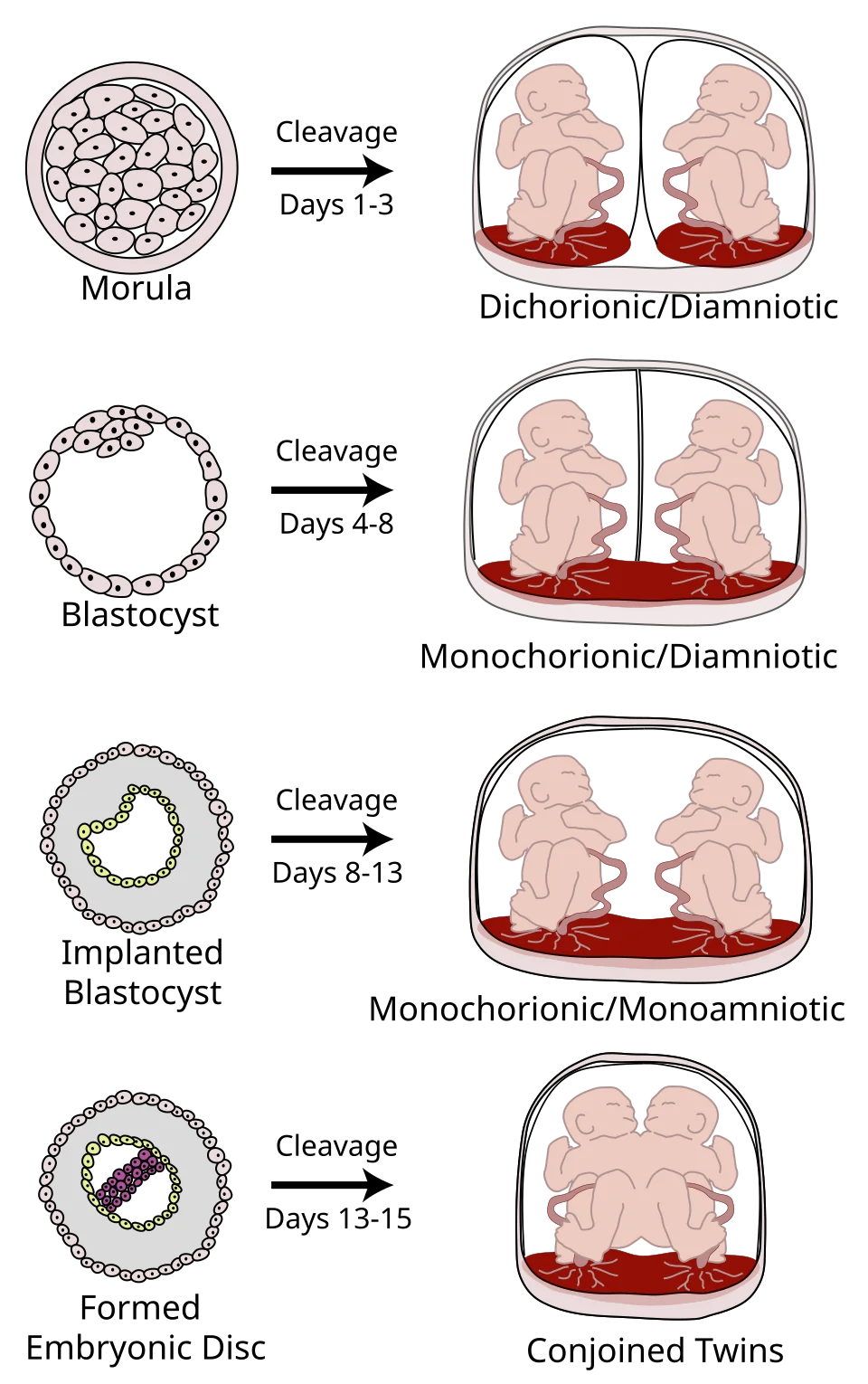
| Day of splitting | Result | Approximate proportion of monozygotic twins |
|---|---|---|
| Days 1-3 (morula) | Dichorionic diamniotic | About 30% |
| Days 4-8 (blastocyst) | Monochorionic diamniotic | About 65-70% |
| Days 8-13 (implanted blastocyst) | Monochorionic monoamniotic | About 1-5% |
| After day 13 (formed embryonic disc) | Conjoined twins | Very rare |
Determining chorionicity
Chorionicity is determined at the first-trimester scan between 11+2 and 14+1 weeks, when it is most accurate. Three findings are used together: the number of placental masses, the sex of the fetuses if visible, and, most importantly, the appearance of the membrane where it meets the placenta.
- Lambda sign (also called the twin peak sign) - a triangular wedge of chorionic tissue extending into the base of the intertwin membrane, indicating dichorionic placentation
- T sign - the intertwin membrane meets the placenta at a right angle with no chorionic wedge, indicating monochorionic placentation
- A thick intertwin membrane, of four layers, suggests dichorionic; a thin two-layer membrane suggests monochorionic
- Discordant fetal sex means dizygotic, and therefore dichorionic, but concordant sex tells you nothing
- No visible intertwin membrane at all raises the possibility of monoamniotic twins, which requires expert confirmation
Complications
Maternal
- Hyperemesis gravidarum - driven by the higher hCG of two placentas
- Anaemia - greater iron and folate demand; check FBC at booking, 20-24 weeks and 28 weeks
- Pre-eclampsia - two to three times the singleton risk; multiple pregnancy is a moderate risk factor for aspirin prophylaxis
- Gestational diabetes
- Polyhydramnios, with its consequent respiratory discomfort and risk of preterm labour
- Antepartum haemorrhage - a larger placental surface makes praevia and abruption more likely
- Postpartum haemorrhage - an overdistended uterus contracts poorly and the placental bed is larger
- Venous thromboembolism, operative delivery, and a higher rate of postnatal depression
Fetal
- Preterm birth - around 60% of twins deliver before 37 weeks; mean gestation at birth is roughly 37 weeks for twins and 33 weeks for triplets
- Fetal growth restriction and selective growth restriction, in which one twin's estimated weight falls well below the other's
- Congenital anomaly - increased in monozygotic twins specifically
- Miscarriage and vanishing twin syndrome - loss of one sac in the first trimester, occurring in a substantial minority of twin pregnancies detected early
- Stillbirth - higher throughout, and particularly in monochorionic pregnancies
- Cerebral palsy - risk is several times that of singletons, driven by prematurity and by monochorionic complications
- Cord entanglement - specific to monoamniotic twins, and the reason they are delivered by caesarean before 34 weeks
Twin-to-twin transfusion syndrome
Twin-to-twin transfusion syndrome affects around 10-15% of monochorionic diamniotic pregnancies. Every monochorionic placenta contains vascular anastomoses between the two fetal circulations. Where these are balanced, nothing happens. Where deep arteriovenous anastomoses produce a net unidirectional flow from one twin to the other, the syndrome develops, usually between 16 and 26 weeks.
| Feature | Donor twin | Recipient twin |
|---|---|---|
| Volume status | Hypovolaemic | Hypervolaemic |
| Urine output | Oliguria with a small or absent bladder | Polyuria with a distended bladder |
| Amniotic fluid | Oligohydramnios; may become a stuck twin, held against the uterine wall by the membrane | Polyhydramnios |
| Growth | Growth restricted | Normal or large |
| Haematology | Anaemic | Polycythaemic |
| Cardiac | Normal | Volume overload, tricuspid regurgitation, cardiomyopathy and hydrops |
Severity is described by the Quintero staging system, running from stage I, in which there is discordant amniotic fluid but the donor bladder is still visible, through absent donor bladder, abnormal Dopplers and hydrops, to stage V, the death of one or both twins. Untreated severe disease carries a mortality approaching 90%.
The definitive treatment is fetoscopic laser ablation of the placental anastomoses, performed in specialist centres between around 16 and 26 weeks. It converts a shared circulation into two functionally separate ones and improves survival substantially compared with serial amnioreduction. Amnioreduction remains useful for symptom relief and where laser is not available or appropriate; selective reduction is occasionally considered in severe cases.1,3
Antenatal care
Care is delivered by a named multidisciplinary team with experience in multiple pregnancy, comprising a specialist obstetrician, a specialist midwife and a sonographer. Women need more appointments than the standard schedule provides, and NICE specifies a minimum number depending on chorionicity.
| Chorionicity | Scanning schedule |
|---|---|
| Dichorionic diamniotic | Chorionicity scan at 11+2 to 14+1, anomaly scan at 18+0 to 20+6, then growth scans at 24, 28, 32 and 36 weeks |
| Monochorionic diamniotic | As above, but with additional scans every 2 weeks from 16 weeks, including amniotic fluid and middle cerebral artery Doppler assessment |
| Monochorionic monoamniotic | Every 2 weeks from 16 weeks in a specialist setting, with planned birth by caesarean at 32+0 to 33+6 weeks |
- Aspirin 75-150 mg daily from 12 weeks where there is one or more additional risk factor for pre-eclampsia
- Check haemoglobin at booking, at 20-24 weeks and at 28 weeks, and treat anaemia promptly
- Assess for gestational diabetes if any risk factor is present
- Assess and record fetal presentation from around 34 weeks, as this determines mode of birth planning
- Discuss the risk of preterm birth, and give a clear plan for what to do if contractions or bleeding occur
- Offer information about feeding twins, and referral to specialist support such as the Twins Trust
- NICE does not recommend routine bed rest, cervical cerclage, oral tocolytics or intramuscular progesterone to prevent preterm birth in multiple pregnancy
Screening for trisomies is offered but is more complex. The combined test is used in twins, with a nuchal translucency measured for each fetus; in monochorionic pregnancies a single risk is reported for the pregnancy since the twins are genetically identical, whereas in dichorionic pregnancies a separate risk is given for each. Serum screening is less accurate than in singletons, and NIPT interpretation in twins is more limited.1
Birth
Timing
| Type | Offer birth from |
|---|---|
| Dichorionic diamniotic twins | 37+0 weeks |
| Monochorionic diamniotic twins | 36+0 weeks, with a course of antenatal corticosteroids |
| Monochorionic monoamniotic twins | Caesarean section between 32+0 and 33+6 weeks, with corticosteroids |
| Triplets | 35+0 weeks, with corticosteroids |
The rationale for delivering monochorionic twins earlier is that the stillbirth risk in a shared placenta rises steeply from around 36 weeks. Continuing an uncomplicated dichorionic pregnancy beyond 37+6 weeks increases fetal death and is not recommended.
Mode of birth
Vaginal birth is a reasonable option where the first twin is cephalic and there is no other contraindication, and around 60% of such women achieve it. Caesarean section is indicated for a non-cephalic first twin, for monoamniotic twins, for triplets and higher-order multiples, and for the usual obstetric indications.
- Continuous cardiotocography of both twins, which usually requires a dual-channel monitor and sometimes a fetal scalp electrode on the first twin
- Epidural analgesia is recommended, because internal manipulation of the second twin may be needed and there is a substantial chance of proceeding to caesarean section
- Deliver in an obstetric unit with immediate access to theatre, a senior obstetrician, an anaesthetist and two neonatal teams
- After delivery of the first twin, confirm the lie of the second by palpation or ultrasound, and stabilise it in a longitudinal lie
- The second twin may need external cephalic version, internal podalic version or breech extraction; a non-cephalic second twin does not by itself mandate caesarean section
- Risks specific to the second twin are cord prolapse, malpresentation, abruption after the uterus decompresses, and a rising rate of hypoxia with a longer inter-twin interval
- Active management of the third stage with an oxytocin infusion, because postpartum haemorrhage should be assumed
Prognosis
Most twin pregnancies end well, but they do so under closer surveillance than any other routine pregnancy. Dichorionic diamniotic twins carry a perinatal mortality several times that of singletons, driven principally by preterm birth, and monochorionic twins carry a higher risk again because of the shared circulation. Recognising chorionicity early and applying the correct surveillance schedule is the intervention that changes that.
Postnatally, the practical and psychological burden is substantial and frequently underestimated. Feeding two babies, one of whom may be in a neonatal unit, is exhausting; rates of postnatal depression are higher than after a singleton birth; and readmission of one or both infants is common. Proactive feeding support, a low threshold for mental health assessment, and signposting to peer support are part of the clinical plan rather than optional extras.1,4
References
- NICE NG137. Twin and triplet pregnancy. 2019 (updated 2024). Available here
- RCOG Green-top Guideline No. 51. Management of monochorionic twin pregnancy. Available here
- Senat MV, Deprest J, Boulvain M et al. Endoscopic laser surgery versus serial amnioreduction for severe twin-to-twin transfusion syndrome. N Engl J Med. 2004. Available here
- Twins Trust. Support and information for families with twins, triplets or more. Available here
This article is written for revision and education. It is not clinical guidance and must not be used to make decisions about the care of a patient. Always check current NICE guidance and local protocols.