
Pre-eclampsia and Eclampsia
Key points
- Definition: new hypertension after 20 weeks with proteinuria, maternal organ dysfunction or uteroplacental dysfunction.
- Pathophysiology: failed remodelling of the spiral arteries produces placental ischaemia, which releases anti-angiogenic factors causing systemic endothelial dysfunction.
- Symptoms: severe headache, visual disturbance, epigastric or right upper quadrant pain, vomiting and sudden swelling of the face and hands; many women are asymptomatic.
- Prophylaxis: aspirin 75-150 mg daily from 12 weeks for women with one high-risk or two moderate-risk factors.
- Blood pressure: treat to below 135/85 mmHg, and treat any reading of 160/110 mmHg or above within one hour.
- Magnesium sulfate: 4 g intravenous loading dose then 1 g per hour for 24 hours, for eclampsia and for seizure prophylaxis in severe disease.
- HELLP: haemolysis, elevated liver enzymes and low platelets; may occur with only modest hypertension.
- Cure: delivery of the placenta is the only definitive treatment, but almost half of eclamptic seizures occur after birth.
Introduction
Pre-eclampsia is a multisystem disorder of pregnancy characterised by new hypertension after 20 weeks together with evidence of maternal organ dysfunction or uteroplacental dysfunction. It complicates 2-8% of pregnancies, and severe disease around 1-2%. Eclampsia, the occurrence of generalised tonic-clonic seizures in a woman with pre-eclampsia, now affects approximately 2.7 per 10,000 births in the UK.1,2
Two ideas make the whole subject coherent. The first is that pre-eclampsia is a disease of the placenta, not of the fetus, which is why it occurs in molar pregnancy where there is no fetus at all, and why delivering the placenta cures it. The second is that the clinical manifestations are those of generalised endothelial dysfunction: hypertension from vasoconstriction, proteinuria from glomerular injury, oedema from capillary leak, and hepatic, cerebral and haematological involvement from the same process in those beds.
The term proteinuric pre-eclampsia has been abandoned as a requirement. A woman may have severe pre-eclampsia with no proteinuria at all, and defining the disease by proteinuria delayed diagnosis in exactly the women who deteriorated fastest. The current definition is deliberately broader.
Pathophysiology
The prevailing model has two stages.
Stage one: defective placentation
In normal pregnancy, extravillous trophoblast invades the maternal spiral arteries in two waves, the second occurring at 14-20 weeks. This invasion destroys the smooth muscle and elastic lamina of the vessels, converting them from narrow, high-resistance, vasoactive arterioles into wide, flaccid, low-resistance conduits that deliver a large and unregulated blood flow to the intervillous space.
In pre-eclampsia the second wave of invasion fails. The spiral arteries retain their muscular walls, remain responsive to vasoconstrictors, and deliver an intermittent, high-velocity, low-volume flow. The consequence is placental ischaemia-reperfusion injury and oxidative stress. This all happens in the second trimester, long before any clinical sign appears, which is why aspirin must be started before 16 weeks to be effective and is useless once disease is established.
Stage two: the maternal syndrome
The stressed placenta releases anti-angiogenic factors into the maternal circulation, principally soluble fms-like tyrosine kinase-1 (sFlt-1) and soluble endoglin. sFlt-1 is a decoy receptor that binds and neutralises vascular endothelial growth factor and placental growth factor, both of which are required to maintain healthy endothelium, particularly the fenestrated endothelium of the glomerulus and liver. The resulting endothelial dysfunction produces vasoconstriction, increased capillary permeability, platelet activation and a procoagulant state.
This mechanism has direct clinical utility. Because placental growth factor falls before symptoms appear, PlGF-based blood testing between 20 and 36+6 weeks has a high negative predictive value and is recommended by NICE to help rule out pre-eclampsia in women with suspected disease, reducing unnecessary admission.1,3
Diagnostic criteria
Pre-eclampsia is diagnosed when hypertension of 140/90 mmHg or above develops after 20 weeks, accompanied by one or more of the following.
| Category | Defining findings |
|---|---|
| Proteinuria | Urinary ACR 8 mg/mmol or above, or PCR 30 mg/mmol or above, or at least 300 mg protein in 24 hours |
| Renal | Serum creatinine 90 micromol/L or above, or acute kidney injury |
| Hepatic | Transaminases above 40 IU/L, with or without right upper quadrant or epigastric pain |
| Neurological | Eclampsia, altered mental state, blindness, stroke, clonus, severe headaches, persistent visual scotomata |
| Haematological | Platelet count below 150 x10^9/L, disseminated intravascular coagulation, or haemolysis |
| Uteroplacental | Fetal growth restriction, abnormal umbilical artery Doppler waveform, or stillbirth |
Risk factors and prevention
| High risk | Moderate risk |
|---|---|
| Hypertensive disease in a previous pregnancy | First pregnancy |
| Chronic kidney disease | Age 40 years or older |
| Autoimmune disease: SLE or antiphospholipid syndrome | Pregnancy interval of more than 10 years |
| Type 1 or type 2 diabetes | BMI of 35 kg/m2 or more at booking |
| Chronic hypertension | Family history of pre-eclampsia |
| - | Multiple pregnancy |
Aspirin 75-150 mg daily from 12 weeks until birth reduces the risk of pre-eclampsia, and in the ASPRE trial reduced preterm pre-eclampsia by around 60% when started before 16 weeks at a dose of 150 mg. Aspirin acts by irreversibly inhibiting platelet cyclo-oxygenase, shifting the thromboxane-prostacyclin balance in favour of vasodilatation at the placental bed. Calcium supplementation reduces risk in populations with low dietary calcium intake but is not routinely required in the UK.1,4
Molar pregnancy, hydrops and triploidy are associated with early-onset pre-eclampsia, sometimes before 20 weeks. Pre-eclampsia arising before 20 weeks should therefore prompt an ultrasound to look for trophoblastic disease.
Clinical features
Most women with pre-eclampsia are asymptomatic when it is detected; this is precisely why blood pressure and urinalysis are checked at every antenatal contact. When symptoms do occur they are ominous and indicate severe disease.
- Headache - severe, frontal, and typically unresponsive to simple analgesia
- Visual disturbance - blurring, flashing lights, scotomata, or in severe cases cortical blindness
- Epigastric or right upper quadrant pain - from stretching of the liver capsule by oedema or subcapsular haemorrhage; frequently mistaken for indigestion
- Nausea and vomiting - new in the second half of pregnancy, which is never normal
- Sudden swelling of the face, hands or feet
- Reduced urine output
- Reduced fetal movements or a fetus small for dates
Examination
Measure blood pressure with an appropriately sized cuff and a device validated for pregnancy. Dipstick the urine and quantify any proteinuria. Look for oedema, particularly facial and periorbital. Elicit the deep tendon reflexes and test for ankle clonus: sustained clonus of more than three beats indicates cerebral irritability and imminent eclampsia. Palpate for right upper quadrant tenderness. Assess symphysis-fundal height and fetal wellbeing, and auscultate the lung bases for pulmonary oedema.
Investigations
- Urinary ACR or PCR to quantify proteinuria, rather than relying on the dipstick
- Full blood count - thrombocytopenia, and a falling haemoglobin with a rising haematocrit reflecting haemolysis and haemoconcentration respectively
- Liver function tests - rising transaminases, and lactate dehydrogenase as a marker of haemolysis
- Urea, electrolytes and creatinine, with urate, which rises early although it is too non-specific to diagnose the condition
- Coagulation screen if the platelet count is below 100 x10^9/L or there is bleeding
- PlGF-based testing between 20 and 36+6 weeks to help rule out pre-eclampsia in suspected disease
- Cardiotocography at diagnosis and repeated if there is any change in condition
- Ultrasound for fetal growth, amniotic fluid volume and umbilical artery Doppler, repeated every two weeks
Validated risk models such as fullPIERS and PREP-S can be used at diagnosis to estimate the probability of adverse maternal outcome and to guide the intensity of monitoring and the threshold for transfer to a unit with appropriate neonatal facilities.1
Management
Management runs on three parallel tracks: controlling blood pressure, preventing and treating seizures, and deciding when to deliver. Only the last is curative.
Blood pressure control
Oral labetalol is first line, with nifedipine modified release where a beta blocker is contraindicated, and methyldopa third line. The target is below 135/85 mmHg. Severe hypertension of 160/110 mmHg or above must be treated within one hour, using oral nifedipine, intravenous labetalol or intravenous hydralazine, with continuous fetal monitoring during titration. Uncontrolled systolic hypertension is what causes maternal intracranial haemorrhage.1,2
Magnesium sulfate
Magnesium sulfate is both the treatment for eclamptic seizures and the prophylaxis against them. The MAGPIE trial established that it halves the risk of eclampsia in women with pre-eclampsia, and the Collaborative Eclampsia Trial showed it is superior to both diazepam and phenytoin for treating and preventing recurrence of seizures. Its mechanism is probably cerebral vasodilatation with membrane stabilisation and NMDA receptor antagonism, rather than a conventional anticonvulsant action.5,6
| Element | Detail |
|---|---|
| Indications | Eclamptic seizure; severe pre-eclampsia where birth is planned within 24 hours; features suggesting imminent eclampsia |
| Loading dose | 4 g intravenously over 5-15 minutes |
| Maintenance | 1 g per hour for 24 hours, continued for 24 hours after birth or after the last seizure, whichever is later |
| Recurrent seizure | A further 2-4 g intravenously over 5-15 minutes |
| Monitoring | Deep tendon reflexes, respiratory rate, oxygen saturations, urine output and conscious level |
| Toxicity | Loss of patellar reflexes first, then respiratory depression, then cardiac conduction abnormalities and arrest |
| Antidote | Calcium gluconate 1 g (10 mL of 10%) intravenously |
Fluid management
Women with severe pre-eclampsia are intravascularly deplete but have leaky capillaries and a low plasma oncotic pressure. Giving generous fluid to correct oliguria therefore produces pulmonary oedema, historically a leading cause of death in this group. Total fluid intake is restricted to around 80 mL/hour, oliguria is tolerated in the absence of rising creatinine, and fluid balance is monitored strictly with an indwelling catheter.
Timing of birth
| Gestation | Approach |
|---|---|
| Before 34 weeks | Conservative management with antenatal corticosteroids, magnesium for neuroprotection, and intensive monitoring, in a unit with neonatal intensive care; deliver if the maternal or fetal condition deteriorates |
| 34 to 36+6 weeks | Individualised: continue surveillance if stable, deliver if there is deterioration, and give corticosteroids if birth is planned before 36 weeks |
| From 37 weeks | Initiate birth within 24-48 hours; there is no fetal benefit in continuing and continued pregnancy only increases maternal risk |
| Any gestation with severe uncontrolled disease | Deliver once the mother is stabilised, regardless of gestation |
Vaginal birth is not contraindicated and is preferred where the maternal and fetal condition allows. Caesarean section is chosen for the usual obstetric reasons, for severe disease requiring urgent delivery, or where induction is likely to fail. Ergometrine and Syntometrine should be avoided for the third stage because they cause hypertension; oxytocin alone is used.1
Eclampsia
Eclampsia is a generalised tonic-clonic seizure in a woman with pre-eclampsia, in the absence of another cause. It may be the first manifestation of the disease, and around a third of cases occur in women who did not have recognised pre-eclampsia beforehand. Timing is instructive: roughly 38% occur antepartum, 18% intrapartum and 44% postpartum, which is why surveillance must continue after birth.
HELLP syndrome
HELLP syndrome comprises Haemolysis, Elevated Liver enzymes and Low Platelets, and complicates around 10-20% of cases of severe pre-eclampsia. It can occur with only modest hypertension and minimal proteinuria, which is why a woman presenting with epigastric pain, nausea and malaise in the third trimester needs bloods rather than an antacid.
- Haemolysis - raised lactate dehydrogenase, raised unconjugated bilirubin, low haptoglobin, and fragmented red cells on the film
- Elevated liver enzymes - transaminases above 70 IU/L are typical
- Low platelets - below 100 x10^9/L, and the count often falls rapidly
- Presents with right upper quadrant or epigastric pain, nausea and vomiting, and a general sense of being unwell
- Around 30% of cases present or worsen postpartum
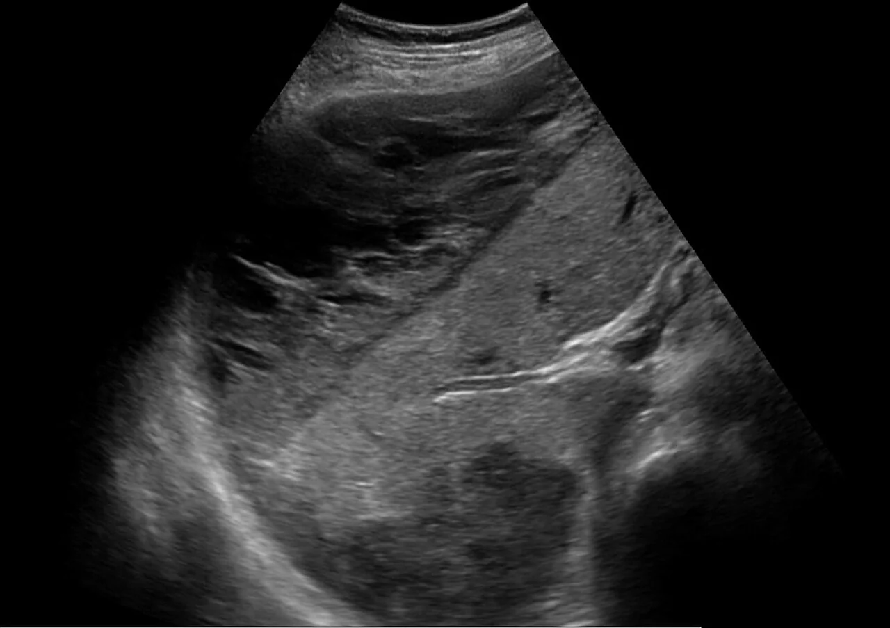
Management is stabilisation and delivery. Magnesium sulfate is given, blood pressure controlled, and platelets transfused if the count is very low or surgery is planned. Corticosteroids do not improve maternal outcomes in HELLP itself, although they are still given for fetal lung maturity where preterm birth is anticipated. The platelet count typically continues to fall for 24-48 hours after birth before recovering.1,7
Complications
| System | Complications |
|---|---|
| Neurological | Eclampsia, intracranial haemorrhage, cerebral oedema, posterior reversible encephalopathy syndrome, cortical blindness |
| Hepatic | HELLP syndrome, subcapsular haematoma, hepatic rupture, hepatic infarction |
| Renal | Acute kidney injury, acute tubular necrosis, rarely cortical necrosis |
| Haematological | Thrombocytopenia, disseminated intravascular coagulation, haemolysis |
| Cardiorespiratory | Pulmonary oedema, most often iatrogenic from fluid overload |
| Obstetric | Placental abruption, postpartum haemorrhage |
| Fetal | Fetal growth restriction, oligohydramnios, iatrogenic preterm birth, stillbirth |
Postnatal care and prognosis
Delivery of the placenta removes the cause but not the disease. Blood pressure typically peaks between days 3 and 5 postpartum, and almost half of eclamptic seizures occur after birth, so monitoring continues rather than stopping at delivery. Magnesium is continued for 24 hours after birth or after the last seizure. Antihypertensives are continued and titrated down as pressure allows, with methyldopa stopped within two days and replaced.
- Measure blood pressure at least four times daily while an inpatient, then on alternate days until normal
- Repeat platelets, transaminases and creatinine as clinically indicated until they normalise
- Ensure proteinuria is rechecked at the postnatal review; persistent proteinuria at 6-8 weeks requires renal referral
- Offer a formal postnatal review with an obstetrician to discuss what happened and plan future pregnancies
- Avoid non-steroidal anti-inflammatory drugs for postnatal analgesia if hypertension is poorly controlled, oliguria persists or platelets are low
The recurrence risk of pre-eclampsia in a subsequent pregnancy is around 16% overall, rising to roughly 25% if the index pregnancy involved severe disease and up to 55% where it required delivery before 28 weeks. Every subsequent pregnancy warrants aspirin from 12 weeks, growth surveillance and increased antenatal contact.
The long-term implications extend well beyond obstetrics. Women who have had pre-eclampsia have roughly double the lifetime risk of ischaemic heart disease, stroke and chronic hypertension, and a substantially increased risk of end-stage renal disease. Pre-eclampsia is now regarded as a sex-specific cardiovascular risk factor and should be recorded as such in the primary care record, with annual blood pressure measurement and attention to conventional risk factors.1,8
References
- NICE NG133. Hypertension in pregnancy: diagnosis and management. 2019 (updated 2023). Available here
- MBRRACE-UK. Saving Lives, Improving Mothers' Care. Available here
- NICE DG49. PLGF-based testing to help diagnose suspected preterm pre-eclampsia. 2022. Available here
- Rolnik DL, Wright D, Poon LC et al. Aspirin versus placebo in pregnancies at high risk for preterm preeclampsia (ASPRE). N Engl J Med. 2017. Available here
- Altman D, Carroli G, Duley L et al. Do women with pre-eclampsia, and their babies, benefit from magnesium sulphate? The Magpie Trial. Lancet. 2002. Available here
- The Eclampsia Trial Collaborative Group. Which anticonvulsant for women with eclampsia? Lancet. 1995. Available here
- Hellerhoff, CC BY-SA 3.0, via Wikimedia Commons. Available here
- Bellamy L, Casas JP, Hingorani AD, Williams DJ. Pre-eclampsia and risk of cardiovascular disease and cancer in later life. BMJ. 2007. Available here
This article is written for revision and education. It is not clinical guidance and must not be used to make decisions about the care of a patient. Always check current NICE guidance and local protocols.