
Miscarriage and Intrauterine Death
Key points
- Definition: loss of a pregnancy before 24 completed weeks; fetal death from 24 weeks onwards is a stillbirth or late intrauterine fetal death.
- Frequency: around 20% of clinically recognised pregnancies miscarry, the great majority in the first trimester.
- Cause: fetal chromosomal abnormality accounts for approximately half of first-trimester losses; most sporadic miscarriages are never explained.
- Types: threatened, inevitable, incomplete, complete, missed, septic; anembryonic pregnancy is a gestational sac with no embryo.
- Ultrasound criteria: a crown-rump length of 7 mm or more with no heartbeat, or a mean sac diameter of 25 mm or more with no fetal pole, confirms miscarriage.
- Management: expectant management for 7-14 days first line, or mifepristone and misoprostol, or surgical evacuation.
- Anti-D: give to RhD negative women having surgical management; not required for expectant or medical management before 12 weeks.
- Recurrent loss: investigate after two or more losses: antiphospholipid antibodies, parental and fetal cytogenetics, and pelvic ultrasound.
Introduction
Miscarriage is the spontaneous loss of a pregnancy before 24 completed weeks, the legal threshold of viability in the UK. It is common: roughly one in five clinically recognised pregnancies ends this way, and the true rate including biochemical losses is considerably higher. Early miscarriage is loss before 13 weeks and accounts for the overwhelming majority; late miscarriage occurs between 13 and 24 weeks and has a different set of causes, weighted towards cervical insufficiency and infection.1
Fetal death from 24 weeks onwards is legally and clinically a different event. It must be registered as a stillbirth, it requires a formal review through the Perinatal Mortality Review Tool, and the investigation and delivery pathway differ substantially. The UK stillbirth rate is approximately 3.5 per 1,000 total births.2,3
Whatever the gestation, this is a bereavement. The clinical work of confirming the diagnosis and arranging management sits alongside an equally important task: giving accurate information, avoiding language that implies fault, and offering the practical choices that let a woman retain some control over what happens next. Terms such as spontaneous abortion, blighted ovum and incompetent cervix should be avoided in conversation with patients.
Classification
| Type | Bleeding and pain | Cervical os | Ultrasound |
|---|---|---|---|
| Threatened | Bleeding, often light; mild or no pain | Closed | Viable intrauterine pregnancy with fetal heart activity |
| Inevitable | Heavy bleeding with cramping pain | Open | Pregnancy still in utero but loss is unavoidable |
| Incomplete | Bleeding continues, pain variable | Open | Retained products of conception within the uterus |
| Complete | Bleeding and pain settling | Closed | Empty uterus with a thin endometrium, in a woman with a previously confirmed intrauterine pregnancy |
| Missed | Often none; loss of pregnancy symptoms | Closed | Fetal pole with no heart activity, or an empty sac meeting size criteria |
| Septic | Bleeding with fever, offensive discharge, pain | Usually open | Retained products, often with an unwell patient |
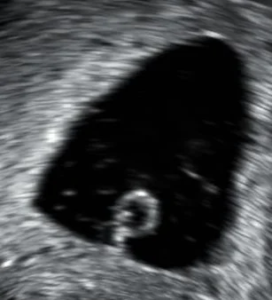
An anembryonic pregnancy, historically called a blighted ovum, is a gestational sac that develops without an embryo, and is a form of missed miscarriage. It is diagnosed when the mean sac diameter reaches 25 mm with no fetal pole visible on scanning through all planes.1
Aetiology and risk factors
Around half of first-trimester miscarriages are due to a fetal chromosomal abnormality, most often an autosomal trisomy, with trisomy 16 the single commonest. These are usually sporadic non-disjunction events and their incidence rises steeply with maternal age, which is why the miscarriage rate climbs from around 10% at age 25 to over 50% by age 42. Explaining this to a woman is often the most useful thing that can be said: the loss was determined at conception and nothing she did caused it.
Maternal factors
- Age - the dominant risk factor, through oocyte aneuploidy; advanced paternal age contributes to a lesser degree
- Endocrine - poorly controlled diabetes, untreated thyroid disease, and thyroid peroxidase antibodies even when euthyroid
- Antiphospholipid syndrome - the only consistently treatable cause of recurrent miscarriage
- Uterine anatomy - septate or bicornuate uterus, submucosal fibroids, intrauterine adhesions (Asherman's syndrome)
- Cervical insufficiency - painless cervical dilatation causing recurrent second-trimester loss
- Infection - ascending bacterial infection, listeria, and severe systemic febrile illness
- Lifestyle - smoking, heavy alcohol use, cocaine, obesity, and very low body weight
Clinical assessment
The typical presentation is per vaginam bleeding with or without suprapubic cramping pain in a woman with a positive pregnancy test. A missed miscarriage may be entirely asymptomatic and discovered at a routine dating scan, or suspected because pregnancy symptoms such as breast tenderness and nausea have abruptly disappeared.
The history should establish the date of the last menstrual period and hence gestation, the quantity of bleeding and whether clots or tissue have passed, the character and site of pain, and any risk factors for ectopic pregnancy. Shoulder-tip pain, unilateral pain, dizziness and syncope point towards an ectopic and change the urgency completely.
Examination
- Observations first: tachycardia and hypotension indicate significant haemorrhage or an unrecognised ectopic
- Abdominal examination for tenderness, guarding, rebound and peritonism
- Speculum examination to assess the volume of bleeding, whether the os is open, and whether products of conception are visible
- Bimanual examination for uterine size, adnexal tenderness and cervical excitation
- Any products sitting in the cervical os should be removed with sponge forceps and sent for histology
Investigations
Ultrasound
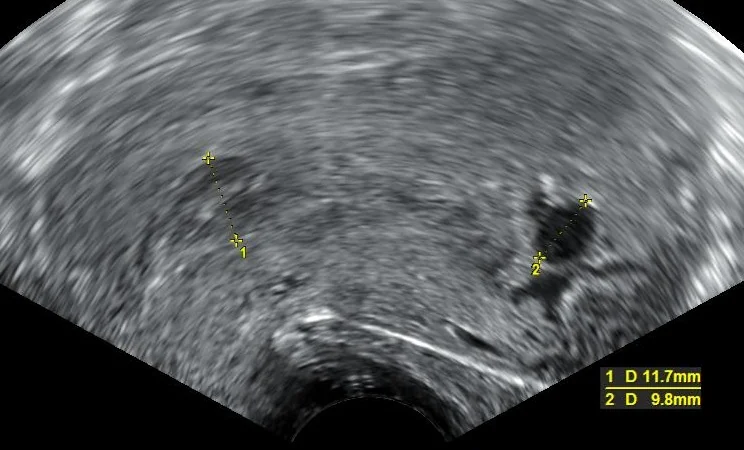
Transvaginal ultrasound is the definitive investigation and should be performed in an early pregnancy assessment unit. The diagnostic thresholds are deliberately conservative, because misdiagnosing a viable pregnancy as a miscarriage is catastrophic and irreversible.
| Finding | Interpretation |
|---|---|
| Crown-rump length 7 mm or more with no fetal heart activity | Miscarriage confirmed, ideally after a second opinion or a repeat scan |
| Crown-rump length under 7 mm with no fetal heart activity | Inconclusive - rescan after at least 7 days |
| Mean sac diameter 25 mm or more with no fetal pole | Anembryonic pregnancy confirmed, with a second opinion or repeat scan |
| Mean sac diameter under 25 mm with no fetal pole | Inconclusive - rescan after at least 7 days |
| No intrauterine pregnancy with a positive test | Pregnancy of unknown location - manage with serial hCG |
Serum hCG
Where the scan shows no intrauterine pregnancy and no adnexal mass, the pregnancy is of unknown location and serial serum hCG measured 48 hours apart guides the next step. A rise of more than 63% suggests a developing intrauterine pregnancy, and a repeat scan is arranged when the level exceeds the discriminatory zone. A fall of more than 50% suggests a failing pregnancy, and a urinary pregnancy test is repeated at 14 days. Anything in between is suspicious for an ectopic pregnancy and requires review in the early pregnancy unit within 24 hours.1
Other tests
Check the full blood count and group and save in significant bleeding, with crossmatch if bleeding is heavy. Determine RhD status in every woman, because it dictates anti-D prophylaxis. Histology of passed tissue confirms products of conception and, importantly, excludes molar pregnancy.
Management of miscarriage
Threatened miscarriage
Where fetal heart activity is present, the pregnancy continues in around three-quarters of cases. Management is expectant, with advice to return if bleeding worsens. NICE recommends vaginal micronised progesterone 400 mg twice daily for women with a threatened miscarriage who have had one or more previous miscarriages, continued until 16 completed weeks; this recommendation derives from the PRISM trial, where benefit was confined to that subgroup.1,4
Confirmed miscarriage
Three options are offered, and the choice is largely the woman's. Expectant management is first line unless there is a reason to avoid it.
| Option | What it involves | Considerations |
|---|---|---|
| Expectant | Wait 7-14 days for spontaneous resolution; repeat pregnancy test at 3 weeks | Avoids drugs and surgery; unpredictable timing and duration of bleeding; not suitable with infection, haemorrhage risk, bleeding disorders or previous traumatic experience |
| Medical | Missed miscarriage: mifepristone 200 mg then misoprostol 800 micrograms 48 hours later. Incomplete miscarriage: misoprostol 600 micrograms alone | Predictable timing; expect pain and heavy bleeding; provide analgesia and antiemetics; repeat pregnancy test at 3 weeks |
| Surgical | Manual vacuum aspiration under local anaesthetic, or surgical management of miscarriage under general anaesthetic | Immediate resolution; risks are infection, haemorrhage, cervical trauma, uterine perforation and Asherman's syndrome |
Septic miscarriage
Retained products with infection produce fever, offensive discharge, uterine tenderness and rising inflammatory markers, and can progress rapidly to septic shock. Management is with the sepsis six, broad-spectrum intravenous antibiotics, and urgent surgical evacuation once the patient is resuscitated. Do not delay evacuation waiting for antibiotics to work: the uterus remains a source of infection until it is emptied.
Follow-up
Advise a urine pregnancy test three weeks after expectant or medical management; a persistently positive test requires review to exclude retained products, molar or ectopic pregnancy. There is no medical need to delay conceiving again, although dating is easier after one normal period. Offer written information and signposting to support organisations, and mention that anniversaries and the expected due date are commonly difficult.
Recurrent miscarriage
Recurrent miscarriage was traditionally defined as three or more consecutive losses, which affects around 1% of couples. RCOG now recommends beginning investigation after two consecutive first-trimester losses, and after a single second-trimester loss, because delaying investigation to a third loss offers no diagnostic advantage and prolongs distress.6
| Investigation | Looking for |
|---|---|
| Lupus anticoagulant, anticardiolipin and anti-beta-2 glycoprotein I antibodies | Antiphospholipid syndrome; must be positive on two occasions at least 12 weeks apart |
| Cytogenetic analysis of products of conception | Aneuploidy, which if present makes an underlying parental cause less likely |
| Parental karyotyping | Balanced translocation in one parent, found in around 2-5% of couples; offered when the fetal result suggests an unbalanced rearrangement |
| Pelvic ultrasound, with 3D or hysteroscopy if abnormal | Congenital uterine anomaly, submucosal fibroid, intrauterine adhesions |
| Thyroid function and thyroid peroxidase antibodies | Overt or subclinical thyroid disease |
| HbA1c | Undiagnosed or poorly controlled diabetes |
Inherited thrombophilia screening is not recommended for recurrent first-trimester loss, because treatment with heparin does not improve live birth rates in that group. Where antiphospholipid syndrome is confirmed, low-dose aspirin plus low molecular weight heparin from a positive pregnancy test substantially improves live birth rates. Cervical cerclage is offered for cervical insufficiency, either as a history-indicated suture or following ultrasound surveillance of cervical length. Even after full investigation around half of couples have no cause identified, and their prognosis with supportive care in a dedicated clinic remains good.6
Late intrauterine fetal death and stillbirth
Fetal death from 24 weeks is diagnosed by real-time ultrasound demonstrating absent fetal cardiac activity, performed by a clinician competent in obstetric ultrasound, with a second opinion offered wherever practicable. Auscultation and cardiotocography are not acceptable methods of confirmation. Most women present with reduced or absent fetal movements; a proportion present with an unrelated complaint.2
Causes
- Placental insufficiency and fetal growth restriction - the largest single group
- Placental abruption
- Congenital anomaly and genetic disease
- Infection, including ascending bacterial infection, listeria, cytomegalovirus and parvovirus B19
- Maternal disease: pre-eclampsia, diabetes, obstetric cholestasis, thrombophilia
- Massive fetomaternal haemorrhage
- Cord accidents and, in monochorionic twins, twin-to-twin transfusion
- Unexplained in a substantial minority even after full investigation
Investigation
A Kleihauer test should be taken before delivery in every case, because fetomaternal haemorrhage is a recognised cause and the fetal cells clear from the maternal circulation with time. Alongside this take FBC, CRP, coagulation screen, U&E, LFTs and bile acids, HbA1c, thyroid function, antiphospholipid antibodies, blood group and antibody screen, and viral serology. After delivery, send the placenta for histology and offer fetal cytogenetic testing and post-mortem examination, discussed sensitively and with written consent.
Delivery
Vaginal birth is recommended for most women: it is safer, recovery is faster, and it preserves options in future pregnancies. Induction is with mifepristone followed by misoprostol, at doses adjusted for gestation, with full analgesia including epidural if wanted. Caesarean section is reserved for obstetric indications such as placenta praevia or transverse lie. Women with an unscarred uterus and no contraindication may reasonably choose to wait for spontaneous labour, but should understand the risks.
Afterwards
- Lactation suppression with cabergoline, offered proactively rather than waiting for the woman to ask
- Memory-making: photographs, hand and foot prints, a lock of hair, and time with the baby as the parents wish
- The stillbirth must be registered, and the medical certificate of stillbirth completed
- Anti-D for RhD negative women, with the dose guided by the Kleihauer result
- Referral to bereavement support and to organisations such as Sands
- Review through the Perinatal Mortality Review Tool, with a follow-up appointment to discuss results and plan future pregnancy care
- Future pregnancies are managed as high risk, with aspirin, serial growth scans and planned birth by around 39 weeks
Prognosis
The prognosis after a single sporadic miscarriage is excellent: the great majority of women go on to have a successful pregnancy, and the risk of a further loss is barely elevated above the population baseline. Even after three consecutive unexplained losses, the chance of a live birth in the next pregnancy is around 60-70% with supportive care alone.
After a stillbirth the recurrence risk depends entirely on the cause. Where growth restriction or a placental cause is identified, the risk in the next pregnancy is meaningfully increased and justifies aspirin, serial growth surveillance and earlier planned birth. Where the death was due to a non-recurring event, the risk is close to baseline, but the anxiety is not, and additional scans and contact with the team have value independent of what they detect.2,3
References
- NICE NG126. Ectopic pregnancy and miscarriage: diagnosis and initial management. 2019 (updated 2023). Available here
- RCOG Green-top Guideline No. 55. Late intrauterine fetal death and stillbirth. Available here
- MBRRACE-UK. Perinatal mortality surveillance reports. Available here
- Coomarasamy A, Devall AJ, Cheed V et al. A randomized trial of progesterone in women with bleeding in early pregnancy (PRISM). N Engl J Med. 2019. Available here
- RCOG Green-top Guideline No. 22. The use of anti-D immunoglobulin for rhesus D prophylaxis. Available here
- RCOG Green-top Guideline No. 17. Recurrent miscarriage. 2023. Available here
This article is written for revision and education. It is not clinical guidance and must not be used to make decisions about the care of a patient. Always check current NICE guidance and local protocols.