
Disseminated Intravascular Coagulation
Key points
- Disseminated intravascular coagulation (DIC): a systemic activation of coagulation triggered by an underlying illness, causing widespread microvascular thrombosis and, through consumption of platelets and clotting factors, simultaneous bleeding.
- It is always secondary: DIC is never a primary diagnosis - it is a complication of something else. The commonest triggers are sepsis, major trauma, malignancy and obstetric emergencies.
- The paradox: patients clot and bleed at the same time. Microthrombi cause ischaemic organ damage, while the resulting depletion of platelets and factors, plus secondary fibrinolysis, causes haemorrhage.
- Classic laboratory picture: everything is deranged: prolonged PT and APTT, low platelets, low fibrinogen, raised D-dimer/fibrin degradation products, and schistocytes on the blood film.
- Fibrinogen is the key discriminator: a low fibrinogen strongly supports DIC. Fibrinogen is an acute phase reactant that should rise in a sick patient - a normal level in severe sepsis may be inappropriately low, and a falling trend is more informative than a single value.
- Acute promyelocytic leukaemia: an important cause of severe DIC. Suspect it in a patient with acute leukaemia and florid bleeding, and start ATRA on clinical suspicion without waiting for genetic confirmation.
- Management: treat the underlying cause - this is the only definitive treatment. Supportive blood product replacement (platelets, fresh frozen plasma, cryoprecipitate) is guided by bleeding, not numbers alone.
- Prognosis: DIC marks severe underlying illness and roughly doubles mortality in sepsis. Outcome depends almost entirely on whether the precipitating condition can be reversed.
Introduction
Disseminated intravascular coagulation is an acquired syndrome in which the normally localised, tightly regulated process of coagulation becomes systemically and inappropriately activated. Fibrin is deposited throughout the microvasculature, causing tissue ischaemia and organ dysfunction, while the platelets and clotting factors consumed in doing so are depleted faster than they can be replaced - producing the characteristic combination of thrombosis and haemorrhage in the same patient.1
The single most important conceptual point is that DIC is always secondary to another condition. It is a marker of severe underlying illness, and its management centres on identifying and treating that illness rather than on correcting the coagulation numbers in isolation.
Pathophysiology
The initiating event is widespread exposure of tissue factor to the circulation - from damaged endothelium, injured tissue, monocytes activated by inflammatory cytokines, or tumour cells. Tissue factor triggers the extrinsic pathway on a systemic rather than a local scale.
- Systemic thrombin generation - tissue factor drives uncontrolled conversion of prothrombin to thrombin throughout the circulation
- Widespread fibrin deposition - thrombin converts fibrinogen to fibrin, producing microthrombi that occlude small vessels and cause ischaemic organ damage (kidneys, lungs, skin, brain)
- Consumption - platelets and clotting factors (particularly fibrinogen, and factors V and VIII) are used up in this process faster than the liver and marrow can replace them, hence the older term "consumption coagulopathy"
- Secondary fibrinolysis - plasmin is activated to break down the excess fibrin, generating fibrin degradation products (including D-dimer). These products are themselves anticoagulant, further impairing haemostasis
- Bleeding - the net result of depleted platelets, depleted factors and circulating fibrin degradation products
- Microangiopathic haemolysis - red cells are physically sheared as they pass through fibrin strands in the microvasculature, producing schistocytes
The balance between thrombosis and bleeding varies: chronic, low-grade DIC (as in malignancy) tends to present with thrombosis, while acute, fulminant DIC (as in sepsis or obstetric catastrophe) usually presents with bleeding.
Causes
| Category | Examples |
|---|---|
| Sepsis and severe infection | The commonest cause - particularly Gram-negative sepsis, but also Gram-positive, meningococcal septicaemia (classically with purpura fulminans), malaria and severe viral infection |
| Trauma and tissue injury | Major trauma, crush injury, extensive burns, major surgery, fat embolism, heat stroke |
| Malignancy | Acute promyelocytic leukaemia (florid, life-threatening DIC), other acute leukaemias, and mucin-secreting adenocarcinomas (typically chronic, thrombotic DIC) |
| Obstetric emergencies | Amniotic fluid embolism, placental abruption, retained dead fetus, HELLP syndrome and severe pre-eclampsia, septic abortion, major postpartum haemorrhage |
| Severe immunological reactions | ABO-incompatible blood transfusion, severe anaphylaxis, transplant rejection |
| Other | Severe liver failure, snake envenomation, severe pancreatitis, giant haemangioma (Kasabach-Merritt syndrome), vasculitis |
Clinical features
The patient is almost always acutely and obviously unwell from the underlying condition, with the features of DIC superimposed.
Bleeding manifestations
- Oozing from venepuncture sites, cannulae, drains and surgical wounds - often the earliest and most characteristic clinical sign
- Widespread petechiae, purpura and ecchymoses
- Mucosal bleeding - epistaxis, gum bleeding, haematuria, gastrointestinal bleeding
- Major haemorrhage, including intracranial bleeding
Thrombotic manifestations
- Acute kidney injury from renal microvascular thrombosis
- Respiratory failure/ARDS
- Confusion, delirium and reduced consciousness from cerebral microthrombi
- Digital ischaemia and peripheral gangrene - particularly in meningococcal sepsis, where purpura fulminans with skin necrosis is a classic presentation
- Hepatic dysfunction, and large vessel thrombosis in some cases
Investigations
There is no single diagnostic test for DIC. The diagnosis rests on recognising a compatible pattern of results in a patient with a known precipitant, and serial testing is more informative than a single set, since a falling trend in platelets and fibrinogen is highly suggestive even when individual values remain within the reference range.
| Test | Result in DIC | Reason |
|---|---|---|
| Platelet count | Low (and falling) | Consumed in widespread microthrombi |
| PT / INR | Prolonged | Consumption of clotting factors |
| APTT | Prolonged | Consumption of clotting factors |
| Fibrinogen | Low | Consumed by thrombin; the most useful discriminating test |
| D-dimer / fibrin degradation products | Markedly raised | Secondary fibrinolysis breaking down excess fibrin |
| Blood film | Schistocytes (red cell fragments) | Mechanical shearing through fibrin strands |
| Haemoglobin | Falling | Bleeding plus microangiopathic haemolysis |
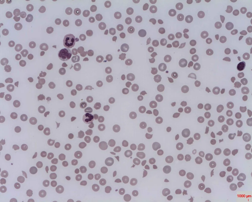
Formal scoring systems (such as the ISTH DIC score, combining platelet count, PT prolongation, fibrinogen and D-dimer) can support the diagnosis and are used in research and some clinical settings, but the diagnosis remains fundamentally clinical and contextual.
Differential diagnosis
| Condition | Platelets | PT/APTT | Fibrinogen | Distinguishing features |
|---|---|---|---|---|
| DIC | Low | Prolonged | Low | Identifiable precipitant; raised D-dimer; schistocytes |
| TTP | Very low | Normal | Normal | Normal clotting with severe thrombocytopenia; neurological signs; renal impairment; ADAMTS13 deficiency |
| HUS | Low | Normal | Normal | Renal failure dominant; often preceded by diarrhoeal illness in children |
| Liver failure | Often low | Prolonged | Low (late) | Factor VIII is normal or high in liver disease but low in DIC; other liver synthetic markers abnormal; less marked D-dimer rise |
| Vitamin K deficiency / warfarin | Normal | Prolonged (PT first) | Normal | Normal platelets and fibrinogen; corrects with vitamin K |
| Dilutional coagulopathy | Low | Prolonged | Low | History of massive transfusion/fluid resuscitation without a DIC trigger |
The distinction from TTP and HUS matters greatly, because these thrombotic microangiopathies also produce thrombocytopenia and schistocytes but have normal coagulation times and fibrinogen - and because TTP requires urgent plasma exchange, an entirely different treatment. Factor VIII levels help separate DIC from liver disease: factor VIII is produced by endothelium rather than hepatocytes, so it is preserved or raised in liver failure but consumed and low in DIC.
Management
Treat the underlying cause
This is the only definitive treatment and takes absolute priority. DIC will not resolve while its trigger persists, and no amount of blood product replacement substitutes for source control - antibiotics and resuscitation in sepsis, delivery of the fetus and placenta in obstetric DIC, ATRA in acute promyelocytic leukaemia, surgical control in trauma.2
Supportive blood product replacement
Replacement is guided principally by active bleeding or an imminent invasive procedure, rather than by laboratory values alone - transfusing to correct numbers in a non-bleeding patient is not beneficial and consumes scarce products:
- Platelet transfusion - for active bleeding with a platelet count below around 50 x10^9/L, or prophylactically at lower thresholds (around 20-30) in high-risk non-bleeding patients per local policy
- Fresh frozen plasma (FFP) - replaces multiple clotting factors, for bleeding with prolonged PT/APTT
- Cryoprecipitate or fibrinogen concentrate - specifically to replace fibrinogen, typically when it falls below around 1.5 g/L in a bleeding patient (a higher threshold, around 2 g/L, is used in obstetric haemorrhage)
- Red cell transfusion as required for anaemia and ongoing blood loss
- Vitamin K if there is coexisting deficiency
Anticoagulation and antifibrinolytics
- Therapeutic heparin is considered only where the thrombotic phenotype dominates - for example chronic DIC in malignancy, or overt large vessel thrombosis - and is generally avoided when bleeding predominates
- Prophylactic-dose LMWH for VTE prevention is appropriate in critically ill non-bleeding patients with DIC
- Tranexamic acid and other antifibrinolytics are generally avoided in DIC, since fibrinolysis is a compensatory response clearing the microthrombi - blocking it risks worsening organ ischaemia. Important exceptions exist (notably major obstetric and trauma haemorrhage, where tranexamic acid is given per established protocols) and its use should follow specialist advice
- Involve haematology early - DIC management frequently requires specialist input on product selection and thresholds
Complications
- Multi-organ failure from widespread microvascular thrombosis - the major cause of death
- Acute kidney injury, often requiring renal replacement therapy
- Acute respiratory distress syndrome
- Major haemorrhage, including intracranial and gastrointestinal bleeding
- Peripheral ischaemia, digital gangrene and skin necrosis - purpura fulminans, particularly in meningococcal sepsis, sometimes requiring amputation
- Adrenal haemorrhage - Waterhouse-Friderichsen syndrome, classically complicating meningococcal septicaemia and causing acute adrenal insufficiency
- Transfusion-related complications from large-volume blood product support
Red flags
Prognosis
DIC is a marker of severe systemic illness, and its presence approximately doubles mortality in sepsis compared with sepsis alone.1 Overall outcome is determined almost entirely by the nature and reversibility of the underlying cause rather than by the coagulopathy itself.
Obstetric DIC often has the best outlook, because the precipitant (placental abruption, retained products, amniotic fluid embolism) can frequently be removed decisively, after which coagulation normalises rapidly. DIC complicating sepsis with established multi-organ failure, or advanced malignancy, carries a substantially worse prognosis. Survivors of severe DIC may be left with lasting sequelae including chronic kidney disease and, in cases of purpura fulminans, tissue loss requiring amputation.
References
- Levi M, Ten Cate H. Disseminated intravascular coagulation. N Engl J Med. 1999. Available here
- Levi M, Toh CH, Thachil J, Watson HG. Guidelines for the diagnosis and management of disseminated intravascular coagulation. Br J Haematol. 2009. Available here
- Erhabor Osaro, CC BY-SA 3.0, via Wikimedia Commons. Available here
- Taylor FB, Toh CH, Hoots WK et al. Towards definition, clinical and laboratory criteria for disseminated intravascular coagulation (ISTH). Thromb Haemost. 2001. Available here
- NICE. Blood transfusion (NG24). 2015. Available here
This article is written for revision and education. It is not clinical guidance and must not be used to make decisions about the care of a patient. Always check current NICE guidance and local protocols.