
Appendicitis: Diagnosis and Management
Key points
- Appendicitis: acute inflammation of the vermiform appendix, usually initiated by obstruction of its lumen. It is the commonest surgical emergency in the UK.
- Lifetime risk: around 7 to 8%, with a peak incidence between the ages of 10 and 20.
- Why the pain migrates: early visceral pain from the midgut appendix is felt periumbilically; once inflammation reaches the parietal peritoneum it localises sharply to the right iliac fossa.
- Classic features: migratory pain, anorexia, nausea, low-grade fever and tenderness at McBurney point with localised guarding.
- Never omit: a pregnancy test in every woman of childbearing age. Ectopic pregnancy is the diagnosis that kills if it is mistaken for appendicitis.
- Scoring: the Alvarado and AIR scores stratify risk. A low score supports discharge or observation; a high score supports theatre without imaging.
- Imaging: contrast CT in adults, ultrasound first in children and women of childbearing age, and ultrasound then MRI in pregnancy.
- Treatment: laparoscopic appendicectomy is the standard. Antibiotics alone succeed in most uncomplicated cases but about one in four recur within a year.
Introduction
Acute appendicitis is inflammation of the vermiform appendix and is the commonest cause of the acute abdomen requiring surgery in the UK, with around 50,000 appendicectomies performed each year.1 The lifetime risk is approximately 7 to 8%, and incidence peaks between the ages of 10 and 20, though it occurs at every age.
It is a clinical diagnosis supported by imaging rather than a radiological one, and this is the source of most of the difficulty. The classic presentation is present in fewer than half of patients, and the diagnosis is hardest precisely in the groups where a missed diagnosis is most dangerous: very young children, older adults, and pregnant women.
Two competing errors define the topic. Operating on a normal appendix exposes a patient to the risks of anaesthesia and surgery for nothing; historically UK negative appendicectomy rates approached 20%, and they have fallen substantially with routine cross-sectional imaging. Failing to operate risks perforation, which raises morbidity sharply. Modern practice tries to reduce both, and the tools for doing so are risk scores and selective imaging rather than clinical instinct alone.
Anatomy
The appendix is a blind-ending tube of variable length, typically 6 to 10 cm, arising from the posteromedial wall of the caecum about 2 cm below the ileocaecal valve. Its base is constant and lies at the point where the three taeniae coli of the caecum converge, which is how a surgeon finds it. Its tip, by contrast, is highly variable, and that variability explains most atypical presentations.
| Position | Approximate frequency | Clinical consequence |
|---|---|---|
| Retrocaecal | About 65% | Tenderness may be mild or absent anteriorly because the caecum shields the appendix from the anterior parietal peritoneum. Psoas irritation causes pain on hip extension and a preference for lying with the right hip flexed. |
| Pelvic | About 30% | Suprapubic pain, diarrhoea, tenesmus and urinary frequency from irritation of the rectum and bladder. Tenderness may be found only on rectal or vaginal examination. |
| Pre-ileal or post-ileal | About 5% | Vomiting and diarrhoea may dominate, and the picture is easily mistaken for gastroenteritis |
| Subhepatic | Uncommon | Right upper quadrant pain mimicking cholecystitis, and the usual position in late pregnancy |
McBurney point lies one third of the way along a line drawn from the right anterior superior iliac spine to the umbilicus, and marks the usual surface position of the appendix base. The blood supply is the appendicular artery, a branch of the ileocolic artery, which runs in the free edge of the mesoappendix. It is an end artery with no collateral supply, which is why thrombosis within it leads rapidly to gangrene and perforation.
The appendix is a midgut structure, so its visceral afferents enter the cord at T10. This is the anatomical basis for the periumbilical pain that opens the classic history.
Pathophysiology
The dominant model is obstruction of the appendiceal lumen, which converts it into a closed loop.
- Luminal obstruction by a faecolith, lymphoid hyperplasia, an impacted seed or, in older patients, a caecal or appendiceal tumour
- Continued mucus secretion proximal to the obstruction raises intraluminal pressure, producing distension and the dull visceral T10 pain
- Bacterial overgrowth of the trapped luminal contents, with mucosal invasion and a neutrophilic infiltrate
- Venous and lymphatic congestion as intraluminal pressure exceeds venous pressure, causing wall oedema and serosal inflammation. Once the inflamed serosa touches the parietal peritoneum the pain becomes somatic and localises to the right iliac fossa.
- Arterial compromise and gangrene as pressure exceeds arterial perfusion pressure in an end-artery territory
- Perforation, releasing pus and faecal material. This is either walled off by omentum and adjacent bowel to form an appendix mass or abscess, or it spills freely to produce generalised peritonitis.
The distinction between uncomplicated appendicitis (inflamed but intact) and complicated appendicitis (gangrenous, perforated, or with an abscess or phlegmon) governs almost every management decision, including whether antibiotics alone can be considered and how long antibiotics are given after surgery.
Clinical features
The classic sequence, present in perhaps half of patients, is: periumbilical pain, followed by anorexia and nausea, followed by migration of the pain to the right iliac fossa, followed by low-grade fever.
- Migratory pain - the most discriminating single symptom. Ask where the pain was when it started, not just where it is now.
- Anorexia - near universal in children and young adults. A patient who is hungry and eating rarely has appendicitis, though this is a soft rule in older people.
- Nausea and one or two episodes of vomiting, which characteristically follow the pain rather than precede it
- Low-grade fever, typically 37.5 to 38.5 °C. A temperature above 39 °C suggests perforation or an alternative diagnosis.
- Constipation is more common than diarrhoea, but a pelvic appendix irritating the rectum causes diarrhoea and tenesmus and is a classic trap
- Urinary frequency and dysuria where the appendix lies against the bladder or ureter
Examination
- The patient lies still and dislikes moving. Ask them to cough, or to hop, and watch their face - both are gentler than testing for rebound.
- Tenderness maximal at McBurney point, with localised guarding and percussion tenderness
- Rovsing sign - palpation of the left iliac fossa produces pain in the right iliac fossa
- Psoas sign - pain on passive extension of the right hip, suggesting a retrocaecal appendix lying on the psoas muscle
- Obturator sign - pain on passive internal rotation of the flexed right hip, suggesting a pelvic appendix
- Digital rectal examination may elicit right-sided tenderness with a pelvic appendix, though it is no longer performed routinely
- A tender mass in the right iliac fossa after several days of symptoms suggests an appendix mass or abscess
- Always examine the hernial orifices and testes, and consider gynaecological examination in women
Risk scores
Scores do not make the diagnosis, but they formalise risk stratification and reduce both negative appendicectomy and unnecessary CT. The Alvarado score is the best known; the Appendicitis Inflammatory Response (AIR) score performs slightly better in European cohorts and is increasingly used in the UK.2
| Component | Points |
|---|---|
| M - Migration of pain to the right iliac fossa | 1 |
| A - Anorexia | 1 |
| N - Nausea or vomiting | 1 |
| T - Tenderness in the right iliac fossa | 2 |
| R - Rebound tenderness | 1 |
| E - Elevated temperature above 37.3 °C | 1 |
| L - Leucocytosis above 10 x10^9/L | 2 |
| S - Shift of white cell count to the left (neutrophilia above 75%) | 1 |
- 0 to 3 - appendicitis unlikely; consider discharge with safety netting or investigate an alternative cause
- 4 to 6 - equivocal; admit for observation and serial examination, or image
- 7 to 10 - appendicitis likely; surgical review with a view to appendicectomy
The score is less reliable in women, in whom gynaecological pathology produces false positives, and in children and older adults. Treat it as a way of documenting and communicating risk rather than as a decision rule.
Differential diagnosis
| System | Diagnoses |
|---|---|
| Gynaecological | Ectopic pregnancy, ovarian cyst rupture or torsion, pelvic inflammatory disease, tubo-ovarian abscess, mittelschmerz, endometriosis |
| Gastrointestinal | Mesenteric adenitis, Crohn disease, Meckel diverticulitis, caecal or appendiceal tumour, gastroenteritis, diverticulitis of a redundant sigmoid loop, constipation |
| Urological | Ureteric colic, urinary tract infection, pyelonephritis, testicular torsion |
| Other | Rectus sheath haematoma, psoas abscess, herpes zoster, right lower lobe pneumonia, diabetic ketoacidosis |
Mesenteric adenitis deserves particular mention in children: it follows a recent upper respiratory tract infection, produces a higher fever with less localised tenderness, and settles without surgery. Meckel diverticulitis is clinically indistinguishable from appendicitis and is usually found at operation, which is why the terminal ileum should be inspected if a normal appendix is found.
Investigations
Bedside and laboratory
- Urinary or serum beta-hCG in every woman of childbearing age
- FBC - neutrophil leucocytosis is present in around 80% but is neither sensitive nor specific. A normal white cell count and a normal CRP together make appendicitis considerably less likely.
- CRP - rises later than the white cell count, so it may be normal in the first 12 hours. A markedly raised CRP suggests complicated disease.
- U&Es - for dehydration and before anaesthesia
- Urine dipstick - to look for an alternative urinary cause, but remember that mild pyuria and haematuria occur in appendicitis itself and do not exclude it
- Group and save and a clotting screen before theatre
Imaging
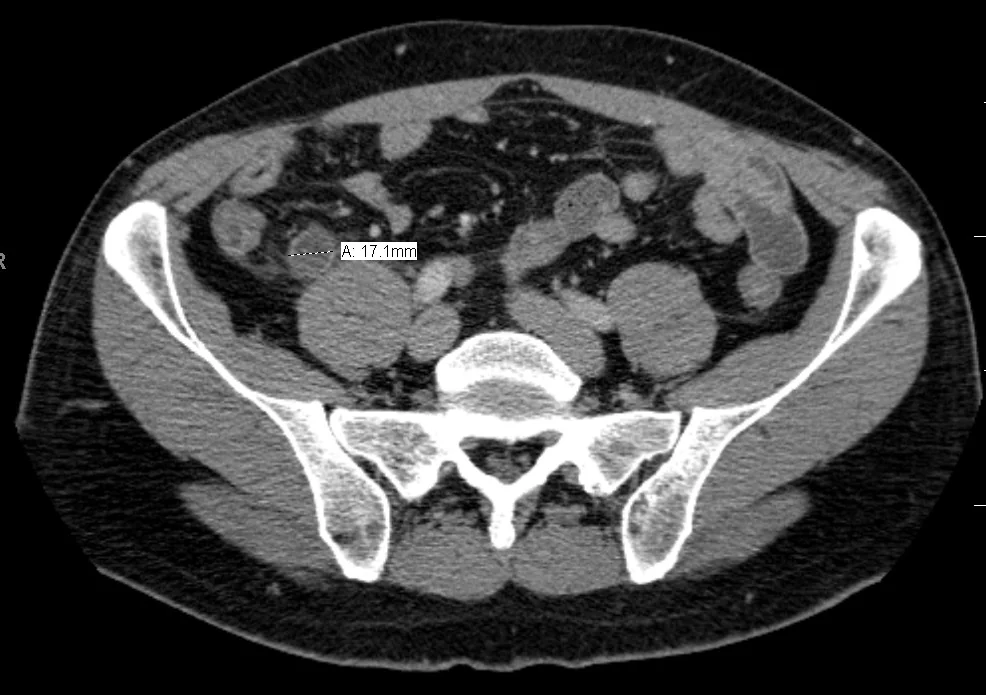
- Contrast-enhanced CT of the abdomen and pelvis - sensitivity and specificity both above 90%. Features are an appendix diameter above 6 mm, wall thickening and enhancement, periappendiceal fat stranding, a faecolith, free fluid, and an abscess or extraluminal gas in complicated disease. CT is the default in adults, particularly those over 40 in whom a caecal tumour must be excluded.3
- Ultrasound - first line in children and in women of childbearing age because it avoids radiation and assesses the ovaries. A non-compressible blind-ending tubular structure over 6 mm in diameter is diagnostic, but a non-visualised appendix is common and does not exclude appendicitis.
- MRI - the second-line modality in pregnancy after an inconclusive ultrasound, avoiding ionising radiation entirely
- Diagnostic laparoscopy - both diagnostic and therapeutic in equivocal cases, particularly in young women
Management
Initial measures
- Nil by mouth, intravenous fluids and analgesia with an antiemetic
- Venous thromboembolism risk assessment and prophylaxis
- Consent, group and save, and prompt listing for theatre
- Prophylactic intravenous antibiotics at induction, following local policy - typically a combination covering Gram-negative and anaerobic organisms, such as co-amoxiclav, or a cephalosporin with metronidazole
Appendicectomy
Laparoscopic appendicectomy is the standard operation in the UK. Compared with open surgery it produces less wound infection, less pain, a shorter hospital stay and a faster return to normal activity, at the cost of a higher rate of intra-abdominal abscess in some series and a longer operating time.4 The open approach through a Lanz or gridiron incision remains appropriate where laparoscopy is unavailable, in significant adhesions, or in some cases of advanced pregnancy.
Post-operative antibiotics are not required after removal of a simple inflamed appendix. In complicated appendicitis, with gangrene, perforation or contamination, a course of intravenous antibiotics is continued, typically for three to five days and converted to oral once the patient improves.
Antibiotics instead of surgery
Non-operative management with antibiotics alone is a genuine option in uncomplicated appendicitis. The CODA trial randomised over 1,500 adults to antibiotics or appendicectomy and found antibiotics non-inferior for 30-day health status, but around 30% of the antibiotic group had undergone appendicectomy by 90 days, rising to about 40% by four years, and the failure rate was substantially higher in those with an appendicolith.5
- Reasonable to offer where the patient wishes to avoid surgery, where operative risk is high, or where theatre capacity is constrained
- Not appropriate in complicated appendicitis, in the presence of an appendicolith, or where a caecal tumour has not been excluded in an older patient
- The patient must be counselled explicitly that the appendix remains in place, that recurrence is common, and that they must return promptly if symptoms recur
Appendix mass and appendix abscess
A patient presenting after several days with a tender right iliac fossa mass has a walled-off inflammatory phlegmon. Operating on this is technically difficult and carries a high complication rate, so the conventional approach is conservative:
- Intravenous antibiotics, fluids and bowel rest, with serial examination and inflammatory markers
- Radiologically guided percutaneous drainage if there is a drainable abscess collection on CT
- Surgery if the patient deteriorates, develops generalised peritonitis, or fails to settle
- Interval imaging by colonoscopy or CT colonography in patients over 40, because a perforated caecal carcinoma can present in exactly this way
- Interval appendicectomy is no longer routine, and is reserved for recurrent symptoms or diagnostic uncertainty
Complications
Of the disease
- Perforation - the principal complication, occurring in around 20% overall but in a much higher proportion of children under five and adults over 65, in whom the diagnosis is delayed
- Generalised peritonitis and sepsis where perforation is not contained
- Appendix mass or abscess where it is contained
- Pelvic abscess - suspect where swinging fever, diarrhoea and tenesmus develop several days after presentation
- Portal pyaemia and liver abscess - rare, from septic thrombophlebitis of the portal venous system
- Adhesional small bowel obstruction and subfertility - long-term consequences of peritoneal inflammation
Of the operation
- Surgical site infection, more common after open surgery and in perforated disease
- Intra-abdominal or pelvic collection, presenting around day five to seven with fever and ileus
- Bleeding from the appendicular artery or mesoappendix
- Appendiceal stump leak, and stump appendicitis if too long a stump is left
- Injury to adjacent structures including the caecum, ileum, bladder and iliac vessels
- Post-operative ileus, and later adhesional obstruction
- Port-site or wound hernia
Histology of every specimen is mandatory. Around 1% contain an unexpected neoplasm, most commonly a neuroendocrine tumour of the appendix tip, which usually needs no further treatment if it is under 1 cm and completely excised, but requires a right hemicolectomy if larger, at the base, or with mesoappendiceal invasion.
Red flags
Prognosis
For uncomplicated appendicitis treated promptly, prognosis is excellent. Mortality is well under 0.1%, most patients go home within 24 to 48 hours of a laparoscopic appendicectomy, and there is no long-term consequence of losing the appendix.
Perforation changes the picture materially. Complicated appendicitis carries a longer stay, a higher rate of intra-abdominal collection and wound infection, and a small but real mortality that rises with age and comorbidity. Because the strongest predictor of perforation is delay, the practical priority is prompt assessment rather than heroic surgery.
One nuance is worth carrying forward. The trend across the last decade has been towards fewer operations, not more: CT before theatre to cut the negative appendicectomy rate, antibiotics alone in selected uncomplicated cases, and conservative management of the appendix mass. The trade-off in each instance is recurrence and a residual diagnostic uncertainty, and the right choice depends on the patient in front of you rather than on a single protocol.
References
- NICE Clinical Knowledge Summaries. Appendicitis. Available here
- Andersson M, Andersson RE. The appendicitis inflammatory response score. World Journal of Surgery. 2008. Available here
- Di Saverio S, Podda M, De Simone B et al. Diagnosis and treatment of acute appendicitis: 2020 update of the WSES Jerusalem guidelines. World Journal of Emergency Surgery. 2020. Available here
- Jaschinski T, Mosch CG, Eikermann M et al. Laparoscopic versus open surgery for suspected appendicitis. Cochrane Database of Systematic Reviews. 2018. Available here
- CODA Collaborative. A randomized trial comparing antibiotics with appendectomy for appendicitis. New England Journal of Medicine. 2020. Available here
This article is written for revision and education. It is not clinical guidance and must not be used to make decisions about the care of a patient. Always check current NICE guidance and local protocols.