
Mesenteric Adenitis
Key points
- Definition: self-limiting inflammatory enlargement of the mesenteric lymph nodes, usually in the right lower quadrant, producing abdominal pain that mimics appendicitis.
- Who gets it: children and adolescents, most often between 5 and 14 years. It is one of the commonest causes of non-surgical acute abdominal pain in children.
- Cause: usually viral, adenovirus most often. Yersinia enterocolitica is the classic bacterial cause and produces a pseudo-appendicitis with terminal ileitis.
- The sequence that helps: in mesenteric adenitis fever tends to come first and pain second; in appendicitis pain comes first and fever follows.
- Examination: tenderness is diffuse, poorly localised and may shift when the child rolls, and guarding is usually absent.
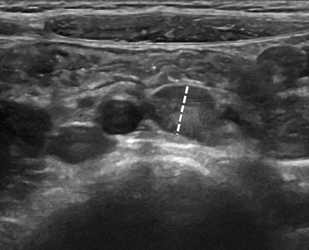
- Ultrasound: three or more clustered mesenteric nodes with a short-axis diameter of 8 mm or more, together with a normal appendix.
- It is a diagnosis of exclusion: the real clinical task is to be confident this is not appendicitis, and no single test achieves that.
- Management: analgesia, fluids, active observation and clear safety netting. Antibiotics are needed only where a bacterial cause is identified and the child is unwell.
Introduction
Mesenteric adenitis - more precisely acute non-specific mesenteric lymphadenitis - is reactive enlargement of the lymph nodes in the small bowel mesentery, particularly the ileocolic group. It causes abdominal pain, fever and tenderness in the right iliac fossa, and it is the condition most often confused with acute appendicitis in children.4
The topic is worth taking seriously despite its benign nature, because the clinical decision it forces is a genuinely difficult one. Diagnosing appendicitis as mesenteric adenitis risks perforation and peritonitis; diagnosing mesenteric adenitis as appendicitis leads to an unnecessary operation. Historically, before imaging was routine, mesenteric adenitis was often found only when a normal appendix was removed at laparotomy.
Two things resolve most cases: an ultrasound that visualises a normal appendix alongside enlarged nodes, and a period of active observation with repeated examination. Neither the white cell count nor a single examination is reliable on its own, and being explicit about that is part of a good answer.
Aetiology and pathophysiology
The mesentery of the terminal ileum contains a dense concentration of lymph nodes draining the most lymphoid-rich part of the gut. Any enteric or systemic infection delivers antigen to these nodes, which respond with reactive follicular hyperplasia. The nodal capsule stretches and the surrounding mesentery becomes inflamed, producing visceral pain that is initially felt centrally and later localises to the right iliac fossa - the same referral pattern as appendicitis, which is why the two are so hard to separate.
Causative organisms
- Adenovirus - the commonest identified cause
- Epstein-Barr virus, coxsackievirus, rhinovirus, influenza and other respiratory viruses, which is why an upper respiratory tract infection so often precedes the pain
- Yersinia enterocolitica and Y. pseudotuberculosis - the classic bacterial causes, producing a pseudo-appendicitis with terminal ileitis and prominent mesenteric adenitis, often with diarrhoea and sometimes with a reactive arthritis or erythema nodosum afterwards
- Campylobacter jejuni, Salmonella and Shigella
- Group A streptococcus, following a recent pharyngitis
- Bartonella henselae (cat scratch disease), where there is animal contact
- Mycobacterium tuberculosis, which causes a chronic rather than acute adenitis and should be considered where there is weight loss, night sweats or relevant contact and travel history
Epidemiology
Mesenteric adenitis is a disease of childhood and adolescence, and its age distribution tracks the size of the gut-associated lymphoid tissue, which is proportionally greatest in the school-age years and involutes after puberty.
- Peak age 5-14 years, and it becomes uncommon in adults, in whom right iliac fossa pain with enlarged nodes should prompt a wider search
- Slight male predominance, mirroring that of appendicitis
- Seasonal clustering in the winter and spring months, following the peaks of respiratory and enteric viral infection
- It is one of the commonest final diagnoses in children admitted for observation with acute abdominal pain, and historically accounted for a large share of the normal appendices removed at operation
- Recurrence occurs in a minority; repeated episodes should trigger a broader assessment rather than a repeated label
Clinical features
- Abdominal pain - central or right iliac fossa, often colicky, and characteristically less severe and less sharply localised than in appendicitis
- Fever, frequently above 38.5°C, and typically preceding the abdominal pain
- A preceding or concurrent upper respiratory tract infection - sore throat, coryza, cough - or a recent diarrhoeal illness
- Nausea, vomiting, anorexia and sometimes diarrhoea, though the child usually retains some appetite
- Cervical lymphadenopathy and pharyngeal inflammation, reflecting the systemic nature of the illness
- A child who looks better than the abdominal signs suggest - reasonably comfortable between episodes, willing to move, and not lying rigidly still
Examination
- Diffuse, poorly localised tenderness, often maximal in the right iliac fossa but not confined to McBurney's point
- Shifting tenderness - the point of maximal tenderness may move when the child rolls onto their side, because the mobile mesentery moves with them. In appendicitis the inflamed appendix is fixed and the tenderness stays put.
- Guarding and rebound tenderness are usually absent, and true rigidity should make you reject the diagnosis
- No pain on coughing or heel drop, in contrast to peritoneal irritation from appendicitis
- Generalised lymphadenopathy may be palpable, particularly cervical nodes
- Examine the throat, the ears, the chest, the groins and the testes, and check a urine dipstick - all of which look for the alternative explanation
Distinguishing it from appendicitis
| Feature | Mesenteric adenitis | Acute appendicitis |
|---|---|---|
| Order of symptoms | Fever first, then abdominal pain | Pain first, then anorexia, nausea and later fever |
| Preceding illness | Common - upper respiratory tract infection or diarrhoea | Usually none |
| Nature of pain | Colicky, central or diffuse, waxing and waning | Constant, migrating from periumbilical to right iliac fossa, progressively worse |
| Fever | Often high, above 38.5°C, early | Low grade initially; high fever suggests perforation |
| Tenderness | Diffuse, poorly localised, may shift with position | Localised and fixed at McBurney's point |
| Guarding and rebound | Usually absent | Present, with percussion tenderness and pain on coughing |
| General appearance | Reasonably well, moving freely, still eating a little | Unwell, lying still, refusing to move or cough |
| Full blood count | Lymphocytosis or a mild neutrophilia | Neutrophilia, rising with progression |
| Ultrasound | Clustered enlarged nodes with a normal appendix | Non-compressible blind-ending tubular structure over 6 mm, free fluid, appendicolith |
| Course | Improves over days without intervention | Progressive deterioration; perforation typically within 48-72 hours |
Differential diagnosis
- Acute appendicitis - the diagnosis that must be excluded before any other consideration1
- Gastroenteritis - diarrhoea predominates, pain is less localised, contact history2,5
- Urinary tract infection - dysuria may be absent in younger children, so dipstick every child with abdominal pain
- Constipation - a chronic history with palpable faecal loading
- Intussusception - younger child, paroxysmal screaming, pallor, lethargy
- Lower lobe pneumonia - referred abdominal pain with tachypnoea and reduced air entry; listen to the chest
- Testicular or ovarian torsion - examine the genitalia in boys and consider pelvic pathology in adolescent girls
- Inflammatory bowel disease - weight loss, chronic diarrhoea, perianal disease, mouth ulcers, raised faecal calprotectin
- Henoch-Schonlein purpura - purpuric rash, arthralgia, haematuria
- Diabetic ketoacidosis - abdominal pain with polyuria, polydipsia and Kussmaul breathing; check a blood glucose and ketones
- Lymphoma - persistent nodes, systemic symptoms, hepatosplenomegaly. Nodal enlargement that does not resolve requires re-evaluation.6
Investigations
- Full blood count - a lymphocytosis is more suggestive of a viral aetiology, a marked neutrophilia of appendicitis, but the overlap is wide and a normal count does not exclude appendicitis
- CRP - typically mildly to moderately raised. Serial measurement is more informative than a single value: a rising CRP with worsening signs points towards a surgical cause.
- Urine dipstick and culture - to exclude urinary tract infection, remembering that pyuria can occur from an inflamed appendix lying against the ureter
- Abdominal ultrasound - the key investigation. It looks for enlarged mesenteric nodes, but crucially it must also visualise a normal appendix; failure to see the appendix leaves the question unanswered.
- Stool culture for Yersinia, Campylobacter and Salmonella where there is diarrhoea
- Throat swab, EBV serology or a monospot where the history suggests them
- Pregnancy test in any adolescent girl of childbearing potential
- CT is generally avoided in children because of the radiation dose; MRI is used in some centres where ultrasound is inconclusive
Management
- Analgesia - paracetamol and ibuprofen. Giving adequate analgesia does not mask a surgical abdomen and should not be withheld for that reason.
- Oral fluids and rest, with intravenous fluids only if the child cannot maintain intake
- Active observation - admission for a period of serial examination by the same clinician, typically over 6-12 hours, is a legitimate and often superior alternative to further investigation in an equivocal case
- Antibiotics are not routinely indicated. They are reserved for a confirmed bacterial cause in a systemically unwell or immunocompromised child; Yersinia infection in a healthy child is self-limiting.
- No antibiotics, no surgery and no imaging are needed in a well child with a convincing history, a benign abdomen and a normal appendix on ultrasound
- Discharge with clear safety netting: return if the pain localises or worsens, if vomiting persists, if the child becomes systemically unwell, or if symptoms have not settled within a few days
The Yersinia subgroup
Yersinia enterocolitica deserves separate mention because it produces the closest imitation of appendicitis. It is acquired from undercooked pork, contaminated milk or untreated water, and causes a triad of fever, right iliac fossa pain and diarrhoea with terminal ileitis and florid mesenteric adenitis - the picture historically called pseudo-appendicitis. In an otherwise healthy child the illness resolves without antibiotics over one to three weeks. Two sequelae are worth knowing: a reactive arthritis, particularly in HLA-B27 positive individuals, and erythema nodosum, either of which may appear a few weeks after the abdominal illness and can be the clue that identifies the original diagnosis retrospectively.
Complications and prognosis
Mesenteric adenitis itself is benign. Complications are rare and include suppuration of a node with abscess formation, secondary peritonitis, and very occasionally an enlarged node acting as a lead point for intussusception.
By far the most important complication is diagnostic: labelling appendicitis as mesenteric adenitis, with the child returning 24-48 hours later with a perforated appendix and peritonitis. This is why the safety netting, and the willingness to reassess, matter more than any of the treatment.
The natural history is of pain settling over a few days and the nodes resolving radiologically over one to four weeks. Recurrent episodes occur in a minority and, where they do, warrant a wider look - for inflammatory bowel disease, for a chronic infection such as tuberculosis, and for lymphoproliferative disease - rather than repeated reassurance.
It is also worth being honest about diagnostic uncertainty in the notes and to the family. Recording that appendicitis was considered, what made it less likely, what was found on ultrasound, and precisely what should prompt return, is both better medicine and better documentation than a bare diagnosis of mesenteric adenitis - and it makes the reassessment straightforward for whoever sees the child next.
For the family, the message is simple and worth giving explicitly: this is a reaction of the abdominal lymph glands to an infection, it needs no specific treatment, it will settle on its own, and the reason to come back is that the pain moves to one spot, becomes constant, or gets worse rather than better.
References
- NICE Clinical Knowledge Summaries. Appendicitis. Available here
- NICE CG84. Diarrhoea and vomiting caused by gastroenteritis in under 5s: diagnosis and management. 2009. Available here
- NICE NG143. Fever in under 5s: assessment and initial management. 2019, updated 2021. Available here
- Helbling R, Conficconi E, Wyttenbach M et al. Acute nonspecific mesenteric lymphadenitis: more than no need for surgery. BioMed Research International. 2017. Available here
- NICE Clinical Knowledge Summaries. Gastroenteritis. Available here
- NICE NG12. Suspected cancer: recognition and referral. 2015, updated 2023. Available here
This article is written for revision and education. It is not clinical guidance and must not be used to make decisions about the care of a patient. Always check current NICE guidance and local protocols.