
Inflammatory Bowel Disease: Crohn's Disease and Ulcerative Colitis
Key points
- IBD: chronic relapsing-remitting inflammation of the gastrointestinal tract, comprising Crohn's disease and ulcerative colitis.
- Crohn's disease: transmural, granulomatous inflammation affecting anywhere from mouth to anus, with skip lesions; the terminal ileum is most often involved.
- Ulcerative colitis: continuous mucosal inflammation starting at the rectum and extending proximally, never beyond the ileocaecal valve.
- Crohn's presentation: abdominal pain, non-bloody diarrhoea, weight loss, mouth ulcers and perianal disease (fistulae, fissures, abscesses).
- UC presentation: bloody diarrhoea with mucus, urgency, tenesmus and lower abdominal pain relieved by defecation.
- Investigations: faecal calprotectin to distinguish inflammatory from functional bowel disease, then colonoscopy with biopsy for definitive diagnosis.
- Inducing remission: corticosteroids for both; in UC, aminosalicylates are first-line for mild-to-moderate disease.
- Acute severe UC: a medical emergency assessed with the Truelove and Witts criteria; treat with IV hydrocortisone and rescue therapy if no response by day 3.
Introduction
Inflammatory bowel disease (IBD) describes chronic, relapsing-remitting inflammatory conditions of the gastrointestinal tract, of which the two principal forms are Crohn's disease and ulcerative colitis.1 Around 1 in 123 people in the UK are affected, and incidence peaks in young adulthood (15-30 years) with a smaller second peak in later life.
The aetiology is multifactorial, involving a dysregulated mucosal immune response to gut microbiota in a genetically susceptible individual, modified by environmental factors. Around 10-15% of colitis cannot initially be classified as either condition and is labelled IBD unclassified.
Comparing Crohn's disease and ulcerative colitis
| Feature | Crohn's disease | Ulcerative colitis |
|---|---|---|
| Site | Anywhere from mouth to anus; terminal ileum most common | Rectum, extending proximally in continuity; never beyond the ileocaecal valve |
| Distribution | Patchy, with skip lesions | Continuous from the rectum |
| Depth | Transmural (full thickness) | Mucosa and submucosa only |
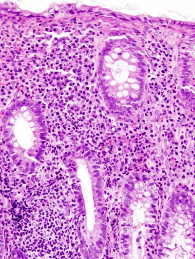
| Histology | Non-caseating granulomas | Crypt abscesses, goblet cell depletion, no granulomas |
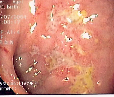
| Endoscopy | Cobblestone mucosa, deep serpiginous and aphthous ulcers | Diffuse erythema, granular friable mucosa, loss of vascular pattern, pseudopolyps |
| Typical symptoms | Abdominal pain, non-bloody diarrhoea, weight loss | Bloody diarrhoea with mucus, urgency, tenesmus |
| Perianal disease | Common: fistulae, fissures, abscesses, skin tags | Rare |
| Barium/imaging | Strictures giving the 'string sign of Kantor', rose-thorn ulcers | Loss of haustra giving a featureless 'lead pipe' colon |
| Smoking | Worsens disease | Protective |
| Surgery | Not curative; disease recurs at the anastomosis | Colectomy is curative for the colonic disease |
| Complications | Strictures, fistulae, abscesses, malabsorption, gallstones, renal stones | Toxic megacolon, colorectal cancer, primary sclerosing cholangitis |
Clinical features
Crohn's disease
Presentation reflects transmural, patchy inflammation and depends on the site involved. Typical features are crampy abdominal pain (classically right lower quadrant with terminal ileal disease, mimicking appendicitis), non-bloody diarrhoea, weight loss and systemic upset with fever and fatigue. Mouth ulceration is common, and perianal disease - fistulae, fissures, abscesses and skin tags - is a strong pointer to Crohn's rather than UC. Malabsorption from small bowel involvement causes deficiencies of iron, vitamin B12 (terminal ileum) and fat-soluble vitamins.
Ulcerative colitis
The hallmark is bloody diarrhoea with mucus, accompanied by urgency and tenesmus, and lower abdominal cramping that is often relieved by defecation. Disease limited to the rectum (proctitis) may cause rectal bleeding and urgency with normal or even constipated bowel habit, whereas extensive colitis causes frequent bloody stools with systemic upset, fever, tachycardia and weight loss. Symptoms are typically more gradual in onset than infective colitis and follow a relapsing-remitting course.
Extraintestinal manifestations
Both conditions have manifestations outside the gut, which occur in up to a third of patients and may precede bowel symptoms.1 Some parallel disease activity and some do not:
- Musculoskeletal: enteropathic arthritis (peripheral arthritis tracks disease activity), sacroiliitis and ankylosing spondylitis (independent of activity), osteoporosis
- Skin: erythema nodosum (tender red shin nodules, tracks activity), pyoderma gangrenosum (painful ulcer with a violaceous undermined edge, independent of activity)
- Eyes: episcleritis (tracks activity), uveitis and scleritis (independent) - uveitis is painful with photophobia and needs urgent ophthalmology review
- Hepatobiliary: primary sclerosing cholangitis, strongly associated with ulcerative colitis and conferring a substantial risk of cholangiocarcinoma and colorectal cancer
- Haematological: anaemia (iron deficiency, anaemia of chronic disease, B12 deficiency) and a markedly increased venous thromboembolism risk during flares
- Renal: oxalate stones in Crohn's disease with fat malabsorption
Investigations
Initial tests
- Bloods: full blood count (anaemia, thrombocytosis), CRP and ESR (raised in active disease), urea and electrolytes, liver function tests, albumin (low in severe disease), haematinics (iron studies, B12, folate)
- Faecal calprotectin: a protein released by neutrophils into the stool, raised in intestinal inflammation. Its main use is distinguishing IBD from irritable bowel syndrome, where it is normal, avoiding unnecessary colonoscopy2
- Stool culture and Clostridioides difficile toxin: essential to exclude infective colitis, which can both mimic and precipitate a flare
- Coeliac serology, where the presentation is compatible
Endoscopy and histology
Colonoscopy with ileal intubation and multiple biopsies is the definitive investigation, allowing assessment of disease distribution and histological confirmation. In suspected acute severe colitis, a flexible sigmoidoscopy without bowel preparation is preferred, as full colonoscopy risks perforation.
Imaging
- MR enterography: the investigation of choice for small bowel Crohn's disease, assessing disease extent, strictures and fistulae without radiation
- MRI pelvis: for perianal fistulising Crohn's disease
- Abdominal X-ray: in acute severe colitis, to look for colonic dilatation (toxic megacolon) and to assess disease extent from faecal loading
- CT abdomen: in the acute setting for suspected perforation, obstruction or abscess
- Capsule endoscopy: for suspected small bowel Crohn's disease not seen on other imaging; contraindicated if a stricture is suspected
Management of Crohn's disease
Inducing remission
Corticosteroids (oral prednisolone, or budesonide for ileocaecal disease with fewer systemic effects) are first-line for inducing remission.3 Exclusive enteral nutrition is an effective steroid-sparing alternative and is preferred first-line in children, where growth is a concern. Aminosalicylates are much less effective in Crohn's disease than in UC and have a limited role.
Maintaining remission
Azathioprine or mercaptopurine are first-line maintenance agents; thiopurine methyltransferase (TPMT) activity must be checked before starting, as deficiency predisposes to severe myelosuppression. Methotrexate is an alternative. Biologics - anti-TNF agents (infliximab, adalimumab), ustekinumab or vedolizumab - are used for severe or refractory disease and for fistulising disease. Screening for latent tuberculosis and hepatitis B is mandatory before starting anti-TNF therapy.
Surgery
Surgery is not curative in Crohn's disease, as inflammation typically recurs at the anastomosis, so it is reserved for complications: strictures (strictureplasty or limited resection), fistulae, abscesses, perforation, or disease refractory to medical therapy. Around half of patients require surgery within 10 years. Repeated small bowel resections risk short bowel syndrome.
Management of ulcerative colitis
Inducing remission in mild to moderate disease
Treatment is stepwise and the route depends on disease extent.4 Aminosalicylates (mesalazine) are first-line: topical (suppository or enema) for proctitis and left-sided disease, oral for more extensive disease, and often both combined. If there is no response, add an oral corticosteroid.
Acute severe ulcerative colitis
This is a medical emergency requiring admission. Severity is assessed with the Truelove and Witts criteria.
| Parameter | Threshold for severe disease |
|---|---|
| Bowel frequency | 6 or more bloody stools per day |
| Temperature | Above 37.8°C |
| Heart rate | Above 90 beats per minute |
| Haemoglobin | Below 105 g/L |
| ESR | Above 30 mm/hour |
Management involves IV corticosteroids (hydrocortisone), fluid and electrolyte correction, VTE prophylaxis (the thrombotic risk is high despite the bleeding), stool cultures to exclude infection, and stopping opioids and antimotility agents, which can precipitate toxic megacolon. Response is assessed at day 3 using stool frequency and CRP; failure to respond triggers rescue therapy with infliximab or ciclosporin, with early surgical involvement throughout.
Maintaining remission and surgery
Maintenance uses aminosalicylates first-line, with azathioprine or mercaptopurine for those with frequent relapses or who needed rescue therapy, and biologics for refractory disease. Colectomy is curative for the colonic disease, and is performed for acute severe colitis failing medical therapy, chronic refractory symptoms, or dysplasia. Options include subtotal colectomy with end ileostomy acutely, and later completion proctectomy with ileo-anal pouch (restorative proctocolectomy).
Colorectal cancer surveillance
Patients with extensive colitis have an increased colorectal cancer risk that rises with disease duration, extent and severity, and is markedly higher with coexisting primary sclerosing cholangitis. Colonoscopic surveillance begins around 10 years after symptom onset, with intervals of 1-5 years based on risk.
Complications
Crohn's disease
- Strictures causing small bowel obstruction
- Fistulae (enterocutaneous, enterovesical, enterovaginal, perianal) and abscesses
- Malabsorption with B12, iron and fat-soluble vitamin deficiency; gallstones and oxalate renal stones
- Short bowel syndrome after repeated resections
- Increased risk of small bowel and colorectal cancer
Ulcerative colitis
- Toxic megacolon: colonic dilatation above 6 cm with systemic toxicity, risking perforation
- Perforation and massive lower GI haemorrhage
- Colorectal cancer, hence surveillance colonoscopy
- Primary sclerosing cholangitis
- Venous thromboembolism during flares
Red flags
Prognosis
Both conditions follow a relapsing-remitting course over a lifetime, and with modern therapy most patients achieve good disease control and a near-normal life expectancy.1 In ulcerative colitis, around half of patients relapse in any given year, and roughly 20-30% eventually require colectomy, which cures the colonic disease. In Crohn's disease, around half require surgery within 10 years and recurrence after resection is common, so the emphasis is on sustained medical maintenance to avoid repeated operations.
Adverse prognostic features in Crohn's disease include young age at diagnosis, extensive small bowel involvement, perianal disease, stricturing or penetrating behaviour, and smoking. Early use of immunomodulators and biologics in high-risk patients has improved long-term outcomes and reduced hospitalisation and surgery rates.
References
- Lamb CA et al. British Society of Gastroenterology consensus guidelines on the management of inflammatory bowel disease in adults. Gut. 2019. Available here
- NICE DG11. Faecal calprotectin diagnostic tests for inflammatory diseases of the bowel. 2013. Available here
- NICE NG129. Crohn's disease: management. 2019. Available here
- NICE NG130. Ulcerative colitis: management. 2019. Available here
- Samir, CC BY-SA 3.0, via Wikimedia Commons. Available here
- User:KGH, CC BY-SA 3.0, via Wikimedia Commons. Available here
- Truelove SC, Witts LJ. Cortisone in ulcerative colitis: final report on a therapeutic trial. BMJ. 1955. Available here
- NHS. Inflammatory bowel disease. 2023. Available here
This article is written for revision and education. It is not clinical guidance and must not be used to make decisions about the care of a patient. Always check current NICE guidance and local protocols.