
Colorectal Cancer: Screening, Diagnosis and Management
Key points
- Colorectal cancer: the fourth most common cancer in the UK, almost all adenocarcinomas arising from adenomatous polyps via the adenoma-carcinoma sequence.
- Presentation by site: left-sided and rectal tumours cause change in bowel habit and rectal bleeding; right-sided tumours cause iron-deficiency anaemia and present later.
- Genetic syndromes: Lynch syndrome (HNPCC, mismatch repair defects) and familial adenomatous polyposis (APC mutation) account for a small but important minority.
- Screening: faecal immunochemical test (FIT) offered every 2 years to adults aged 50-74 in England, with colonoscopy if positive.
- FIT in symptomatic patients: used to triage who needs urgent referral; a FIT of 10 µg Hb/g or more warrants a suspected cancer pathway referral.
- Diagnosis: colonoscopy with biopsy is the gold standard; CT colonography is an alternative for frail patients.
- Staging: CT chest-abdomen-pelvis for all, plus MRI rectum for rectal tumours to assess the circumferential resection margin.
- Management: surgical resection determined by tumour site, with chemotherapy for node-positive disease and neoadjuvant chemoradiotherapy for rectal cancer.
Introduction
Colorectal cancer is a malignancy of the colon or rectum, of which the overwhelming majority are adenocarcinomas. It is the fourth most common cancer in the UK, with around 44,000 new cases each year, and the second most common cause of cancer death.1
Most cases arise sporadically through the adenoma-carcinoma sequence, in which a benign adenomatous polyp accumulates genetic mutations over roughly 10 years and progresses to invasive carcinoma. This long, detectable premalignant phase is precisely what makes screening and polypectomy so effective at preventing the disease.
Aetiology and risk factors
Modifiable and non-modifiable risk factors
- Age: the dominant risk factor, with incidence rising steeply after 50
- Diet: high consumption of red and processed meat, low fibre intake
- Obesity and physical inactivity
- Smoking and alcohol
- Inflammatory bowel disease: particularly ulcerative colitis with extensive, long-standing disease, and Crohn colitis
- Family history of colorectal cancer in a first-degree relative
- Previous adenomatous polyps or colorectal cancer
- Type 2 diabetes and acromegaly
- Protective factors include aspirin, dietary fibre, and physical activity
Hereditary syndromes
Around 5% of cases arise from an inherited syndrome. Recognising these matters because they change surveillance, surgical planning and family screening.2
| Feature | Lynch syndrome (HNPCC) | Familial adenomatous polyposis (FAP) |
|---|---|---|
| Genetics | Autosomal dominant; DNA mismatch repair genes (MLH1, MSH2, MSH6, PMS2) | Autosomal dominant; APC tumour suppressor gene on chromosome 5 |
| Polyps | Few polyps, but rapid progression to cancer | Hundreds to thousands of adenomatous polyps from adolescence |
| Site | Predominantly right-sided (proximal) colon | Throughout the colon and rectum |
| Lifetime cancer risk | Around 50-80% if untreated | Essentially 100% by age 40 if untreated |
| Extracolonic cancers | Endometrial (second commonest), ovarian, gastric, urothelial, small bowel | Duodenal and thyroid tumours, desmoid tumours, congenital hypertrophy of the retinal pigment epithelium |
| Management | Regular colonoscopic surveillance from age 25, consider aspirin, hysterectomy discussion | Prophylactic total colectomy, typically in the late teens or early twenties |
Lynch syndrome is identified using the Amsterdam criteria clinically, and confirmed by testing tumour tissue for microsatellite instability or loss of mismatch repair proteins on immunohistochemistry, followed by germline testing. All new colorectal cancers in the UK are now tested for mismatch repair deficiency.
Clinical features
Presentation depends substantially on the location of the tumour, because the calibre of the lumen and the consistency of the stool differ along the colon.
| Feature | Right-sided (caecum, ascending) | Left-sided (descending, sigmoid) and rectal |
|---|---|---|
| Lumen and stool | Wide lumen, liquid stool - obstruction is late | Narrow lumen, formed stool - obstruction is earlier |
| Typical presentation | Iron-deficiency anaemia, fatigue, weight loss, vague abdominal pain, occasionally a palpable mass | Change in bowel habit, rectal bleeding, tenesmus, colicky pain, obstruction |
| Stage at diagnosis | Often later, as symptoms are non-specific | Often earlier, as symptoms are more obvious |
Additional features to elicit include the duration and nature of any change in bowel habit (particularly increased frequency or looser stool persisting beyond 6 weeks), the presence of blood mixed within the stool rather than on its surface, tenesmus, unintentional weight loss, and a full family history.
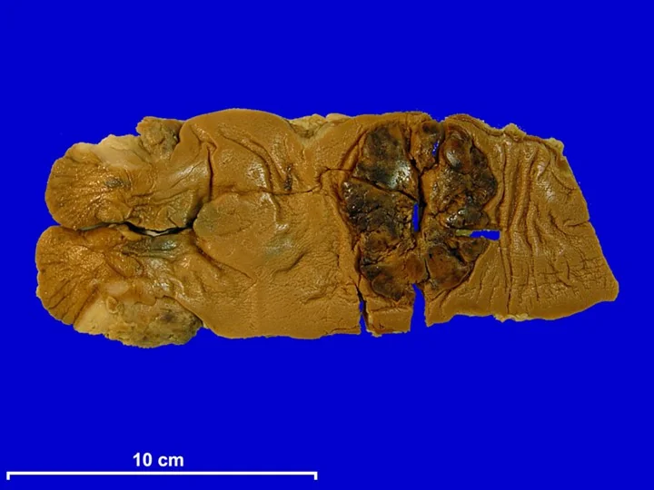
Around 20% of patients present as an emergency with large bowel obstruction (absolute constipation, distension, vomiting), perforation with peritonitis, or rarely with an acute lower GI bleed. Examination should always include abdominal palpation and a digital rectal examination, as a substantial proportion of rectal cancers are palpable.
Screening and FIT
The NHS bowel cancer screening programme
England offers a home faecal immunochemical test (FIT) every 2 years to adults aged 50-74 (the programme having been extended down from 60).3 FIT uses antibodies specific to human haemoglobin, so unlike the older guaiac test it is not affected by dietary meat or peroxidase-containing vegetables. A positive result triggers an invitation for colonoscopy.
Screening works because it detects both early-stage cancers and premalignant adenomas, which can be removed at colonoscopy, preventing cancer developing at all. Higher-risk groups (Lynch syndrome, FAP, long-standing colitis, previous adenomas) enter separate, more intensive colonoscopic surveillance programmes rather than FIT screening.
FIT in symptomatic patients
FIT is also used to triage symptomatic patients in primary care. NICE recommends offering FIT to guide referral, with a result of 10 µg haemoglobin per gram of faeces or above prompting a suspected cancer pathway referral.4 A negative FIT does not entirely exclude cancer, and patients with strong ongoing symptoms, a rectal mass or unexplained anaemia should still be referred regardless of the result.
Investigations
Diagnosis
Colonoscopy with biopsy is the gold standard. It visualises the whole colon, allows biopsy for histological confirmation, and permits therapeutic polypectomy at the same sitting. CT colonography is an alternative for frail patients or where colonoscopy is incomplete, and flexible sigmoidoscopy may be used for distal disease, though it does not exclude a synchronous proximal tumour.
Staging and baseline tests
- CT chest, abdomen and pelvis: for all patients, to detect liver and lung metastases and assess local extent
- MRI rectum: essential for rectal tumours, to assess depth of invasion, nodal status and the circumferential resection margin, which determines whether neoadjuvant therapy is needed
- Endoanal ultrasound: for early rectal tumours where local excision is being considered
- Carcinoembryonic antigen (CEA): a baseline tumour marker used for monitoring response and detecting recurrence, not for diagnosis
- Mismatch repair / microsatellite instability testing on the tumour, to identify Lynch syndrome and guide immunotherapy
- Full blood count (iron-deficiency anaemia), urea and electrolytes, and liver function tests
Staging uses the TNM system. The older Dukes classification is still occasionally encountered: Dukes A is confined to the bowel wall, B penetrates through it, C involves regional lymph nodes, and D denotes distant metastases.
Management
Management is decided by a colorectal multidisciplinary team.2
Surgery
The operation is determined by the tumour's site and its blood supply, since the resection must include the draining lymph nodes:
| Tumour site | Operation |
|---|---|
| Caecum and ascending colon | Right hemicolectomy |
| Transverse colon | Extended right hemicolectomy |
| Descending colon | Left hemicolectomy |
| Sigmoid colon | Sigmoid colectomy (high anterior resection) |
| Upper and middle rectum | Anterior resection with total mesorectal excision, often with a defunctioning stoma |
| Low rectum and anal verge | Abdominoperineal resection, resulting in a permanent end colostomy |
| Emergency obstructing left-sided tumour | Hartmann procedure (resection with end colostomy) or stenting as a bridge to surgery |
Oncological treatment
- Adjuvant chemotherapy (typically fluorouracil- or capecitabine-based, often with oxaliplatin) for node-positive (stage III) and high-risk stage II colon cancer
- Neoadjuvant chemoradiotherapy for locally advanced rectal cancer, to downstage the tumour and reduce local recurrence by achieving a clear circumferential margin
- Palliative chemotherapy and biological agents (e.g. anti-EGFR therapy in RAS wild-type tumours) for metastatic disease
- Immunotherapy for mismatch repair-deficient or microsatellite-instability-high tumours, which respond well to checkpoint inhibitors
- Metastasectomy: resection of isolated liver or lung metastases can be curative in selected patients
Follow-up after curative treatment includes regular CEA measurement, surveillance CT imaging, and colonoscopy to detect metachronous tumours.
Complications
- Large bowel obstruction, presenting acutely with distension, vomiting and absolute constipation
- Perforation with faecal peritonitis, carrying high mortality
- Iron-deficiency anaemia from chronic occult blood loss
- Fistula formation into bladder, vagina or small bowel
- Metastatic disease, most commonly to the liver (via the portal vein), then lung, peritoneum and bone
- Venous thromboembolism
- Surgical: anastomotic leak, stoma complications, and for rectal surgery, sexual and urinary dysfunction from pelvic nerve injury and low anterior resection syndrome
Red flags
Prognosis
Prognosis depends overwhelmingly on stage at diagnosis. Around 90% of patients diagnosed at stage I survive 5 years, falling to roughly 10% at stage IV.1 Overall 5-year survival in the UK is around 55-60%, and has improved substantially with screening, better surgery (particularly total mesorectal excision for rectal cancer) and more effective chemotherapy.
Adverse prognostic factors include nodal involvement, lymphovascular or perineural invasion, tumour perforation, an involved circumferential resection margin in rectal cancer, and a persistently raised postoperative CEA. Notably, colorectal cancer differs from many solid tumours in that resection of isolated liver metastases can still achieve long-term cure, so metastatic disease is not automatically incurable.
References
- Cancer Research UK. Bowel cancer statistics. Available here
- NICE NG151. Colorectal cancer. 2020 (updated 2021). Available here
- NHS. Bowel cancer screening. 2024. Available here
- NICE DG56. Quantitative faecal immunochemical testing to guide colorectal cancer pathway referral in primary care. 2023. Available here
- Deilson Elgui de Oliveira, CC BY-SA 4.0, via Wikimedia Commons. Available here
- NICE NG12. Suspected cancer: recognition and referral. 2015 (updated 2023). Available here
- Dekker E et al. Colorectal cancer. Lancet. 2019. Available here
This article is written for revision and education. It is not clinical guidance and must not be used to make decisions about the care of a patient. Always check current NICE guidance and local protocols.