
Gastric Cancer: Diagnosis, Staging and Management
Key points
- Gastric cancer: over 90% are adenocarcinomas, arising from the gastric mucosa; rarer types include lymphoma (MALT) and gastrointestinal stromal tumours.
- Main risk factor: Helicobacter pylori infection, which drives a sequence of chronic gastritis, atrophy, intestinal metaplasia, dysplasia and carcinoma.
- Other risk factors: smoking, salted and smoked foods, pernicious anaemia, previous gastric surgery, male sex, and a family history.
- Presentation: typically late and non-specific: dyspepsia, early satiety, weight loss, anorexia and iron-deficiency anaemia.
- Classic signs: Virchow's node (left supraclavicular), Sister Mary Joseph nodule (umbilical), and Troisier's sign - all indicate advanced disease.
- Diagnosis: upper GI endoscopy with biopsy, then CT chest-abdomen-pelvis and staging laparoscopy to detect peritoneal disease.
- Management: gastrectomy with perioperative chemotherapy for operable disease; palliative chemotherapy, stenting or surgery for advanced disease.
- Prognosis: poor in the UK, with 5-year survival around 20%, because most present at an advanced stage.
Introduction
Gastric cancer is a malignancy arising from the stomach wall, of which over 90% are adenocarcinomas derived from the glandular epithelium of the gastric mucosa.1 Less common types include gastric MALT lymphoma, gastrointestinal stromal tumours (GISTs) arising from the interstitial cells of Cajal, and neuroendocrine tumours.
Around 6,500 people are diagnosed each year in the UK. Global incidence is falling, largely attributed to declining H. pylori prevalence and better food preservation, but it remains a major cause of cancer death worldwide and is particularly common in East Asia, where screening programmes exist. The UK has no screening programme, which contributes to late presentation.
Aetiology and risk factors
The dominant modifiable risk factor is chronic Helicobacter pylori infection, classified by the WHO as a Group 1 carcinogen.2 It drives the Correa cascade: chronic active gastritis progresses to atrophic gastritis, then intestinal metaplasia, then dysplasia, and finally invasive adenocarcinoma - a sequence typically unfolding over decades.
Other recognised risk factors:
- Smoking and high alcohol intake
- Diet: high intake of salted, smoked, pickled or nitrosamine-rich foods; low fruit and vegetable intake
- Pernicious anaemia and autoimmune atrophic gastritis
- Previous partial gastrectomy (risk in the gastric remnant, typically 15-20 years later)
- Male sex and increasing age
- Blood group A
- Family history and hereditary syndromes: hereditary diffuse gastric cancer (CDH1 mutation, causing diffuse-type cancer at a young age and often prompting prophylactic gastrectomy), Lynch syndrome, familial adenomatous polyposis
Histological classification
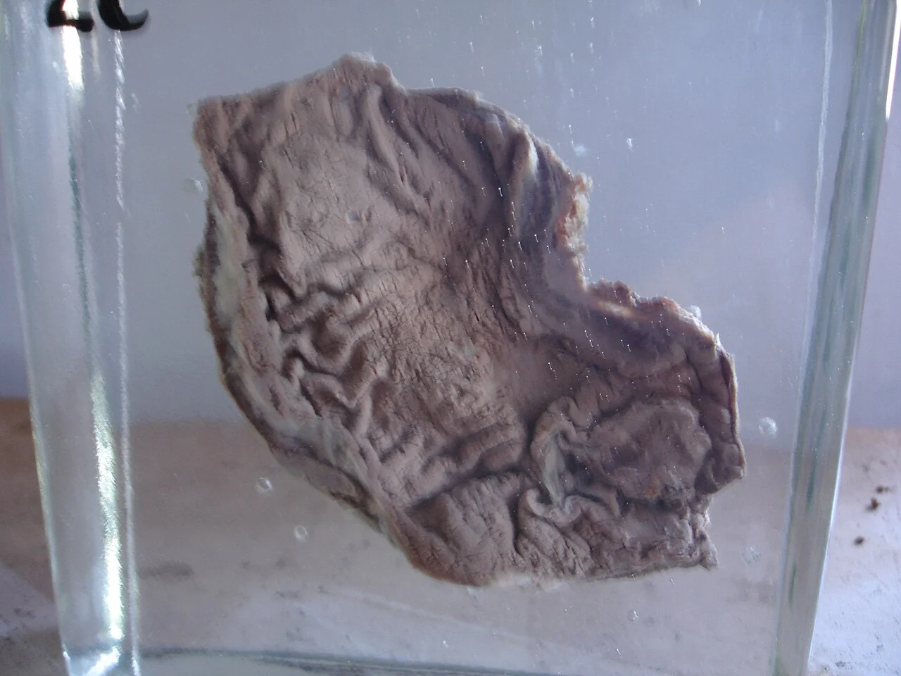
The Lauren classification divides gastric adenocarcinoma into two types. The intestinal type forms glandular structures, is associated with H. pylori and the Correa cascade, occurs in older patients and tends to be better differentiated. The diffuse type infiltrates the wall without forming a discrete mass, contains signet ring cells (with mucin displacing the nucleus to the periphery), occurs in younger patients, is associated with CDH1 mutations, and carries a worse prognosis. Extensive diffuse infiltration produces a rigid, thickened, non-distensible stomach known as linitis plastica ("leather bottle stomach").
Clinical features
Early gastric cancer is typically asymptomatic or produces vague, non-specific symptoms indistinguishable from benign dyspepsia, which is the central reason for late diagnosis. Symptoms include:
- Dyspepsia or epigastric pain, often persistent and unresponsive to acid suppression
- Early satiety and a sensation of fullness, particularly with linitis plastica where the stomach cannot distend
- Unintentional weight loss and anorexia
- Nausea and vomiting; persistent vomiting suggests gastric outlet obstruction from a distal (antral or pyloric) tumour
- Dysphagia, with tumours at the gastro-oesophageal junction
- Iron-deficiency anaemia from chronic occult blood loss, or overt haematemesis and melaena
- Fatigue and malaise
Examination findings
Examination is often normal in early disease. Classic findings, all of which indicate advanced, usually metastatic disease, include:
| Sign | Finding and significance |
|---|---|
| Virchow's node | Enlarged left supraclavicular lymph node, receiving thoracic duct drainage; its presence is Troisier's sign |
| Sister Mary Joseph nodule | Metastatic nodule at the umbilicus, spreading along the falciform ligament |
| Krukenberg tumour | Ovarian metastases, classically containing signet ring cells, from transcoelomic spread |
| Blumer shelf | Palpable mass in the rectovesical or rectouterine pouch on rectal examination, from peritoneal deposits |
| Epigastric mass | Palpable primary tumour |
| Hepatomegaly, jaundice, ascites | Liver or peritoneal metastatic disease |
| Acanthosis nigricans | Paraneoplastic velvety hyperpigmentation of the flexures |
Investigations
Diagnosis
Upper GI endoscopy with biopsy is the diagnostic investigation of choice and should be arranged via the urgent suspected cancer pathway.3 Multiple biopsies (typically at least six) are taken from the ulcer edge or suspicious area, because diffuse-type tumours infiltrate submucosally and superficial biopsies can be falsely negative. This is the reason every gastric ulcer is biopsied and re-scoped after treatment to confirm healing.
Staging
- CT chest, abdomen and pelvis: first-line for detecting distant metastases and assessing local extent
- Staging laparoscopy: particularly important in gastric cancer, as CT poorly detects small-volume peritoneal metastases; peritoneal washings are taken for cytology
- Endoscopic ultrasound (EUS): assesses depth of invasion (T stage) and local nodes, useful where the distinction between early and locally advanced disease will change management
- PET-CT: may be used selectively, though it is less reliable in diffuse-type and mucinous tumours, which are often not FDG-avid
- HER2 testing on the biopsy specimen, since HER2-positive tumours can be treated with trastuzumab
Baseline bloods include full blood count (iron-deficiency anaemia), urea and electrolytes, liver function tests and nutritional assessment.
Differential diagnosis
- Peptic ulcer disease and gastritis: the principal differential, and the reason for biopsying every gastric ulcer
- Functional dyspepsia: symptoms with no structural cause at endoscopy
- GORD: predominantly retrosternal burning
- Gastric lymphoma or GIST: distinguished on histology and immunohistochemistry (GISTs are typically CD117/KIT positive)
- Pancreatic cancer: epigastric pain radiating to the back, obstructive jaundice, weight loss
- Oesophageal cancer: particularly for tumours at the gastro-oesophageal junction, where the two overlap
Management
Management is directed by an upper GI multidisciplinary team and depends on stage and fitness.1
Curative treatment
- Endoscopic mucosal resection or submucosal dissection: for very early tumours confined to the mucosa with no nodal involvement
- Subtotal (partial) gastrectomy: for distal (antral) tumours
- Total gastrectomy: for proximal or diffusely infiltrating tumours, with reconstruction typically by Roux-en-Y oesophagojejunostomy
- D2 lymphadenectomy: removal of perigastric and second-tier nodes, now the standard extent of nodal dissection
- Perioperative chemotherapy: chemotherapy given both before and after surgery (e.g. the FLOT regimen) improves survival and is standard for resectable disease beyond the earliest stages
Palliative treatment
For advanced or metastatic disease, treatment aims at symptom control and prolonging life: palliative chemotherapy, with trastuzumab added for HER2-positive tumours; endoscopic stenting or a palliative bypass for gastric outlet obstruction; radiotherapy for bleeding or pain; and early palliative care involvement. Nutritional support is a central and often overlooked component, given the high prevalence of malnutrition.
After gastrectomy
Patients require lifelong vitamin B12 replacement after total gastrectomy, because intrinsic factor is no longer produced. They also need iron and calcium monitoring, dietetic support with small frequent meals, and monitoring for dumping syndrome.
Complications
- Gastric outlet obstruction from distal tumours, causing vomiting of undigested food and weight loss
- Upper GI bleeding, acute or chronic, causing iron-deficiency anaemia
- Perforation, a surgical emergency
- Malnutrition and cachexia
- Metastatic disease: liver, lung, peritoneum (with malignant ascites), ovaries (Krukenberg tumour) and bone
- Venous thromboembolism, to which gastrointestinal malignancy strongly predisposes
- Post-surgical: anastomotic leak, dumping syndrome, vitamin B12 and iron deficiency, and osteoporosis
Red flags
Prognosis
Prognosis in the UK is poor, with overall 5-year survival of around 20%, driven almost entirely by stage at diagnosis.4 Early gastric cancer confined to the mucosa or submucosa has a 5-year survival exceeding 90% and can often be cured endoscopically, whereas the majority of UK patients present with locally advanced or metastatic disease, where median survival is around a year even with chemotherapy.
The contrast with Japan and South Korea, where population screening detects a much higher proportion of early-stage disease and survival is correspondingly better, illustrates how far outcomes are determined by the stage at which the cancer is found rather than by the treatment given. Diffuse-type tumours and those with signet ring cells carry a worse prognosis than intestinal-type tumours.
References
- NICE NG83. Oesophago-gastric cancer: assessment and management in adults. 2018. Available here
- IARC Working Group. Schistosomes, liver flukes and Helicobacter pylori. IARC Monographs, Vol. 61. 1994. Available here
- NICE NG12. Suspected cancer: recognition and referral. 2015 (updated 2023). Available here
- Cancer Research UK. Stomach cancer statistics. Available here
- Netha Hussain, CC BY-SA 3.0, via Wikimedia Commons. Available here
- NHS. Stomach cancer. 2022. Available here
- Smyth EC et al. Gastric cancer. Lancet. 2020. Available here
This article is written for revision and education. It is not clinical guidance and must not be used to make decisions about the care of a patient. Always check current NICE guidance and local protocols.