
Osteoporosis and Fragility Fractures
Key points
- Osteoporosis: reduced bone mineral density and disrupted bone microarchitecture, increasing the risk of fracture from minimal trauma. It is asymptomatic until a fracture occurs.
- Fragility fracture: a fracture occurring from a fall from standing height or less - classic sites are the hip, vertebrae, distal radius (Colles') and proximal humerus.
- Diagnosis: a DEXA T-score of -2.5 or below at the hip or lumbar spine; a T-score between -1.0 and -2.5 is osteopenia.
- Risk assessment: FRAX or QFracture estimates 10-year fracture probability and, combined with NOGG thresholds, determines who needs a DEXA scan and who needs treatment.
- Secondary causes: screen for hyperthyroidism, hyperparathyroidism, coeliac disease, myeloma and hypogonadism, particularly in younger patients or unexpectedly severe disease.
- First-line treatment: an oral bisphosphonate (alendronic acid or risedronate), with IV zoledronic acid as an alternative where oral bisphosphonates are unsuitable or poorly tolerated.
- Denosumab: stopping it abruptly causes a rebound increase in vertebral fracture risk - a follow-on bisphosphonate is needed if it is discontinued.
- Falls prevention: runs alongside bone-specific treatment, since most fragility fractures result from a fall, not from bone fragility alone.
Introduction
Osteoporosis is a systemic skeletal disease characterised by low bone mass and disrupted bone microarchitecture, resulting in increased bone fragility and susceptibility to fracture. It causes no symptoms of its own - it is entirely silent until the moment a fragility fracture occurs, which is why proactive risk assessment, rather than waiting for symptoms, is central to its management.1
A fragility fracture is one occurring from a fall from standing height or less, or with trauma that would not normally break a healthy bone. The classic sites - vertebrae, hip (proximal femur), distal radius (Colles' fracture) and proximal humerus - reflect where trabecular bone loss and mechanical loading combine to make fracture most likely.
This topic is heavily examined because it sits at the intersection of prevention (who to screen and treat before a fracture happens) and acute medicine (managing the fracture and its complications once it has), and because the drug side effects - atypical femoral fracture, osteonecrosis of the jaw, denosumab rebound - are classic safety-netting content.
Pathophysiology and risk factors
Bone is continuously remodelled by osteoblasts (bone formation) and osteoclasts (bone resorption). Osteoporosis results when resorption outpaces formation over time - most dramatically after the menopause, when falling oestrogen removes a key brake on osteoclast activity, but also with ageing, immobility, and a range of secondary causes.
- Increasing age and postmenopausal status in women
- Low body mass index
- Smoking and alcohol excess
- Previous fragility fracture - the single strongest predictor of future fracture
- Family history of hip fracture
- Long-term oral or high-dose inhaled glucocorticoid use
- Prolonged immobility
- Early menopause or hypogonadism (in men or women)
- Malabsorption - coeliac disease, inflammatory bowel disease
- Chronic kidney or liver disease
- Endocrine disease - hyperthyroidism, hyperparathyroidism, Cushing syndrome
- Chronic inflammatory disease - rheumatoid arthritis and other conditions requiring long-term corticosteroids compound risk through both the disease and its treatment
- Certain drugs - long-term proton pump inhibitors, aromatase inhibitors, androgen deprivation therapy
Assessing fracture risk
NICE recommends assessing fracture risk in all women aged 65 and over and men aged 75 and over, and in younger patients with risk factors - previous fragility fracture, current or frequent glucocorticoid use, a history of falls, a family history of hip fracture, other causes of secondary osteoporosis, low BMI, smoking or alcohol excess.2
FRAX or QFracture are validated tools that estimate an individual's 10-year probability of a major osteoporotic fracture and of hip fracture specifically, using clinical risk factors with or without a BMD value. The result is interpreted against NOGG (National Osteoporosis Guideline Group) thresholds to decide whether to arrange a DEXA scan, start treatment directly, or simply advise on lifestyle and reassess in future.
Investigations
DEXA (dual-energy X-ray absorptiometry)
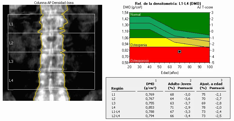
DEXA measures bone mineral density, usually at the lumbar spine and hip, and expresses the result as a T-score (the number of standard deviations from the mean BMD of a young healthy adult of the same sex) used for diagnosis in postmenopausal women and men over 50, and a Z-score (compared with an age-matched population) used in younger patients, where a low Z-score should prompt a search for a secondary cause.
| T-score | Classification |
|---|---|
| -1.0 or above | Normal |
| Between -1.0 and -2.5 | Osteopenia |
| -2.5 or below | Osteoporosis |
| -2.5 or below, plus a fragility fracture | Severe (established) osteoporosis |
Screening for secondary causes
- FBC, U&Es, LFTs (including ALP), calcium and phosphate - a baseline metabolic screen
- TFTs - to exclude hyperthyroidism
- Vitamin D - deficiency is common and correctable, and affects both fracture risk and treatment planning
- Coeliac serology - malabsorption is an important and treatable secondary cause
- Serum protein electrophoresis and urinary Bence Jones protein - if myeloma is a concern, particularly with anaemia, renal impairment or an unexpectedly severe presentation
- Testosterone - in men where hypogonadism is suspected
- PTH - if calcium is abnormal, to assess for primary hyperparathyroidism
- Lateral spine X-ray or vertebral fracture assessment (VFA) on DEXA - to identify silent vertebral fractures, which are often asymptomatic but change management
Management
Lifestyle measures
- Adequate dietary calcium and vitamin D, with supplementation if dietary intake is insufficient or sun exposure is limited
- Regular weight-bearing and muscle-strengthening exercise
- Smoking cessation and reduced alcohol intake
- Falls risk assessment and prevention - reviewing home hazards, vision, medication (sedatives, antihypertensives), and strength/balance training, since most fragility fractures follow a fall
Pharmacological treatment
Oral bisphosphonates (alendronic acid or risedronate, taken weekly) are first-line for most patients.3 They must be taken on an empty stomach with a full glass of water, and the patient should remain upright for at least 30 minutes afterwards, to reduce the risk of oesophageal irritation.
| Drug | Mechanism | Key points |
|---|---|---|
| Alendronic acid / risedronate (oral bisphosphonate) | Inhibits osteoclast-mediated bone resorption | First-line; contraindicated in significant oesophageal disease and severe renal impairment |
| Zoledronic acid (IV bisphosphonate) | Same as above, given annually by infusion | Alternative where oral bisphosphonates are not tolerated or adherence is a concern |
| Denosumab | Monoclonal antibody against RANKL, reducing osteoclast formation | 6-monthly subcutaneous injection; useful in renal impairment; stopping it causes rebound bone loss |
| Teriparatide | Recombinant PTH analogue - anabolic, stimulates new bone formation | Specialist-initiated for severe osteoporosis; maximum treatment duration around 24 months |
| Romosozumab | Anti-sclerostin monoclonal antibody - anabolic | Newer option for very high fracture risk, specialist-initiated |
| HRT | Oestrogen replacement | Can be considered for bone protection in early menopause, alongside its other indications and risks |
Complications
Hip fracture carries the greatest morbidity and mortality of any osteoporotic fracture, with roughly 20-30% one-year mortality in some cohorts and a substantial proportion of survivors losing independence. Vertebral fractures cause acute pain, and cumulatively cause height loss, kyphosis and, in severe disease, restrictive lung impairment from a reduced chest cavity volume. Distal radius (Colles') fractures, while less life-threatening, are common, painful, and can impair hand function. Each fragility fracture substantially increases the risk of a further fracture, which is why secondary fracture prevention services exist to ensure every fragility fracture triggers a bone health assessment.
Red flags
Prognosis
Osteoporosis treatment substantially reduces fracture risk - bisphosphonates typically reduce vertebral fracture risk by around 40-50% and hip fracture risk meaningfully as well, when taken consistently. Adherence is a genuine practical challenge given the long, largely symptom-free treatment course, which is why patient understanding of the 'silent disease, real consequence' framing matters. Once a fragility fracture has occurred, the risk of a further fracture is significantly elevated, making secondary prevention - assessment and treatment triggered by the first fracture - one of the highest-yield interventions in this area of medicine.
References
- NICE CG146. Osteoporosis: assessing the risk of fragility fracture. 2012, updated 2017. Available here
- NICE Clinical Knowledge Summaries. Osteoporosis - prevention of fragility fractures. Available here
- National Osteoporosis Guideline Group (NOGG). Clinical guideline for the prevention and treatment of osteoporosis. Available here
- Kanis JA, Harvey NC, McCloskey E et al. Algorithm for the management of patients at low, high and very high risk of osteoporotic fractures. Osteoporosis International. 2020. Available here
- Royal Osteoporosis Society. Clinical guidance for healthcare professionals. Available here
This article is written for revision and education. It is not clinical guidance and must not be used to make decisions about the care of a patient. Always check current NICE guidance and local protocols.