
Rheumatoid Arthritis: Diagnosis and Management
Key points
- Rheumatoid arthritis: a chronic, symmetrical, small-joint polyarthritis driven by autoimmune synovitis, which erodes cartilage and bone if left untreated.
- Pattern: MCPs, PIPs, wrists and MTPs - the DIPs are characteristically spared, which is the single most useful discriminator from osteoarthritis.
- Morning stiffness: lasting more than 30-60 minutes and easing with activity points to an inflammatory arthropathy rather than a mechanical one.
- Serology: rheumatoid factor is sensitive but not specific; anti-CCP is more specific and can predate symptoms by years.
- Treat-to-target: early, aggressive DMARD therapy aiming for remission or low disease activity, because the window to prevent erosive damage is early.
- First-line DMARD: methotrexate, usually with a short bridging course of corticosteroid while it takes effect.
- Escalation: a biologic, typically an anti-TNF, is added when disease activity remains high (DAS28 > 5.1) despite two conventional DMARDs.
- Extra-articular disease: rheumatoid nodules, interstitial lung disease, scleritis, and vasculitis all indicate more severe, seropositive disease.
Introduction
Rheumatoid arthritis (RA) is a chronic systemic autoimmune disease whose dominant feature is a symmetrical, erosive, small-joint polyarthritis. It affects around 1% of the UK population, is two to three times commoner in women, and typically presents between the ages of 30 and 60, though it can occur at any age.1
The underlying process is synovitis: the synovium becomes thickened and inflamed, forming an invasive pannus of proliferating tissue that erodes adjacent cartilage and subchondral bone. Left untreated, this produces irreversible joint destruction and deformity within a few years of onset - which is why RA is one of the clearest examples in medicine of a disease where early, aggressive treatment changes the long-term outcome.
It is a favourite exam topic because the diagnosis rests on recognising a pattern - which joints, how symmetrical, how long the stiffness lasts - and because the management pathway (DMARDs, then biologics) is testable at every stage.
Aetiology and pathophysiology
RA is an autoimmune disease in a genetically susceptible individual, triggered by an environmental exposure - classically smoking - that leads to citrullination of self-proteins. In people carrying the shared epitope of HLA-DRB1, these citrullinated peptides are presented to T cells, breaking tolerance and driving B cells to produce anti-citrullinated protein antibodies (anti-CCP) and, in most patients, rheumatoid factor.2
The resulting immune complexes and cytokine cascade - chiefly TNF-alpha, IL-6 and IL-1 - drive synovial inflammation, angiogenesis and the proliferation of fibroblast-like synoviocytes that form the pannus. This is precisely why anti-TNF and anti-IL-6 biologics are effective: they interrupt the cytokines actually driving the disease.
Risk factors
- Female sex (2-3x)
- Family history and the HLA-DRB1 shared epitope
- Smoking - the strongest modifiable risk factor, especially in anti-CCP-positive disease
- Obesity
- Silica exposure
- Periodontal disease (Porphyromonas gingivalis can citrullinate proteins directly)
Clinical features
RA usually presents with an insidious, symmetrical polyarthritis developing over weeks to months, though a minority present acutely or with a migratory pattern.
- Pain and swelling of the small joints of the hands and feet - metacarpophalangeal (MCP), proximal interphalangeal (PIP), wrists and metatarsophalangeal (MTP) joints
- Symmetry - not perfect, but the same joint groups on both sides are typically affected
- Morning stiffness lasting more than 30-60 minutes, easing with movement - the key discriminator from mechanical joint pain
- Systemic features - fatigue, low-grade fever, malaise, weight loss
- DIP joints and the axial skeleton (except the atlanto-axial joint) are spared
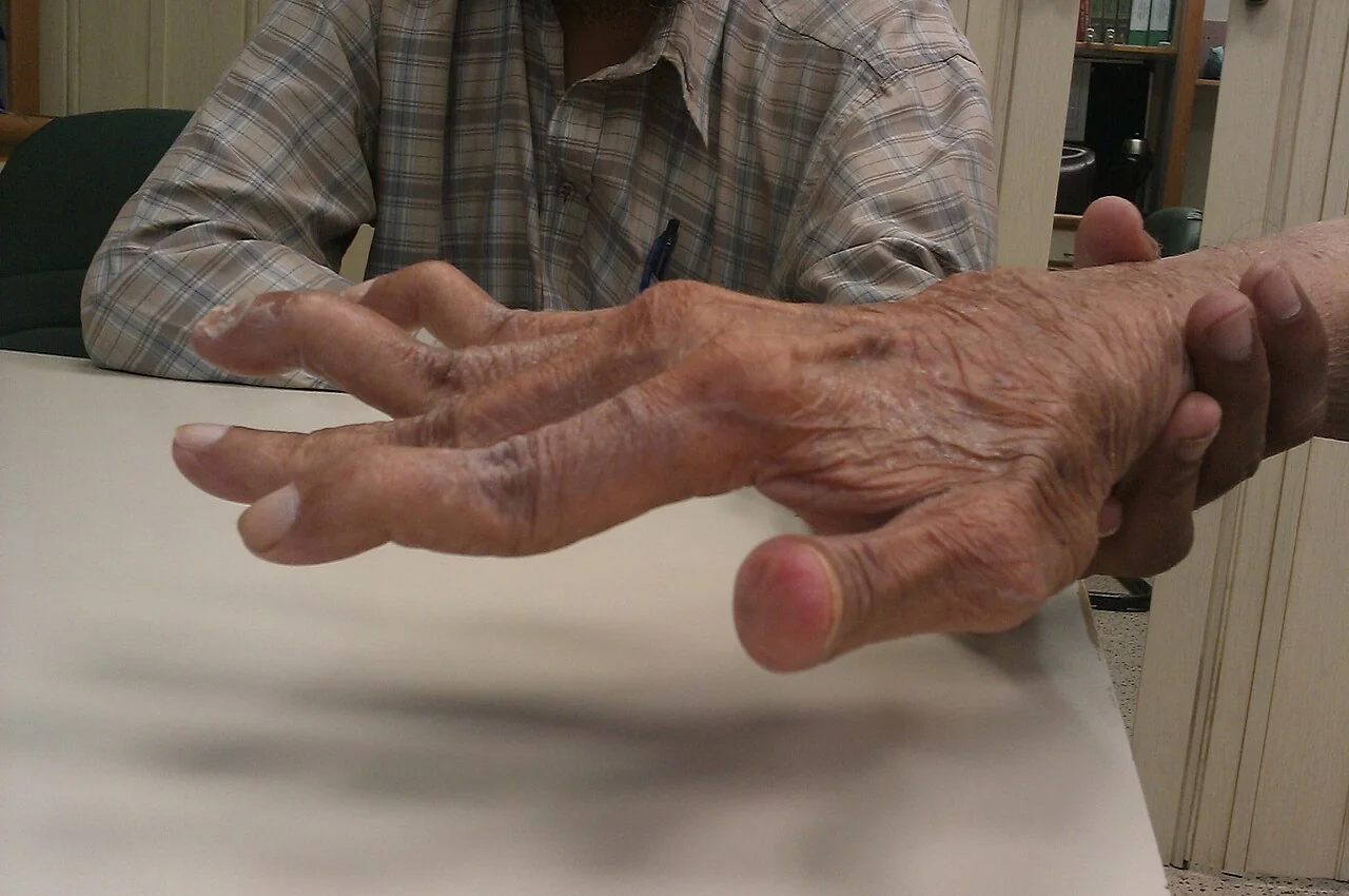
Late deformities
These develop after years of uncontrolled disease and are now seen far less often thanks to early DMARD therapy, but remain a common exam image:
- Ulnar deviation of the fingers at the MCP joints
- Swan neck deformity - PIP hyperextension with DIP flexion
- Boutonniere deformity - PIP flexion with DIP hyperextension
- Z-thumb deformity
- Volar subluxation of the wrist
Extra-articular features
These occur predominantly in seropositive, long-standing disease and signal more severe illness:
- Rheumatoid nodules - firm, non-tender subcutaneous nodules over extensor surfaces such as the elbow
- Pulmonary - interstitial lung disease (usually usual interstitial pneumonia pattern), pleural effusion, pulmonary nodules, obliterative bronchiolitis
- Ocular - episcleritis (painless, self-limiting) and scleritis (painful, sight-threatening)
- Haematological - anaemia of chronic disease, and Felty syndrome (RA + splenomegaly + neutropenia)
- Vasculitis - nail-fold infarcts, digital ischaemia, mononeuritis multiplex in severe disease
- Neurological - carpal tunnel syndrome from tenosynovitis, cervical myelopathy from atlanto-axial subluxation
- Cardiovascular - accelerated atherosclerosis, pericarditis
- Renal - amyloidosis (AA type) in longstanding uncontrolled disease
- Secondary Sjögren syndrome - dry eyes and dry mouth
Clinical examination
- Look - symmetrical swelling of MCPs/PIPs, muscle wasting of the small hand muscles, deformities described above, rheumatoid nodules
- Feel - synovitis feels boggy/spongy rather than bony-hard (contrast with the bony swelling of osteoarthritis), warmth, tenderness on squeezing across the MCPs (the 'MCP squeeze test')
- Move - reduced grip strength, reduced range of movement
- Function - ask the patient to make a fist, do up a button, or hold a cup - functional assessment matters as much as the joint count
- Extra-articular examination - eyes for scleritis, chest for basal crackles of fibrosis, abdomen for splenomegaly, peripheral nerves for carpal tunnel and mononeuritis
Differential diagnosis
- Osteoarthritis - bony swelling, DIPs and first CMC joint affected, stiffness brief and worse after activity, asymmetrical
- Psoriatic arthritis - DIP involvement, dactylitis, nail changes, asymmetrical oligoarthritis pattern possible, psoriasis history
- Reactive arthritis and other seronegative spondyloarthropathies - asymmetrical, lower-limb-predominant, associated enthesitis
- SLE - polyarthralgia is often non-erosive, plus other systemic features (rash, renal, serositis, cytopenias)
- Viral arthritis - parvovirus B19, hepatitis B/C - can closely mimic early RA but is typically self-limiting
- Polymyalgia rheumatica - proximal shoulder and hip girdle stiffness in an older patient, rather than small-joint synovitis
- Crystal arthropathy - gout and pseudogout are usually monoarticular or oligoarticular and more acute in onset
- Haemochromatosis arthropathy - characteristically affects the 2nd and 3rd MCPs with hook-like osteophytes
Investigations
Bedside and laboratory
- Rheumatoid factor - sensitive (~70%) but not specific
- Anti-CCP (anti-cyclic citrullinated peptide) - more specific (~95%), and useful when RF is negative but suspicion is high
- FBC - normochromic normocytic anaemia of chronic disease is common; thrombocytosis reflects inflammation
- CRP and ESR - raised, used both to support diagnosis and to track disease activity
- U&Es and LFTs - baseline before starting DMARDs, which require monitoring
- Urinalysis - baseline before starting drugs that can affect the kidneys, and to screen for amyloid-related proteinuria in longstanding disease
Imaging
Plain radiographs of the hands and feet are first-line. Early disease may show only soft tissue swelling and periarticular osteopenia; established disease shows symmetrical joint space narrowing, marginal bony erosions, and eventually subluxation and deformity.
Ultrasound or MRI are more sensitive than plain films for detecting early synovitis and erosions, and are increasingly used where the diagnosis or disease activity is unclear on clinical grounds alone.
Classification criteria
The 2010 ACR/EULAR classification criteria are used in practice and in exams to formalise the diagnosis in patients with at least one joint with definite synovitis not better explained by another disease. A score of 6 or more out of 10 across the four domains below classifies definite RA.3
| Domain | Scoring |
|---|---|
| Joint involvement | 1 large joint = 0; 2-10 large joints = 1; 1-3 small joints = 2; 4-10 small joints = 3; >10 joints (at least 1 small) = 5 |
| Serology | Negative RF and anti-CCP = 0; low-positive = 2; high-positive = 3 |
| Acute phase reactants | Normal CRP and ESR = 0; abnormal CRP or ESR = 1 |
| Duration of symptoms | < 6 weeks = 0; >= 6 weeks = 1 |
Management
Management follows a treat-to-target strategy: start a DMARD as early as possible, review frequently (roughly every 1-3 months until target is met), and escalate therapy until remission or low disease activity is achieved.4 Every newly suspected case should be referred urgently for specialist assessment - within 3 working days for a persistently swollen joint that is not explained by something else - because early treatment is what prevents erosive damage.
Bridging symptom control
While a DMARD takes effect (which can take 6-12 weeks), a short course of corticosteroid (oral or intramuscular) is used to control symptoms and suppress inflammation. NSAIDs, with gastroprotection, help with pain but do not alter disease progression and should not be relied upon alone.
Conventional synthetic DMARDs
Methotrexate is first-line, given once weekly with folic acid to reduce toxicity, and is usually started as monotherapy or in combination with another conventional DMARD (sulfasalazine, hydroxychloroquine or leflunomide) for a faster initial response.
| Drug | Key toxicities | Monitoring |
|---|---|---|
| Methotrexate | Hepatotoxicity, bone marrow suppression, pneumonitis, teratogenic | FBC, U&E, LFT every 1-2 weeks initially, then every 2-3 months |
| Sulfasalazine | Bone marrow suppression, hepatotoxicity, oligospermia | FBC, LFT regularly, especially in first few months |
| Hydroxychloroquine | Retinopathy (dose-dependent) | Baseline and then annual ophthalmological screening |
| Leflunomide | Hepatotoxicity, hypertension, teratogenic (long washout needed) | FBC, LFT, blood pressure |
Biologic and targeted synthetic DMARDs
If disease activity remains high (DAS28 > 5.1) despite trials of at least two conventional DMARDs (usually including methotrexate), a biologic DMARD is added - almost always in combination with methotrexate, which reduces the formation of anti-drug antibodies and improves efficacy.5
- Anti-TNF agents (adalimumab, etanercept, infliximab, certolizumab) - usually first choice among the biologics
- Rituximab - anti-CD20, depletes B cells, often used where anti-TNF has failed or is contraindicated
- Tocilizumab / sarilumab - anti-IL-6 receptor
- Abatacept - blocks T-cell co-stimulation
- JAK inhibitors (baricitinib, tofacitinib, upadacitinib) - oral targeted synthetic DMARDs, used when biologics are unsuitable or have failed
Non-pharmacological management
- Physiotherapy and occupational therapy - joint protection, splinting, aids for activities of daily living
- Podiatry for foot involvement
- Smoking cessation - reduces disease activity and improves drug response as well as cardiovascular risk
- Patient education and access to a specialist nurse-led helpline for flares
- Surgery (joint replacement, tendon repair, synovectomy) for deformity or damage that has already occurred
Complications
Uncontrolled RA leads to progressive joint destruction, deformity and disability. Beyond the joints, chronic inflammation accelerates atherosclerosis, making cardiovascular disease the leading cause of excess mortality in RA - cardiovascular risk should be assessed and managed as actively as in diabetes.6 Amyloidosis, though now rare with modern treatment, can occur in longstanding poorly controlled disease and presents with proteinuria and renal impairment.
Treatment itself carries risk: immunosuppression increases infection risk, methotrexate and leflunomide are teratogenic, and long-term corticosteroid use causes osteoporosis, diabetes and adrenal suppression - another reason bridging steroid courses are kept short.
Red flags
Prognosis
Outcomes have been transformed by early treat-to-target DMARD therapy: many patients now achieve sustained remission or low disease activity and avoid the deformities that were once the hallmark of the disease. Poor prognostic markers include high titre RF and anti-CCP, early erosive change on imaging, high disease activity at presentation, and extra-articular disease - these patients are treated more aggressively from the outset.
Because cardiovascular disease is the leading cause of premature death in RA, prognosis is not just about the joints: risk factor modification and treating inflammation to target both contribute to life expectancy, not only quality of life.
References
- NICE NG100. Rheumatoid arthritis in adults: management. 2018, updated 2020. Available here
- NICE Clinical Knowledge Summaries. Rheumatoid arthritis. Available here
- Aletaha D, Neogi T, Silman AJ et al. 2010 Rheumatoid arthritis classification criteria: an American College of Rheumatology/European League Against Rheumatism collaborative initiative. Arthritis & Rheumatism. 2010. Available here
- Smolen JS, Landewe RBM, Bergstra SA et al. EULAR recommendations for the management of rheumatoid arthritis with synthetic and biological disease-modifying antirheumatic drugs. Annals of the Rheumatic Diseases. 2023. Available here
- British Society for Rheumatology. Guideline for the management of rheumatoid arthritis with DMARDs. Available here
- England BR, Thiele GM, Anand VM et al. Cardiovascular risk in rheumatoid arthritis. BMJ. 2018. Available here
This article is written for revision and education. It is not clinical guidance and must not be used to make decisions about the care of a patient. Always check current NICE guidance and local protocols.