
Seronegative Spondyloarthropathies
Key points
- Seronegative spondyloarthropathies: a family of inflammatory arthritides - ankylosing spondylitis, psoriatic arthritis, reactive arthritis and enteropathic arthritis - unified by HLA-B27 association, negative rheumatoid factor, enthesitis, and a shared extra-articular pattern.
- Inflammatory back pain: onset before 40, insidious onset, improves with exercise, no improvement with rest, and pain at night that improves on getting up - this pattern should prompt investigation for axial spondyloarthritis.
- HLA-B27: present in about 90% of ankylosing spondylitis, but also in ~8% of the healthy population - it supports rather than confirms the diagnosis.
- Best early investigation: MRI of the sacroiliac joints, which shows bone marrow oedema years before plain radiographs show any change.
- Extra-articular association: remembered with the 'A's - Anterior uveitis, Aortic regurgitation, Apical lung fibrosis, AV block, Amyloidosis, and Achilles enthesitis.
- First-line treatment: physiotherapy and regular exercise plus NSAIDs - these remain central even once biologic therapy is added.
- Biologic escalation: anti-TNF or anti-IL-17 therapy for axial disease that remains active despite NSAIDs, since conventional DMARDs do not work on the spine.
- Reactive arthritis: sterile arthritis 1-4 weeks after a GI or GU infection - the classic triad is conjunctivitis, urethritis and arthritis.
Introduction
The seronegative spondyloarthropathies (SpA) are a family of related inflammatory arthritides that share a set of features distinct from rheumatoid arthritis: a strong association with HLA-B27, an absence of rheumatoid factor and anti-CCP (hence 'seronegative'), a tendency to affect the axial skeleton and entheses rather than only the synovium, and a characteristic set of extra-articular associations.1
The family comprises ankylosing spondylitis (axial spondyloarthritis), psoriatic arthritis, reactive arthritis and enteropathic arthritis (associated with inflammatory bowel disease), plus a residual category of undifferentiated spondyloarthritis. Psoriatic arthritis is covered in its own article given its distinct clinical patterns; this article focuses on axial spondyloarthritis as the prototype of the group, with reactive and enteropathic arthritis covered alongside it.
The exam-relevant thread that ties the family together is enthesitis - inflammation where tendons and ligaments insert into bone - rather than synovitis alone. This explains the Achilles pain, plantar fasciitis and dactylitis ('sausage digit') seen across the group, and why the joint pattern differs so much from rheumatoid arthritis.
Ankylosing spondylitis: aetiology
Ankylosing spondylitis (AS), now more often termed axial spondyloarthritis to capture the disease before radiographic change has occurred, is a chronic inflammatory disease of the sacroiliac joints and spine. Around 90% of patients carry HLA-B27, which is thought to present arthritogenic peptides (possibly of microbial origin) that trigger an autoimmune response, though the precise mechanism remains incompletely understood.
It typically presents in young adults, with a male predominance historically reported as high as 3:1, though this gap narrows once milder, non-radiographic disease in women (which is more often missed) is accounted for. Onset is usually before age 45.
Ankylosing spondylitis: clinical features
The hallmark presentation is inflammatory back pain, which is clinically distinguishable from the far commoner mechanical back pain:
| Feature | Inflammatory | Mechanical |
|---|---|---|
| Age of onset | Usually under 40 | Any age |
| Onset | Insidious | Often sudden, after activity or injury |
| Morning stiffness | Over 30 minutes | Brief |
| Effect of exercise | Improves | Worsens |
| Effect of rest | No improvement, or worsens | Improves |
| Night pain | Common, improves on getting up | Uncommon |
As disease progresses, spinal mobility becomes progressively restricted from the lumbar spine upward, with loss of the normal lumbar lordosis, and in advanced untreated disease a fixed thoracic kyphosis and 'question mark posture'. Peripheral joints - hips and shoulders particularly - are involved in a minority, and enthesitis produces Achilles tendon pain and plantar fasciitis.
Extra-articular features - the 'A's
- Anterior uveitis - the commonest extra-articular feature, occurring in up to a third of patients over time
- Aortic regurgitation, from aortitis affecting the aortic root
- Atrioventricular block and other conduction abnormalities
- Apical pulmonary fibrosis (rare, late)
- Amyloidosis (AA type, in longstanding uncontrolled disease)
- Achilles enthesitis and plantar fasciitis
- Association with IBD and, less classically remembered by the mnemonic, psoriasis
Ankylosing spondylitis: examination
- Posture - loss of lumbar lordosis, increased thoracic kyphosis in established disease
- Schober's test - a line is marked 5 cm below and 10 cm above the L5 spinous process (a 15 cm span) with the patient standing; on maximal forward flexion, the distance should increase to more than 20 cm. An increase of less than 5 cm (i.e. to under 20 cm) indicates restricted lumbar flexion
- Chest expansion - reduced (normally more than 5 cm), from costovertebral joint involvement
- Occiput-to-wall distance - increased where cervical kyphosis has developed
- Peripheral joints and entheses - hip and shoulder movement, Achilles and plantar fascia tenderness
Investigations
- CRP/ESR - often raised, but a normal level does not exclude active disease
- HLA-B27 - present in around 90% of AS, but also in ~8% of the general population; used to support the diagnosis in the context of the clinical picture, not to make it alone
- Rheumatoid factor and anti-CCP - negative, by definition of 'seronegative'
- MRI of the sacroiliac joints - the most sensitive early investigation, showing bone marrow oedema (active sacroiliitis) that can precede radiographic change by years
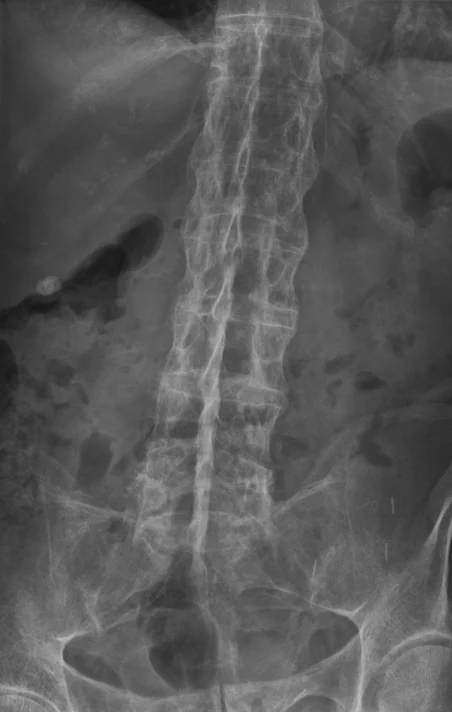
- Plain radiograph of the sacroiliac joints and spine - sacroiliitis (erosion, sclerosis, and eventually fusion), squaring of vertebral bodies, syndesmophytes bridging vertebrae producing the 'bamboo spine' in advanced disease. This is diagnostic once present but represents established, often irreversible change
- Spirometry - a restrictive pattern can develop from reduced chest wall expansion in advanced disease
Ankylosing spondylitis: management
Physiotherapy and a structured, regular exercise programme are central to management at every stage and are not simply an adjunct to drug treatment - they maintain spinal mobility and function directly.2
NSAIDs are first-line pharmacological treatment and are often used regularly (not just as needed) in active disease, since they have a genuine effect on axial inflammation.
Conventional synthetic DMARDs (methotrexate, sulfasalazine) are not effective for axial disease, but sulfasalazine can help peripheral joint involvement.
If disease activity remains high despite at least two NSAIDs trialled, a biologic is added - anti-TNF agents (adalimumab, etanercept) or anti-IL-17 agents (secukinumab, ixekizumab) are both effective for axial disease, unlike conventional DMARDs.
- Smoking cessation - smoking worsens disease activity and radiographic progression
- Postural exercises and hydrotherapy
- Screen for and treat anterior uveitis promptly
- Bone health assessment - osteoporosis is common even in a young population, related to inflammation and reduced mobility
- Surgery (hip replacement, and rarely spinal osteotomy) for severe structural damage
Reactive arthritis
Reactive arthritis is a sterile synovitis triggered by an infection elsewhere in the body, typically arising 1-4 weeks after a gastrointestinal or genitourinary infection. Common triggers are Chlamydia trachomatis (sexually acquired) and Salmonella, Shigella, Campylobacter or Yersinia (enteric).3
It classically produces an asymmetrical oligoarthritis of the lower limb, often with enthesitis and dactylitis. The historical triad - sometimes remembered as 'can't see, can't pee, can't climb a tree' - is conjunctivitis, urethritis and arthritis, though the full triad is only present in a minority.
- Circinate balanitis - painless serpiginous plaques on the glans penis
- Keratoderma blenorrhagicum - waxy, brown, hyperkeratotic plaques on the palms and soles, which can look identical to pustular psoriasis
- Conjunctivitis, and less commonly anterior uveitis
- Enthesitis and dactylitis, as in the rest of the family
Most cases are self-limiting within a few months. Management is with NSAIDs and, if needed, intra-articular corticosteroid; a chronic or relapsing course may need sulfasalazine or methotrexate. If a triggering infection (particularly chlamydia) is still active, it should be treated with appropriate antibiotics, both to prevent transmission and complications, though this does not shorten the arthritis itself.
Enteropathic arthritis
Arthritis occurs in up to 20% of patients with inflammatory bowel disease and takes two recognisable patterns. Peripheral (type 1) enteropathic arthritis is an asymmetrical, large-joint, pauciarticular arthritis whose activity broadly parallels bowel disease activity - it improves as the IBD is controlled. Axial enteropathic arthritis behaves like ankylosing spondylitis and runs its own course independent of bowel disease activity.
Differential diagnosis
- Mechanical low back pain - by far the commonest cause of back pain, distinguished by the pattern in the table above
- Rheumatoid arthritis - symmetrical small-joint polyarthritis, seropositive, spares the axial skeleton (except the atlanto-axial joint)
- Psoriatic arthritis - overlapping features, but with psoriasis, nail changes and a more variable joint pattern
- Diffuse idiopathic skeletal hyperostosis (DISH) - flowing spinal calcification in older patients, without sacroiliitis or a raised CRP
- Fibromyalgia - diffuse pain without objective inflammatory markers or imaging change
Complications
Untreated axial disease leads to progressive spinal fusion, kyphosis and a markedly increased risk of vertebral fracture even from minor trauma, because a rigid, osteoporotic spine fractures like a long bone rather than absorbing force. Cardiac conduction disease and aortic regurgitation can develop insidiously. Reactive and enteropathic arthritis can, in a minority, become chronic and destructive rather than self-limiting.
Red flags
Prognosis
Outcomes in axial spondyloarthritis have improved substantially with earlier diagnosis (via MRI and the ASAS criteria), sustained exercise therapy, and biologic treatment for those who need it - many patients now maintain good function and never progress to the classic bamboo spine. Reactive arthritis is usually self-limiting, though a minority go on to develop chronic axial or peripheral spondyloarthritis. Enteropathic arthritis largely tracks the course of the underlying bowel disease for the peripheral pattern, while the axial pattern behaves like AS regardless of bowel control.
References
- NICE Clinical Knowledge Summaries. Ankylosing spondylitis. Available here
- van der Heijde D, Ramiro S, Landewe R et al. 2016 update of the ASAS-EULAR management recommendations for axial spondyloarthritis. Annals of the Rheumatic Diseases. 2017. Available here
- NICE TA383. Secukinumab for active ankylosing spondylitis. Available here
- Sieper J, Poddubnyy D. Axial spondyloarthritis. The Lancet. 2017. Available here
- British Society for Rheumatology. Reactive arthritis clinical guidance. Available here
This article is written for revision and education. It is not clinical guidance and must not be used to make decisions about the care of a patient. Always check current NICE guidance and local protocols.