
Crystal Arthropathy: Gout and Pseudogout
Key points
- Gout: deposition of monosodium urate crystals, driven by chronic hyperuricaemia. Classically affects the first MTP joint (podagra).
- Pseudogout: deposition of calcium pyrophosphate crystals (CPPD), typically in older patients and classically affecting the knee.
- Crystal identification: the definitive test is joint aspiration and polarised light microscopy - urate crystals are needle-shaped and negatively birefringent; CPPD crystals are rhomboid and positively birefringent.
- Serum urate: can be normal or even low during an acute gout attack - a normal level does not exclude the diagnosis.
- Acute management: NSAIDs or colchicine first-line for both conditions; oral corticosteroids if these are contraindicated.
- Do not start allopurinol acutely: starting or changing urate-lowering therapy during an acute attack can prolong or worsen it. Start once the attack has settled, with prophylactic cover.
- Urate-lowering therapy: allopurinol first-line, titrated to a target urate below 360 micromol/L (300 if tophi or ongoing attacks), for anyone with recurrent attacks, tophi, urate arthropathy, or CKD.
- Radiographic distinction: gout produces punched-out erosions with overhanging edges; pseudogout produces chondrocalcinosis - linear calcification within cartilage.
Introduction
Gout and pseudogout are the two crystal arthropathies: conditions in which crystals deposit within a joint and trigger an intense acute inflammatory response. Both present in a similar way - a sudden, severe, hot, swollen, exquisitely tender joint - and both are common causes of the 'hot joint' presentation that must always be distinguished from septic arthritis.1
Gout is caused by monosodium urate crystals and is closely linked to hyperuricaemia, diet, alcohol and metabolic syndrome. Pseudogout is caused by calcium pyrophosphate dihydrate (CPPD) crystals, is commoner with increasing age, and is often associated with an underlying metabolic disorder. They are considered together here because they are managed similarly in the acute setting but differ in their long-term prevention.
Gout: aetiology and pathophysiology
Gout results from supersaturation of the extracellular fluid with uric acid, the end product of purine metabolism. When serum urate exceeds its solubility threshold (around 380 micromol/L), monosodium urate crystals precipitate, preferentially in cooler, peripheral joints - which is why the first MTP joint of the foot is the classic site.
Hyperuricaemia arises from under-excretion of urate (about 90% of cases, often related to renal impairment or diuretic use) or overproduction (increased purine turnover, as in myeloproliferative disease or after chemotherapy - tumour lysis syndrome). Crystals themselves can be present for years without symptoms; an acute attack is triggered when crystals shed from cartilage into the joint space and activate the NLRP3 inflammasome, driving a rapid neutrophilic response.
- Male sex and post-menopausal women (oestrogen is uricosuric)
- Obesity and metabolic syndrome
- Alcohol, particularly beer (high in purines and impairs renal urate excretion)
- Purine-rich diet - red meat, shellfish, offal
- Diuretics - thiazides and loop diuretics reduce renal urate excretion
- Chronic kidney disease
- High cell turnover states - myeloproliferative/lymphoproliferative disease, psoriasis, cytotoxic chemotherapy
- Family history
- Low-dose aspirin and ciclosporin
Gout: clinical features
- Rapid onset, often overnight, of severe pain peaking within 12-24 hours
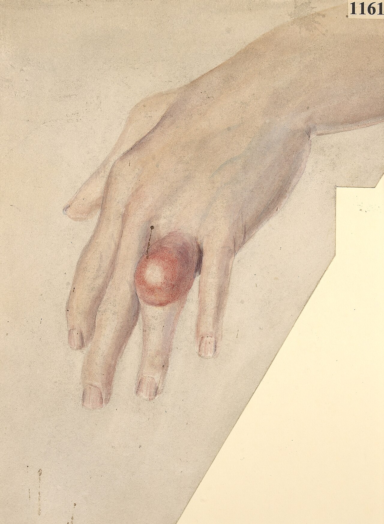
- First metatarsophalangeal (MTP) joint - 'podagra' - is the classic and commonest site, but the ankle, knee, wrist and small joints of the hand can also be affected
- Hot, red, swollen, exquisitely tender joint - even light contact (a bedsheet) can be intolerable
- Self-limiting if untreated, typically resolving over 1-2 weeks, with the overlying skin sometimes desquamating as it settles
- Recurrent attacks become more frequent over time if hyperuricaemia is not addressed
Chronic tophaceous gout
With longstanding, poorly controlled hyperuricaemia, urate crystals aggregate into tophi - firm, chalky white nodules that deposit in the pinna of the ear, the fingers, the olecranon bursa and the Achilles tendon. Established tophi can ulcerate, discharge chalky material, and cause chronic joint damage and deformity.
Pseudogout: aetiology and clinical features
Pseudogout is caused by deposition of calcium pyrophosphate dihydrate (CPPD) crystals within joint cartilage, again with acute attacks triggered by crystal shedding into the joint. It becomes much commoner with age, and when it occurs in a younger patient an underlying metabolic cause should be sought.2
- Older age - the single strongest risk factor
- Pre-existing osteoarthritis
- Hyperparathyroidism
- Haemochromatosis
- Hypomagnesaemia and hypophosphataemia
- Hypothyroidism
Clinically it presents like gout - acute, hot, swollen, painful joint - but classically affects the knee (the commonest site) or the wrist, rather than the first MTP joint. Some patients have a chronic, low-grade pattern resembling osteoarthritis rather than discrete acute attacks.
Differential diagnosis
- Septic arthritis - the diagnosis that must never be missed in an acute hot joint; fever and systemic upset are more marked, and aspiration with Gram stain and culture is mandatory if there is any doubt
- Cellulitis - overlying skin infection without true joint involvement
- Reactive arthritis - typically post-infective, often with additional features (conjunctivitis, urethritis)
- Trauma or haemarthrosis
- Acute flare of rheumatoid or psoriatic arthritis
Investigations
Joint aspiration - the definitive test
Aspiration with polarised light microscopy distinguishes the two crystals and excludes infection at the same time.
| Gout (monosodium urate) | Pseudogout (CPPD) | |
|---|---|---|
| Crystal shape | Needle-shaped | Rhomboid |
| Birefringence | Negatively birefringent (yellow when parallel to the axis of polarisation) | Positively birefringent (blue when parallel) |
| Fluid appearance | Turbid, yellow | Turbid, may be blood-stained |
Blood tests
- Serum urate - supports the diagnosis of gout but can be normal or low during an acute attack, as the inflammatory response itself is uricosuric. Recheck 4-6 weeks after the attack has settled if the diagnosis is uncertain.
- FBC, CRP - leucocytosis and a raised CRP occur in both crystal arthropathies and in sepsis, so do not use them alone to exclude infection
- U&Es - renal function affects urate excretion and drug choice
- Bone profile (calcium) and TFTs - screen for hyperparathyroidism and hypothyroidism in pseudogout, particularly if it occurs in a patient under 60 or affects an atypical joint
- Ferritin/transferrin saturation - screen for haemochromatosis if pseudogout is recurrent or occurs at a young age
Imaging
| Condition | Radiographic findings |
|---|---|
| Gout | Punched-out, well-defined periarticular erosions with sclerotic margins and overhanging edges; joint space relatively preserved until late disease |
| Pseudogout | Chondrocalcinosis - fine linear calcification within articular cartilage, classically the meniscus of the knee or the triangular fibrocartilage complex of the wrist |
Management of the acute attack
Acute management is similar for both conditions.3
- NSAIDs (with gastroprotection) - first-line if no contraindication
- Colchicine - an alternative first-line agent, particularly useful in renal impairment or when NSAIDs are contraindicated; nausea, vomiting and diarrhoea are dose-limiting side effects, and the dose must be reduced in renal impairment
- Oral corticosteroids - used if NSAIDs and colchicine are both contraindicated or ineffective, or as intra-articular corticosteroid injection once septic arthritis has been excluded
- Rest and elevation of the affected joint, with ice
Long-term management
Gout - urate-lowering therapy
Allopurinol is first-line, started at a low dose and titrated upward, aiming for a serum urate below 360 micromol/L (below 300 in patients with tophi, chronic gouty arthritis or ongoing attacks despite treatment).
- Indications for urate-lowering therapy: two or more attacks in 12 months, tophi, chronic gouty joint damage or urate arthropathy on imaging, chronic kidney disease, or urate renal stones
- Febuxostat is used second-line, when allopurinol is not tolerated or is contraindicated
- Cover the start of urate-lowering therapy with a low-dose NSAID or colchicine for the first 3-6 months, since lowering urate can itself trigger attacks by mobilising crystals
- Lifestyle measures - weight loss, reduced alcohol (particularly beer), reduced intake of red meat and shellfish, adequate hydration, and reviewing diuretic therapy where possible
Pseudogout
There is no equivalent of urate-lowering therapy for CPPD disease. Management is aimed at treating any identified underlying cause (correcting hyperparathyroidism, haemochromatosis, hypomagnesaemia), and NSAIDs, colchicine or intra-articular steroid for recurrent flares. Frequent recurrent attacks may occasionally warrant low-dose prophylactic colchicine.
Complications
Recurrent gout causes progressive joint damage, chronic tophaceous deposits, and secondary osteoarthritis. Urate nephropathy and renal stones can complicate longstanding hyperuricaemia. Both gout and pseudogout are associated with cardiovascular disease and metabolic syndrome, which should be assessed and managed alongside the joint disease. Pseudogout can occasionally present with a more destructive arthropathy resembling severe osteoarthritis.
Red flags
Prognosis
With effective urate-lowering therapy and sustained target urate levels, gout attacks become infrequent or stop altogether, and existing tophi can slowly resolve. Poor adherence to urate-lowering therapy is the commonest reason for ongoing attacks, and addressing this is usually more productive than escalating acute treatment alone. Pseudogout tends to follow a more variable course dictated by the underlying joint (often osteoarthritic) and any correctable metabolic driver.
References
- NICE Clinical Knowledge Summaries. Gout. Available here
- NICE NG219. Gout: diagnosis and management. 2022. Available here
- British Society for Rheumatology. Guideline for the management of gout. Rheumatology. 2017. Available here
- NICE Clinical Knowledge Summaries. Calcium pyrophosphate deposition (pseudogout). Available here
- Dalbeth N, Choi HK, Joosten LAB et al. Gout. Nature Reviews Disease Primers. 2019. Available here
This article is written for revision and education. It is not clinical guidance and must not be used to make decisions about the care of a patient. Always check current NICE guidance and local protocols.