
Hyperparathyroidism: Primary, Secondary and Tertiary Disease
Key points
- Primary hyperparathyroidism: autonomous excess PTH secretion, usually from a single parathyroid adenoma (over 80%), causing a raised or inappropriately normal PTH with raised calcium.
- Secondary hyperparathyroidism: an appropriate physiological rise in PTH in response to hypocalcaemia, classically from chronic kidney disease or vitamin D deficiency - low or normal calcium with high PTH.
- Tertiary hyperparathyroidism: autonomous PTH secretion that develops after prolonged secondary hyperparathyroidism, usually in long-standing CKD or dialysis - high calcium and high PTH, as the glands no longer respond to correction of the calcium.
- Bones, stones, groans and psychiatric moans: the mnemonic for the features of hypercalcaemia in primary hyperparathyroidism, plus polyuria, polydipsia and hypertension.
- Key biochemical clue: phosphate is typically low in primary hyperparathyroidism, because PTH promotes renal phosphate excretion.
- Definitive treatment: parathyroidectomy for primary disease meeting surgical criteria, curative in around 95% of cases when performed by an experienced surgeon.
- Localisation before surgery: neck ultrasound and sestamibi scintigraphy to identify the adenoma, allowing minimally invasive, targeted surgery.
- Medical alternative: cinacalcet, a calcimimetic, for patients unfit for or declining surgery.
Introduction
Parathyroid hormone (PTH) is secreted by the four parathyroid glands in response to a fall in serum calcium, sensed via the calcium-sensing receptor. It raises calcium by three mechanisms: increasing osteoclastic bone resorption, increasing renal calcium reabsorption while promoting phosphate excretion, and stimulating renal 1-alpha-hydroxylase to increase production of active vitamin D (calcitriol), which in turn increases intestinal calcium absorption.
Hyperparathyroidism is classified into three distinct types according to the underlying driver, and the distinction is central to both diagnosis and management.
| Type | Mechanism | Calcium | Phosphate | PTH |
|---|---|---|---|---|
| Primary | Autonomous PTH secretion - adenoma, hyperplasia or (rarely) carcinoma | Raised | Low | Raised or inappropriately normal |
| Secondary | Appropriate physiological response to hypocalcaemia - usually CKD or vitamin D deficiency | Low or normal | Raised in CKD (reduced excretion); low in vitamin D deficiency | Raised |
| Tertiary | Autonomous secretion developing after prolonged secondary hyperparathyroidism - glands become unresponsive to feedback | Raised | Raised (underlying CKD) | Markedly raised |
Causes of primary hyperparathyroidism
- Solitary parathyroid adenoma - over 80% of cases
- Multigland hyperplasia - around 10 to 15%, more often seen in MEN1 and MEN2A
- Multiple adenomas - a minority
- Parathyroid carcinoma - rare (under 1%), suggested by a very high calcium, a palpable neck mass, and a markedly elevated PTH
Causes of secondary and tertiary hyperparathyroidism
- Chronic kidney disease - the commonest cause; reduced renal 1-alpha-hydroxylase activity lowers active vitamin D, and phosphate retention further drives PTH secretion
- Vitamin D deficiency - dietary, malabsorptive or from reduced sun exposure (see the vitamin D deficiency and osteomalacia article)
- Malabsorption of calcium or vitamin D, e.g. coeliac disease
- Tertiary hyperparathyroidism develops when secondary hyperparathyroidism is prolonged (classically in long-standing dialysis-dependent CKD) and the parathyroid glands undergo autonomous hyperplasia, continuing to secrete PTH even after the calcium has been corrected
Clinical features
Many cases of primary hyperparathyroidism are now detected incidentally on routine bloods, before symptoms develop. When symptomatic, the features reflect hypercalcaemia and, in longstanding disease, its effects on bone.
- Polyuria and polydipsia - hypercalcaemia impairs the renal concentrating mechanism, producing a form of nephrogenic diabetes insipidus
- Hypertension - common, though the mechanism is not fully understood
- Muscle weakness and fatigue
- Pruritus
- Corneal calcification (band keratopathy) in longstanding severe disease
- Many patients are entirely asymptomatic, with hypercalcaemia found incidentally
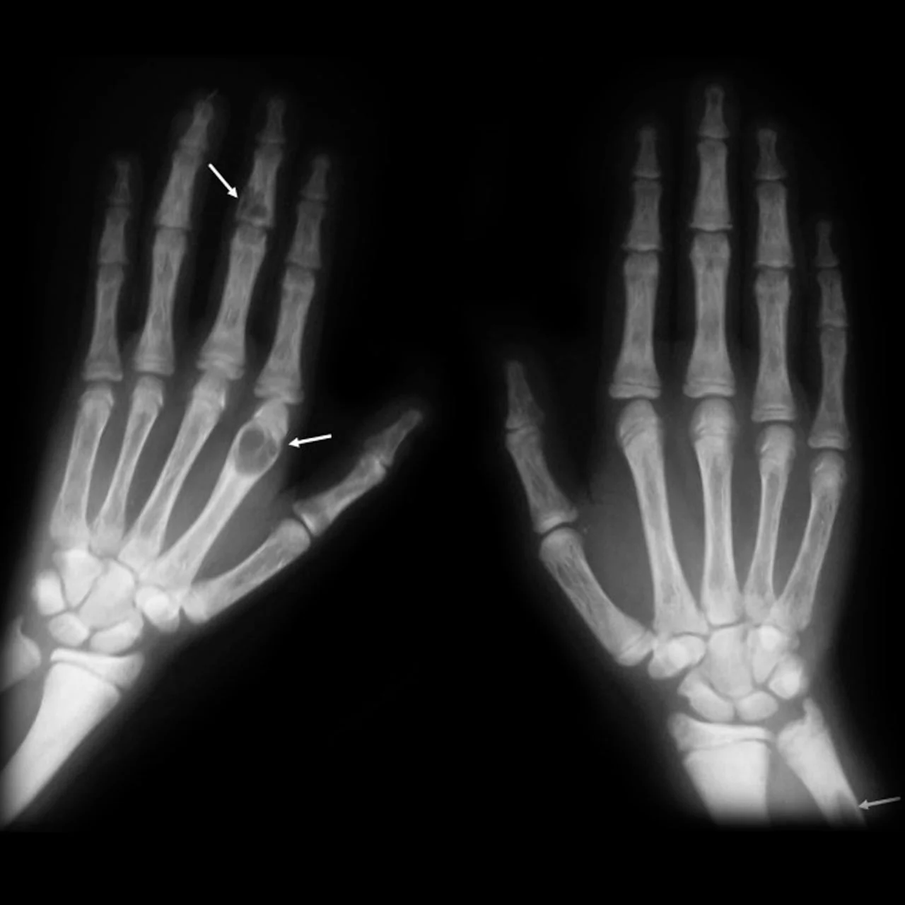
Secondary hyperparathyroidism in CKD more often presents with the features of renal osteodystrophy - bone and joint pain, and increased fracture risk - rather than the hypercalcaemic picture above, since calcium is typically low or normal until the tertiary stage develops.
Investigations
Biochemistry
- Corrected (albumin-adjusted) serum calcium - raised in primary and tertiary disease; low or normal in secondary disease
- PTH - measured alongside calcium. In primary hyperparathyroidism a raised or inappropriately "normal" PTH in the presence of raised calcium is diagnostic, since a normal gland would suppress PTH to near zero in response to hypercalcaemia
- Phosphate - characteristically low in primary hyperparathyroidism, reflecting the phosphaturic action of PTH; typically high in secondary/tertiary disease associated with CKD
- Vitamin D (25-hydroxyvitamin D) - to identify or exclude vitamin D deficiency as a driver of secondary hyperparathyroidism, and because coexisting deficiency can mask the severity of primary disease
- Renal function (U&Es, eGFR) - to assess for CKD as a cause, and because primary hyperparathyroidism itself can impair renal function over time
- Alkaline phosphatase - raised where bone turnover is high
- 24-hour urinary calcium - helps exclude familial hypocalciuric hypercalcaemia (FHH), an important differential caused by an inactivating mutation in the calcium-sensing receptor, in which urinary calcium excretion is inappropriately low despite hypercalcaemia and PTH may be mildly raised. FHH does not benefit from parathyroidectomy, so distinguishing it is essential.
Imaging and localisation
- DEXA bone density scan - assesses osteoporosis, with particular attention to the distal third of the radius, a site rich in cortical bone that is preferentially affected in primary hyperparathyroidism
- Renal ultrasound - to look for nephrocalcinosis or renal calculi
- Neck ultrasound - first-line for localising an adenoma before surgery
- Sestamibi (99mTc-MIBI) scintigraphy - functional imaging that identifies hyperfunctioning parathyroid tissue; often combined with ultrasound or SPECT-CT to guide minimally invasive parathyroidectomy
- 4D-CT - increasingly used, particularly where first-line imaging is discordant or an adenoma is not localised
Management
Primary hyperparathyroidism
Parathyroidectomy is curative and is the treatment of choice where indicated. Criteria for surgery (broadly following consensus/NICE-aligned guidance) include:
- Serum calcium more than 0.25 mmol/L above the upper limit of normal
- Age under 50
- Reduced eGFR (below 60 mL/min/1.73m²)
- Osteoporosis or a previous fragility fracture (T-score -2.5 or below at any site, including the distal radius)
- Nephrocalcinosis, renal calculi, or a 24-hour urinary calcium suggesting a high stone risk
- Symptomatic disease - any of the classical bones/stones/groans/psychiatric moans features
- All patients meeting any one of these criteria, and any symptomatic patient, should be offered surgery; asymptomatic patients not meeting criteria may reasonably be monitored
- Minimally invasive (targeted) parathyroidectomy where a single adenoma is localised - a focused operation with a small incision and shorter recovery
- Bilateral neck exploration where localisation fails or multigland disease is suspected (e.g. MEN syndromes)
- Intraoperative PTH monitoring confirms adequate resection - a fall of more than 50% from baseline within 10 to 15 minutes predicts cure
- Cure rate is around 95% in experienced hands
- Complications include recurrent laryngeal nerve injury (voice change) and transient or permanent hypoparathyroidism, with "hungry bone syndrome" - a sharp fall in calcium as bone rapidly re-mineralises after years of PTH-driven resorption - requiring calcium and vitamin D supplementation postoperatively, particularly in those with severe preoperative bone disease
Conservative management when surgery is not indicated or not wanted
- Maintain adequate hydration and avoid dehydration, which worsens hypercalcaemia
- Avoid factors that raise calcium further - thiazide diuretics, high-dose vitamin D or calcium supplements, and prolonged immobilisation
- Regular monitoring of calcium, renal function and bone density
- Encourage physical activity and adequate but not excessive dietary calcium intake
- Cinacalcet - a calcimimetic that increases the sensitivity of the calcium-sensing receptor, lowering PTH and calcium; used where surgery is declined, not indicated, or not possible (e.g. significant comorbidity), and in parathyroid carcinoma. It lowers calcium but does not improve bone density, so is not a substitute for surgery when bone disease is the indication.
- Bisphosphonates may be used to protect bone density where surgery is deferred
Secondary hyperparathyroidism
- Treat the underlying cause - vitamin D replacement for deficiency, and management of the phosphate and vitamin D axis in CKD
- Phosphate binders and active vitamin D analogues (alfacalcidol or calcitriol) in CKD, since impaired renal 1-alpha-hydroxylation means native vitamin D cannot be adequately activated
- Dietary phosphate restriction in CKD
- Cinacalcet may be used in CKD-associated secondary hyperparathyroidism, particularly in dialysis patients, to reduce PTH
Tertiary hyperparathyroidism
- Cinacalcet first-line in many cases, particularly where the patient is not a surgical candidate
- Parathyroidectomy (usually subtotal, or total with autotransplantation of a small amount of parathyroid tissue) where medical management fails or the hypercalcaemia is severe, especially after renal transplantation when autonomous gland function may persist
Complications
- Osteoporosis and fragility fracture, particularly at cortical bone sites such as the distal radius
- Nephrolithiasis and nephrocalcinosis, with progressive renal impairment
- Peptic ulcer disease and pancreatitis
- Cardiovascular disease - hypertension and, in some studies, increased cardiovascular risk
- Hypercalcaemic crisis - severe hypercalcaemia with confusion, dehydration and renal failure, requiring urgent treatment (aggressive intravenous fluids, bisphosphonates, and consideration of urgent parathyroidectomy)
- Hungry bone syndrome after parathyroidectomy - profound hypocalcaemia, hypophosphataemia and hypomagnesaemia requiring active monitoring and replacement
- Recurrent laryngeal nerve injury at surgery
- Parathyroid carcinoma - rare, but should be suspected with a very high calcium, markedly elevated PTH and a palpable neck mass
Red flags
Prognosis
Primary hyperparathyroidism treated with parathyroidectomy is curable in around 95% of cases, with resolution of hypercalcaemia, improvement in bone density over 1 to 2 years, and reduced risk of further renal stones. Patients managed conservatively because they do not meet surgical criteria generally do well with monitoring, though a proportion will progress and eventually require surgery.
Secondary hyperparathyroidism improves with correction of the underlying driver where possible (vitamin D replacement, control of CKD-related phosphate and calcium abnormalities), but in advanced CKD the course is often progressive, predisposing to tertiary disease and renal osteodystrophy despite treatment.
Tertiary hyperparathyroidism may persist even after renal transplantation restores normal renal function, because the parathyroid glands have undergone structural hyperplasia and become autonomous; a proportion of these patients ultimately require parathyroidectomy.
References
- NICE Clinical Knowledge Summaries. Hyperparathyroidism - primary. Available here
- NICE NG132. Hyperparathyroidism (primary): diagnosis, assessment and initial management. 2019. Available here
- Bilezikian JP, Khan AA, Silverberg SJ et al. Evaluation and management of primary hyperparathyroidism: summary statement from the Fifth International Workshop. J Bone Miner Res. 2022. Available here
- Society for Endocrinology. Clinical guidance on calcium and parathyroid disorders. Available here
- NICE NG203. Chronic kidney disease: assessment and management. 2021. Available here
- BNF. Cinacalcet. Available here
- Frank Gaillard, CC BY-SA 4.0, via Wikimedia Commons. Available here
This article is written for revision and education. It is not clinical guidance and must not be used to make decisions about the care of a patient. Always check current NICE guidance and local protocols.