
Hypocalcaemia
Key points
- Hypocalcaemia: corrected calcium below 2.2 mmol/L. Always correct for albumin before acting: corrected Ca²⁺ = measured Ca²⁺ + 0.02 × (40 − albumin g/L).
- Commonest causes: vitamin D deficiency (much the commonest), chronic kidney disease, hypoparathyroidism - especially post-thyroidectomy - and hypomagnesaemia.
- Phosphate splits the differential: HIGH phosphate = hypoparathyroidism or chronic kidney disease. LOW phosphate = vitamin D deficiency or secondary hyperparathyroidism (PTH is phosphaturic).
- The classic signs: Trousseau's sign - carpopedal spasm on inflating a BP cuff above systolic for 3 minutes (more sensitive and specific). Chvostek's sign - facial twitching on tapping over the facial nerve.
- Symptoms: perioral and peripheral paraesthesiae, muscle cramps and tetany, and in severe cases carpopedal spasm, laryngospasm, bronchospasm and seizures.
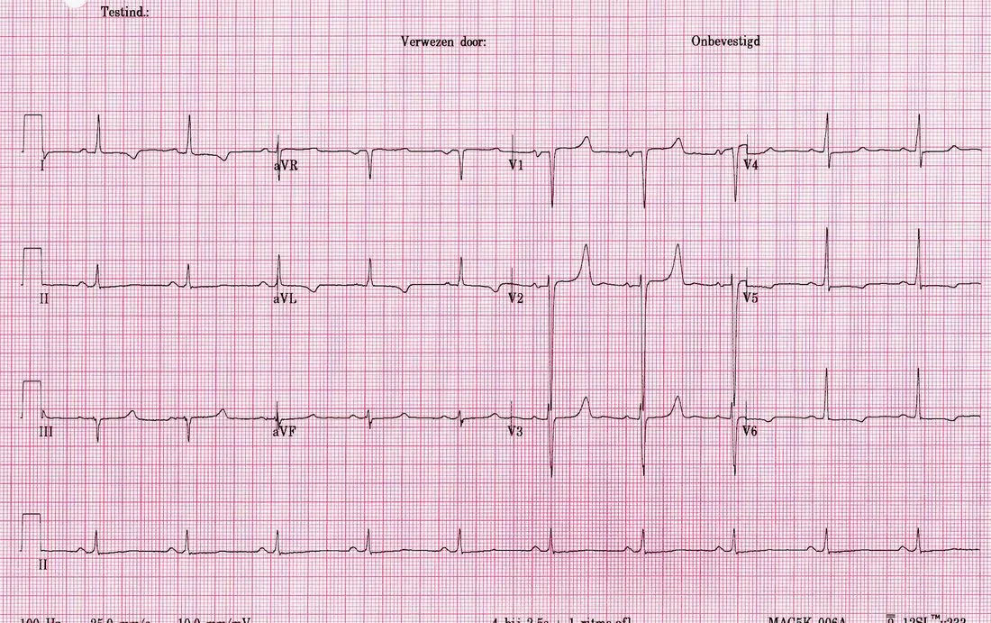
- ECG: a prolonged QT interval, risking torsades de pointes; severe hypocalcaemia can also cause heart failure through reduced myocardial contractility.
- Always check magnesium: hypomagnesaemia causes hypocalcaemia by impairing PTH secretion and action. The calcium will not correct until magnesium is replaced - a recurring clinical trap.
- Severe or symptomatic disease: IV calcium gluconate 10%, 10-20 mL over 10 minutes with cardiac monitoring, then an infusion. Prefer gluconate over chloride peripherally - chloride is far more irritant.
Introduction
Hypocalcaemia is a corrected serum calcium below 2.2 mmol/L. It is common in hospital practice - present in a substantial proportion of critically ill patients - and ranges from an asymptomatic biochemical finding to a genuine emergency with laryngospasm, seizures and arrhythmia.1
Physiologically, calcium is regulated by PTH (which raises calcium by mobilising bone, increasing renal reabsorption and activating vitamin D) and calcitriol (which increases gut absorption). Almost every cause of hypocalcaemia is a failure of one of these two axes, a problem with phosphate, or chelation of calcium out of the circulation - and that framework makes the causes far easier to recall.
Causes
Vitamin D related
- Vitamin D deficiency - much the commonest cause; from poor dietary intake, limited sun exposure, darker skin pigmentation, covering clothing, older age and institutionalisation. Causes osteomalacia in adults and rickets in children
- Malabsorption - coeliac disease, Crohn's disease, chronic pancreatitis, bariatric surgery, cholestatic liver disease
- Chronic kidney disease - impaired 1-alpha-hydroxylation of vitamin D, together with phosphate retention
- Liver disease - impaired 25-hydroxylation
- Anticonvulsants - phenytoin, carbamazepine and phenobarbital induce hepatic enzymes and accelerate vitamin D metabolism
Parathyroid related
- Post-surgical hypoparathyroidism - the commonest cause of true hypoparathyroidism, following total thyroidectomy, parathyroidectomy or radical neck surgery. May be transient or permanent, and calcium must be monitored closely after such operations
- Autoimmune hypoparathyroidism - isolated, or as part of autoimmune polyglandular syndrome type 1 (with Addison's disease and mucocutaneous candidiasis)
- DiGeorge syndrome (22q11 deletion) - congenital absence of the parathyroids, with thymic aplasia, cardiac defects and cleft palate
- Infiltration - haemochromatosis, Wilson disease, metastases
- Radiation to the neck
- Pseudohypoparathyroidism - end-organ resistance to PTH, so calcium is low and PTH is HIGH. Albright hereditary osteodystrophy gives the classic phenotype: short stature, round face, shortened fourth and fifth metacarpals, obesity and learning difficulties. Pseudopseudohypoparathyroidism has the same phenotype but normal biochemistry
- Hungry bone syndrome - profound, prolonged hypocalcaemia after parathyroidectomy (or thyroidectomy for thyrotoxicosis), as demineralised bone avidly takes up calcium, phosphate and magnesium
Other causes
- Hypomagnesaemia - impairs both PTH secretion and its peripheral action. Common with diuretics, proton pump inhibitors, alcohol dependence, diarrhoea and refeeding
- Acute pancreatitis - calcium is saponified by free fatty acids released by fat necrosis; hypocalcaemia is a marker of severity in scoring systems
- Tumour lysis syndrome - massive phosphate release binds calcium
- Rhabdomyolysis - calcium deposits in damaged muscle (with rebound hypercalcaemia during recovery)
- Massive blood transfusion - citrate in stored blood chelates calcium; a recognised issue in major haemorrhage protocols
- Drugs - bisphosphonates, denosumab (particularly if vitamin D deficient), calcitonin, cinacalcet, foscarnet, and phosphate-containing enemas
- Sepsis and critical illness
- Respiratory alkalosis from hyperventilation - lowers ionised calcium without changing the total
- Osteoblastic metastases - prostate and breast cancer, where calcium is deposited into bone
Clinical features
Hypocalcaemia causes neuromuscular irritability, because a low extracellular calcium lowers the threshold for nerve and muscle depolarisation. Mild hypocalcaemia is frequently asymptomatic; the rate of fall matters as much as the absolute level, and a rapid drop after thyroid surgery may be far more symptomatic than a chronically low calcium in CKD.
- Perioral paraesthesiae and tingling of the fingers and toes - typically the earliest symptom
- Muscle cramps, twitching and carpopedal spasm
- Tetany - sustained involuntary muscle contraction
- Laryngospasm and bronchospasm - potentially life-threatening stridor and airway compromise
- Seizures - hypocalcaemia is a genuinely reversible cause and should be excluded in new-onset seizures
- Anxiety, irritability, depression, confusion and, in chronic cases, cognitive impairment
- Cardiac - prolonged QT interval predisposing to torsades de pointes, and reduced myocardial contractility causing heart failure and hypotension that is refractory to inotropes until calcium is corrected
- Chronic hypocalcaemia - cataracts, dry skin, brittle nails, coarse hair, basal ganglia calcification (occasionally with parkinsonism), dental hypoplasia and papilloedema
- Features of the cause - the scar of a recent thyroidectomy, bowed legs and growth failure in rickets, proximal myopathy and bone pain in osteomalacia
The two eponymous signs
| Sign | How to elicit it | What it shows | Reliability |
|---|---|---|---|
| Trousseau's sign | Inflate a blood pressure cuff above systolic pressure for 3 minutes | Carpopedal spasm - flexion at the wrist and metacarpophalangeal joints with extended fingers and an adducted thumb ('main d'accoucheur', the obstetrician's hand) | More sensitive and specific; present in around 94% of hypocalcaemic patients and only ~1% of normocalcaemic people |
| Chvostek's sign | Tap over the facial nerve just anterior to the ear, below the zygomatic arch | Twitching of the ipsilateral facial muscles, especially at the corner of the mouth | Less specific - positive in around 10-25% of people with normal calcium, so a positive sign alone is weak evidence |
Investigations
- Corrected calcium - repeat to confirm, and always correct for albumin
- Serum magnesium - check in every case; hypomagnesaemia causes refractory hypocalcaemia
- Phosphate - the key discriminator (see above)
- PTH - low or inappropriately normal confirms hypoparathyroidism; raised indicates vitamin D deficiency, CKD or PTH resistance
- 25-hydroxyvitamin D - to diagnose deficiency
- U&Es and creatinine - chronic kidney disease
- Alkaline phosphatase - raised in osteomalacia and rickets, and normal in hypoparathyroidism
- ECG - looking for a prolonged QT interval
- Amylase or lipase - if acute pancreatitis is suspected
- Creatine kinase, urate, potassium and phosphate - for rhabdomyolysis and tumour lysis syndrome
- Coeliac serology and malabsorption screen - where vitamin D deficiency is unexplained
- Blood gas - to assess pH, since alkalosis lowers ionised calcium
- Genetic testing and specialist assessment - for suspected pseudohypoparathyroidism or DiGeorge syndrome
- Post-operative calcium monitoring - routine after total thyroidectomy, typically at intervals over the first 24-48 hours
Management
Severe or symptomatic hypocalcaemia
Treat urgently if there is tetany, carpopedal spasm, laryngospasm, seizures, a prolonged QT or arrhythmia, or if the corrected calcium is below about 1.9 mmol/L even without symptoms.2
- Secure the airway if there is laryngospasm or stridor, and call for senior and anaesthetic help
- IV calcium gluconate 10%, 10-20 mL (1-2 ampoules, containing 2.25 mmol calcium per 10 mL) diluted in 50-100 mL of 5% dextrose or 0.9% saline, given over 10-20 minutes, with continuous cardiac monitoring
- Follow with a calcium gluconate infusion - typically 100 mL of 10% calcium gluconate in 1 litre of 5% dextrose or 0.9% saline over 24 hours, titrated to serum calcium
- Recheck calcium every 4-6 hours initially
- Correct magnesium simultaneously - typically magnesium sulphate 20 mmol IV; without this the calcium will not stay corrected
- Treat the underlying cause and start oral replacement once stable
Chronic and mild hypocalcaemia
| Cause | Treatment |
|---|---|
| Vitamin D deficiency | Colecalciferol - a loading regimen followed by maintenance, with calcium supplementation if dietary intake is inadequate. Recheck calcium and vitamin D after treatment |
| Hypoparathyroidism | Active vitamin D analogues - alfacalcidol or calcitriol - are required, because these patients cannot 1-alpha-hydroxylate vitamin D without PTH. Plain colecalciferol is ineffective, which is a key examination point. Add calcium supplements and monitor for hypercalciuria and nephrocalcinosis; recombinant PTH is available in selected refractory cases |
| Chronic kidney disease | Alfacalcidol or calcitriol, phosphate binders and dietary phosphate restriction; manage as CKD-mineral and bone disorder |
| Hypomagnesaemia | Replace magnesium - oral or IV depending on severity; identify and stop contributing drugs (PPIs, diuretics) |
| Post-thyroidectomy | Monitor calcium closely post-operatively; treat with calcium and, if hypoparathyroidism is confirmed, an active vitamin D analogue. Many cases are transient and recover over weeks to months |
| Hungry bone syndrome | Large doses of calcium and active vitamin D, sometimes for weeks, with magnesium and phosphate replacement; anticipate it after parathyroidectomy in severe hyperparathyroidism |
| Pseudohypoparathyroidism | Active vitamin D analogues and calcium, with specialist endocrine follow-up |
| Drug-induced (bisphosphonates, denosumab) | Ensure vitamin D repletion BEFORE giving these drugs - failure to do so is a recognised cause of severe symptomatic hypocalcaemia |
Complications
- Laryngospasm and airway obstruction - the most immediately life-threatening consequence
- Seizures - hypocalcaemia is a reversible cause and should always be excluded
- Prolonged QT and torsades de pointes, and other ventricular arrhythmias
- Heart failure and hypotension - from impaired myocardial contractility, characteristically refractory to inotropes until calcium is corrected
- Tetany and carpopedal spasm with significant distress
- Cataracts - a recognised consequence of chronic hypocalcaemia
- Basal ganglia calcification - occasionally causing extrapyramidal features
- Papilloedema and raised intracranial pressure
- Osteomalacia and rickets - with bone pain, proximal myopathy, fractures, bowed legs and growth failure in children
- Dental hypoplasia and enamel defects in childhood-onset disease
- Complications of treatment - hypercalciuria, nephrocalcinosis and renal stones from long-term calcium and active vitamin D therapy (so urinary calcium must be monitored), tissue necrosis from extravasated calcium chloride, and metastatic calcification if calcium is given in hyperphosphataemic states
Red flags
Prognosis
Acute symptomatic hypocalcaemia responds rapidly and completely to intravenous calcium, with tetany, paraesthesiae and ECG changes resolving within minutes to hours. Provided the airway is protected and arrhythmia avoided, there is usually no lasting consequence, and the prognosis then depends entirely on the underlying cause.
Most causes are readily and durably treatable. Vitamin D deficiency, by far the commonest, corrects fully with colecalciferol, with resolution of biochemical abnormalities within weeks and of osteomalacic bone pain and proximal myopathy over months. Hypomagnesaemia-related hypocalcaemia corrects promptly once magnesium is replaced, and post-thyroidectomy hypoparathyroidism is transient in the majority, with parathyroid function recovering over weeks to months.
Permanent hypoparathyroidism is the exception, and carries a genuine long-term burden. It requires lifelong treatment with active vitamin D analogues and calcium, and unlike most endocrine deficiencies it cannot be replaced with the missing hormone in routine practice, so control is often imperfect. The main long-term hazard comes from treatment rather than the disease: because PTH normally promotes renal calcium reabsorption, patients lacking it develop hypercalciuria when calcium is raised towards normal, with consequent nephrocalcinosis, renal stones and progressive chronic kidney disease. For this reason the therapeutic target is deliberately a low-normal serum calcium - enough to abolish symptoms rather than to normalise the number - and urinary calcium and renal function must be monitored long term. Other recognised long-term complications include cataracts and basal ganglia calcification, and quality of life is often reported as poorer than the biochemistry alone would suggest.
References
- NICE Clinical Knowledge Summaries. Hypocalcaemia. Available here
- Society for Endocrinology. Emergency Endocrine Guidance: Acute hypocalcaemia in adults. Available here
- Bollerslev J, Rejnmark L, Marcocci C et al. European Society of Endocrinology Clinical Guideline: treatment of chronic hypoparathyroidism in adults. Available here
- Cooper MS, Gittoes NJL. Diagnosis and management of hypocalcaemia. BMJ. 2008. Available here
- NICE NG203. Chronic kidney disease: assessment and management (CKD-mineral and bone disorder). 2021. Available here
- BNF. Calcium gluconate, alfacalcidol and colecalciferol. Available here
- User:Drj (ECGpedia), CC BY-SA 4.0, via Wikimedia Commons. Available here
This article is written for revision and education. It is not clinical guidance and must not be used to make decisions about the care of a patient. Always check current NICE guidance and local protocols.