
Chronic Pancreatitis: Diagnosis and Management
Key points
- Chronic pancreatitis: progressive, irreversible inflammatory fibrosis of the pancreas, causing permanent loss of exocrine and endocrine function.
- Commonest cause: chronic alcohol excess accounts for around 70-80% of UK cases; smoking is a strong independent and dose-related risk factor.
- Classic triad: abdominal pain, steatorrhoea and diabetes - though the full triad is a late finding and indicates over 90% loss of exocrine function.
- Pain: epigastric, radiating to the back, relieved by sitting forward; often the dominant symptom, and it may paradoxically diminish as the gland 'burns out'.
- Amylase caveat: amylase and lipase are usually normal in chronic pancreatitis, because there is insufficient residual acinar tissue to release them.
- Exocrine testing: faecal elastase-1 is the practical test - a low level confirms pancreatic exocrine insufficiency.
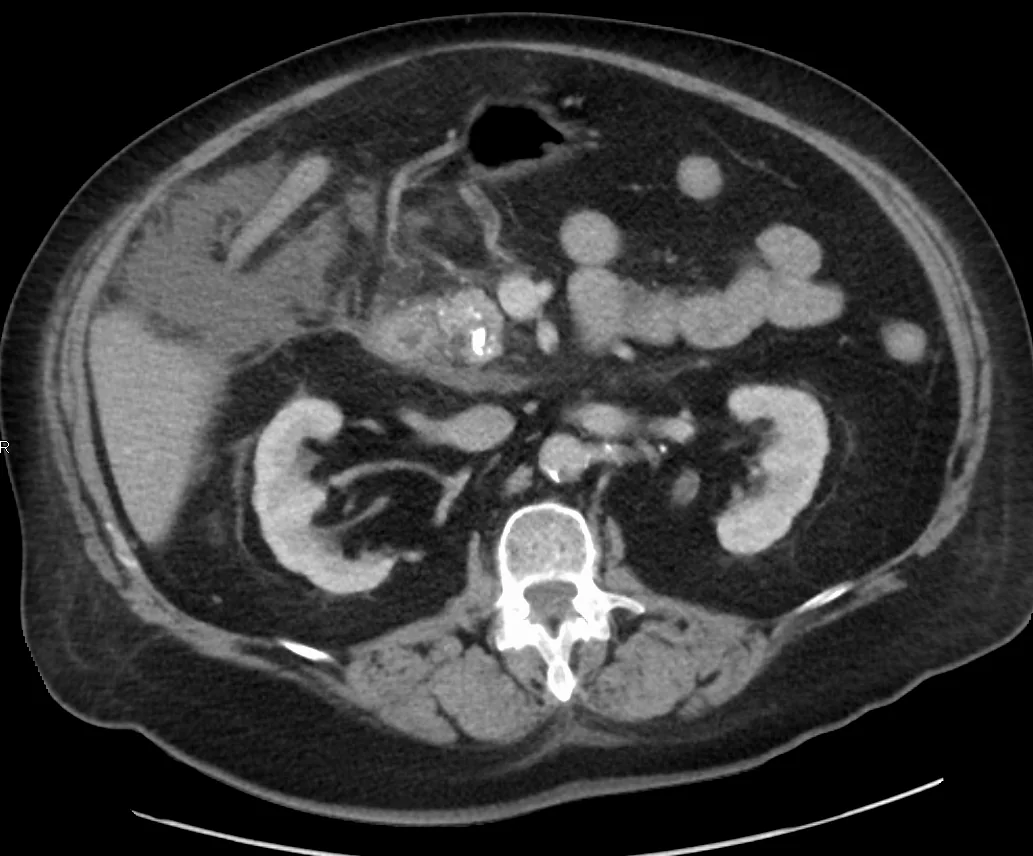
- Imaging: CT showing pancreatic calcification, ductal dilatation and atrophy; endoscopic ultrasound and MRCP are more sensitive in early disease.
- Management: alcohol and smoking cessation, pancreatic enzyme replacement therapy with a PPI, fat-soluble vitamin replacement, and a structured approach to pain.
Introduction
Chronic pancreatitis is a progressive inflammatory condition in which repeated or sustained injury causes irreversible fibrosis of the pancreas, with destruction of acinar tissue and, later, of the islets of Langerhans. The result is permanent loss of both exocrine function (digestive enzymes) and endocrine function (insulin and glucagon).1
It differs fundamentally from acute pancreatitis: acute disease is a self-limiting event from which the gland usually recovers completely, whereas chronic disease represents established structural damage that does not reverse. It carries substantial morbidity through chronic pain, malnutrition and diabetes, and is associated with a markedly increased risk of pancreatic cancer.
Causes and pathophysiology
Causes
The TIGAR-O classification is widely used:2
| Category | Causes |
|---|---|
| T - Toxic-metabolic | Alcohol (around 70-80% of UK cases), smoking (a strong independent, dose-related risk factor), hypercalcaemia, hypertriglyceridaemia, chronic kidney disease, drugs |
| I - Idiopathic | Around 10-20%; has early-onset and late-onset forms |
| G - Genetic | PRSS1 (hereditary pancreatitis, autosomal dominant, high cancer risk), SPINK1, CFTR (cystic fibrosis and CFTR-related disease), CTRC |
| A - Autoimmune | IgG4-related autoimmune pancreatitis (type 1, with other organ involvement) and type 2 (associated with inflammatory bowel disease). Both respond dramatically to corticosteroids |
| R - Recurrent acute pancreatitis | Repeated attacks of severe acute pancreatitis, or post-necrotic disease |
| O - Obstructive | Pancreatic or ampullary tumour, ductal strictures, pancreas divisum, sphincter of Oddi dysfunction, trauma |
Notably, gallstones cause acute but not chronic pancreatitis - a common point of confusion. In children and young adults, always consider cystic fibrosis and hereditary causes.
Pathophysiology
Repeated acinar injury triggers activation of pancreatic stellate cells, which deposit collagen and extracellular matrix, replacing functional parenchyma with fibrous tissue. Protein plugs form within the ducts and subsequently calcify, producing the intraductal calculi characteristic of the disease, which in turn obstruct outflow and perpetuate injury - a self-sustaining cycle.
Functional consequences follow the order in which tissue is lost. Exocrine insufficiency produces symptoms only once around 90% of acinar function is lost, which is why steatorrhoea is a late feature. Endocrine insufficiency occurs later still, as islets are relatively preserved, giving rise to type 3c (pancreatogenic) diabetes - which is distinctive in that both insulin and glucagon are deficient, making these patients unusually prone to hypoglycaemia on treatment ('brittle' diabetes).
Clinical features
The classic triad is abdominal pain, steatorrhoea and diabetes, but the full triad is a late presentation.
Pain
- Epigastric pain radiating to the back, often severe, and characteristically relieved by sitting forward and worsened by eating and alcohol
- May be episodic with pain-free intervals, or constant and unremitting
- Frequently leads to opioid dependence, which is a major management challenge
- Pain may diminish or disappear over years as the gland 'burns out' and fibrosis becomes complete - so an apparent improvement in pain in a patient developing steatorrhoea is not necessarily good news
- Food avoidance from pain contributes substantially to weight loss
Exocrine insufficiency
- Steatorrhoea - pale, bulky, greasy, offensive stools that float and are difficult to flush, from fat malabsorption
- Weight loss despite a preserved or increased appetite
- Bloating, flatulence and abdominal discomfort after fatty meals
- Deficiency of fat-soluble vitamins A, D, E and K, causing night blindness, osteomalacia and osteoporosis, neuropathy and coagulopathy
- Vitamin B12 deficiency, since pancreatic proteases are needed to release B12 from R-binder protein
Endocrine insufficiency and other features
- Type 3c diabetes mellitus - occurs in around half of patients with long-standing disease and is characteristically brittle, with a high risk of hypoglycaemia because glucagon is also deficient
- Malnutrition and sarcopenia, with a low BMI
- Jaundice, from compression of the intrapancreatic common bile duct by fibrosis or a pseudocyst - always exclude malignancy
- Epigastric mass if a pseudocyst has formed
- Features of the underlying cause, particularly stigmata of chronic alcohol use
Investigations
Assessing pancreatic function
- Faecal elastase-1 - the practical first-line test for exocrine insufficiency. This enzyme is not degraded during intestinal transit, so a low stool concentration reflects reduced pancreatic output. It is unaffected by concurrent enzyme replacement therapy. Note it can be falsely low in watery stool
- HbA1c or fasting glucose - to detect diabetes; repeat annually thereafter
- Faecal fat estimation and direct pancreatic function tests (secretin stimulation) are more accurate but rarely used in practice
Imaging
- CT abdomen with contrast - the usual first-line imaging, showing the diagnostic triad of pancreatic calcification, ductal dilatation and glandular atrophy, and identifying complications such as pseudocysts and, importantly, malignancy
- Abdominal ultrasound - widely available but insensitive; may show calcification or duct dilatation
- MRI with MRCP - excellent for ductal anatomy, showing the 'chain of lakes' appearance of alternating dilatation and stricture. Secretin-enhanced MRCP improves sensitivity
- Endoscopic ultrasound (EUS) - the most sensitive test for early chronic pancreatitis, before calcification appears, and allows fine-needle aspiration of any suspicious mass
- Abdominal X-ray - may show pancreatic calcification, though this is neither sensitive nor routinely used
- ERCP is now used therapeutically rather than diagnostically, given the risk of provoking acute pancreatitis
Other tests
- Full blood count, urea and electrolytes, liver function tests, albumin, calcium and magnesium
- Fat-soluble vitamins (A, D, E), vitamin B12 and INR (for vitamin K status)
- Serum IgG4 if autoimmune pancreatitis is suspected - it responds to steroids, so it must not be missed
- Fasting lipids and calcium to identify metabolic causes
- Genetic testing (PRSS1, SPINK1, CFTR) and a sweat test in young patients or those with a family history
- CA 19-9 and cross-sectional imaging if malignancy is suspected - though CA 19-9 can be raised in benign chronic pancreatitis, limiting its usefulness
- DEXA scan for osteoporosis, which is common and under-recognised
Differential diagnosis
- Pancreatic cancer - the crucial differential, since the two share symptoms, imaging appearances and risk factors, and chronic pancreatitis predisposes to cancer. Any new mass, rapid deterioration, or new jaundice requires urgent investigation
- Peptic ulcer disease and functional dyspepsia
- Biliary colic, cholecystitis and choledocholithiasis
- Coeliac disease and other causes of malabsorption
- Small intestinal bacterial overgrowth, which commonly coexists and also causes steatorrhoea
- Irritable bowel syndrome
- Mesenteric ischaemia - postprandial pain with weight loss and food fear in an arteriopath
- Autoimmune pancreatitis - important to identify because it is steroid-responsive and can mimic malignancy radiologically
Management
Management addresses four areas: removing the cause, replacing lost function, controlling pain, and detecting complications.1
Modifying the cause
- Complete alcohol abstinence - this reduces pain, slows progression and improves survival, and should be supported with specialist alcohol services
- Smoking cessation - equally important, as smoking independently accelerates progression and is the strongest modifiable risk factor for pancreatic cancer in this group
- Corticosteroids for autoimmune pancreatitis, which typically produces a rapid and striking response
- Treat hypertriglyceridaemia or hypercalcaemia where present
Pancreatic enzyme replacement therapy (PERT)
PERT is the cornerstone of treating exocrine insufficiency and is frequently under-prescribed and under-dosed.3
- Use a pancreatin preparation (e.g. Creon), taken with every meal and snack - not before or after, but distributed throughout the meal so it mixes with food
- Typical starting doses are 40,000-50,000 units of lipase with meals and half that with snacks, titrated upwards against symptoms, weight and stool consistency
- Co-prescribe a proton pump inhibitor: gastric acid degrades the enzymes and, because bicarbonate secretion is also impaired, the duodenal pH is too low for the enteric coating to dissolve properly
- Do not restrict dietary fat - this was formerly advised but worsens malnutrition. Instead give adequate enzymes to allow a normal diet
- Dietitian referral is important, with small frequent meals and oral nutritional supplements as needed
Nutritional and endocrine management
- Replace fat-soluble vitamins A, D, E and K, and vitamin B12, monitoring levels annually
- Bone health: calcium and vitamin D, DEXA scanning, and bisphosphonates where indicated - osteoporosis is common
- Diabetes: most patients with type 3c diabetes ultimately require insulin. Manage with care because glucagon deficiency makes hypoglycaemia both more likely and harder to counter-regulate - specialist diabetes input is valuable
- Annual review of nutritional status, weight, glycaemic control and bone density
Pain management
Pain is often the most difficult aspect of care and benefits from a multidisciplinary approach including a pain specialist:
- Alcohol and smoking cessation as the foundation
- Simple analgesia - paracetamol and NSAIDs, escalating through the analgesic ladder
- Neuropathic agents - pregabalin has evidence of benefit and is recommended by NICE
- Opioids where necessary, but with caution given the high risk of dependence in this population; involve pain services early
- Antioxidant therapy may help some patients, though the evidence is modest
- Endoscopic therapy - ERCP with pancreatic sphincterotomy, stone extraction, lithotripsy or stenting for obstructive pain from ductal stones or strictures
- Coeliac plexus block for refractory pain, giving temporary relief
- Surgery for selected patients: drainage procedures such as the lateral pancreaticojejunostomy (Puestow procedure) where the duct is dilated, or resection (Whipple, Beger or Frey procedures) where disease is localised to the head. Total pancreatectomy with islet autotransplantation is used in specialist centres
Complications
- Pancreatic exocrine insufficiency with steatorrhoea, malnutrition and vitamin deficiency
- Type 3c diabetes mellitus, characteristically brittle with hypoglycaemia risk
- Chronic pain and opioid dependence
- Pancreatic pseudocyst - may cause pain, early satiety, gastric outlet or biliary obstruction, or become infected
- Biliary obstruction from fibrosis or pseudocyst compressing the common bile duct, causing obstructive jaundice and secondary biliary cirrhosis
- Duodenal obstruction
- Splenic vein thrombosis with left-sided (sinistral) portal hypertension and isolated gastric varices - a classic association, treatable by splenectomy
- Pseudoaneurysm formation, particularly of the splenic artery, with risk of catastrophic haemorrhage
- Pancreatic ascites and pleural effusion from a disrupted duct
- Osteoporosis and fragility fractures
- Pancreatic adenocarcinoma - the most serious long-term complication
Red flags
Prognosis
Chronic pancreatitis is a progressive and irreversible condition, but its course can be substantially modified. Alcohol and smoking cessation slow progression, reduce pain and improve survival, and are the interventions with the greatest impact. Pain often follows an unpredictable course, and in some patients it diminishes over 5-10 years as the gland becomes fully fibrotic, though usually at the cost of complete exocrine and endocrine failure.2
Overall life expectancy is reduced, with 10-year survival around 70% and 20-year survival around 45%. Much of this excess mortality is not directly due to the pancreatitis but to the consequences of continued alcohol and tobacco use - cardiovascular disease, liver disease and extrapancreatic malignancy - which reinforces the importance of addressing these behaviours.
The risk of pancreatic adenocarcinoma is increased roughly 15-16 fold, and is far higher still in hereditary pancreatitis (PRSS1 mutations), where the lifetime risk approaches 40% and surveillance is offered in specialist centres. With good enzyme replacement, nutritional support and diabetes care, most patients can maintain a reasonable weight and quality of life, and the modifiable nature of the main drivers means there is a genuinely worthwhile message to give patients.
References
- NICE NG104. Pancreatitis. 2018 (updated 2020). Available here
- Beyer G et al. Chronic pancreatitis. Lancet. 2020. Available here
- Löhr JM et al. United European Gastroenterology evidence-based guidelines for the diagnosis and therapy of chronic pancreatitis (HaPanEU). United European Gastroenterol J. 2017. Available here
- James Heilman, MD, CC BY-SA 4.0, via Wikimedia Commons. Available here
- NICE Clinical Knowledge Summaries (CKS). Pancreatitis - chronic. 2023. Available here
- BNF. Pancreatin. Available here
- British Society of Gastroenterology. Guidelines for the investigation of chronic diarrhoea in adults. Gut. 2018. Available here
This article is written for revision and education. It is not clinical guidance and must not be used to make decisions about the care of a patient. Always check current NICE guidance and local protocols.