
Acute Cholecystitis
Key points
- Acute cholecystitis: inflammation of the gallbladder, in 90 to 95% of cases caused by a stone persistently impacted in the cystic duct or Hartmann pouch.
- Distinguishing it from colic: pain lasting more than six hours, fever, a positive Murphy sign, and raised inflammatory markers. Biliary colic has none of these.
- Murphy sign: palpation below the right costal margin during inspiration causes pain and arrest of the breath, with no equivalent finding on the left.
- Ultrasound findings: gallstones, wall thickness above 3 mm, pericholecystic fluid, gallbladder distension and a sonographic Murphy sign.
- Bilirubin: usually normal or only mildly raised. Frank jaundice suggests a duct stone, Mirizzi syndrome or cholangitis rather than simple cholecystitis.
- Acalculous cholecystitis: 5 to 10% of cases, in critically ill, fasted or septic patients. It progresses faster to gangrene and perforation and carries a much higher mortality.
- Initial management: admission, nil by mouth, intravenous fluid, analgesia and intravenous antibiotics covering enteric Gram-negatives and anaerobes.
- Definitive treatment: laparoscopic cholecystectomy during the same admission, ideally within 72 hours of symptom onset. Percutaneous cholecystostomy is the fallback in unfit patients.
Introduction
Acute cholecystitis is inflammation of the gallbladder wall, and it is the commonest complication of gallstone disease. It develops when a stone remains impacted at the gallbladder neck or in the cystic duct rather than falling back, so that the obstruction persists instead of resolving after an hour or two.
That single mechanical difference explains everything that distinguishes it from biliary colic. Continuous obstruction means continuous distension, and continuous distension produces wall ischaemia, an inflammatory response, and eventually infection. Where biliary colic is a purely mechanical event with no fever and normal blood tests, cholecystitis is an inflammatory illness with a raised temperature, a raised white cell count and a tender abdomen.
The management question has shifted over the last two decades. It used to be conventional to treat the acute episode with antibiotics and bring the patient back for surgery six weeks later. The evidence now favours operating during the index admission, and understanding why is worth more marks than memorising a drug regimen.
Pathophysiology
- Persistent obstruction of the cystic duct or Hartmann pouch by a stone, so that bile cannot leave the gallbladder
- Continued mucus secretion by the gallbladder epithelium raises intraluminal pressure and distends the wall
- Chemical inflammation as concentrated bile salts and lysolecithin injure the mucosa, producing an inflammatory infiltrate. This early phase is sterile.
- Venous and lymphatic congestion as pressure rises, leading to mural oedema and ischaemia, particularly at the fundus, which is the part of the gallbladder furthest from the cystic artery
- Secondary bacterial infection in around half of cases, most often with Escherichia coli, Klebsiella, Enterococcus and anaerobes ascending from the duodenum
- Progression to empyema, gangrene or perforation if the obstruction is not relieved and the pressure continues to rise
Because the initial insult is chemical rather than infective, antibiotics alone do not treat the disease. They control the secondary infection and its systemic consequences, but the obstruction is what needs relieving, which is why definitive treatment is removal of the gallbladder.
Acalculous cholecystitis
In 5 to 10% of cases there is no stone. The mechanism is gallbladder stasis with mucosal ischaemia in a patient who is already critically ill, and the population is quite different from that with calculous disease.
- Critically ill patients on intensive care, particularly after major trauma, burns, cardiac surgery or prolonged mechanical ventilation
- Prolonged fasting and total parenteral nutrition, which abolish the cholecystokinin-driven emptying that normally keeps the gallbladder clear
- Sepsis and hypoperfusion, causing low-flow ischaemia in the gallbladder wall
- Diabetes mellitus and vasculitis, through microvascular disease
- Immunosuppression and HIV, where cytomegalovirus and cryptosporidium can cause a specific acalculous cholecystitis
Clinical features
- Right upper quadrant or epigastric pain lasting more than six hours, constant rather than colicky, often radiating to the right scapula. The duration is the key discriminator from biliary colic.
- Fever, usually low grade at 37.5 to 38.5 °C. High swinging fever with rigors suggests empyema or cholangitis.
- Nausea, vomiting and anorexia
- The patient lies still, unlike in biliary colic, because the parietal peritoneum over the gallbladder is now involved
- Right upper quadrant tenderness with localised guarding, and a positive Murphy sign
- A palpable tender mass in the right upper quadrant in around a fifth, representing the distended gallbladder wrapped in omentum
- Mild jaundice in a minority, from adjacent oedema. Deep jaundice points elsewhere.
| Biliary colic | Acute cholecystitis | Choledocholithiasis | Ascending cholangitis | |
|---|---|---|---|---|
| Pain | 30 minutes to a few hours, then resolves | Continuous, over 6 hours | Variable, may be painless | Right upper quadrant pain |
| Fever | Absent | Present, low grade | Absent | Present, often with rigors |
| Murphy sign | Negative | Positive | Negative | May be positive |
| Jaundice | Absent | Absent or mild | Present | Present |
| White cell count and CRP | Normal | Raised | Normal | Markedly raised |
| ALP and bilirubin | Normal | Normal or mildly raised | Markedly raised | Markedly raised |
| Urgency | Outpatient management | Admit, antibiotics, cholecystectomy this admission | MRCP then ERCP | Emergency - resuscitate and drain the biliary tree |
Investigations
Blood tests
- FBC - neutrophil leucocytosis, typically 12 to 15 x10^9/L. A very high count suggests empyema or gangrene.
- CRP - raised, and useful for tracking the response to treatment
- LFTs - usually normal or mildly deranged. A significantly raised bilirubin or ALP should prompt a search for a duct stone, Mirizzi syndrome or cholangitis rather than being accepted as part of cholecystitis.
- Amylase or lipase - to exclude concurrent gallstone pancreatitis
- U&Es, clotting, group and save in anticipation of surgery
- Blood cultures if febrile, and a venous gas with lactate if systemically unwell
Imaging
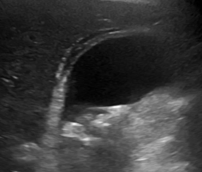
- Transabdominal ultrasound is first line. Look for gallstones, a gallbladder wall thicker than 3 mm, pericholecystic fluid, gallbladder distension above about 4 cm in transverse diameter, an impacted stone at the neck, and a sonographic Murphy sign where maximal tenderness is elicited directly over the gallbladder under the probe
- Common bile duct diameter should be reported. A duct above 6 mm raises the possibility of choledocholithiasis.
- CT of the abdomen - not the first-line test for stones, which are usually radiolucent, but valuable where the diagnosis is uncertain or a complication such as perforation, emphysematous cholecystitis or an abscess is suspected
- MRCP - where LFTs or duct diameter suggest a common bile duct stone that ultrasound has not visualised
- HIDA cholescintigraphy - the most accurate single test, with failure of the gallbladder to fill indicating cystic duct obstruction. It is rarely needed but is useful in equivocal cases, particularly suspected acalculous disease.
Management
Initial treatment
- Admit, with regular observations and a NEWS2 score
- Nil by mouth initially, with intravenous fluid resuscitation and correction of electrolytes
- Analgesia - a non-steroidal anti-inflammatory such as diclofenac, with intravenous opioid and an antiemetic as required
- Intravenous antibiotics covering enteric Gram-negatives and anaerobes, according to local microbiology policy. Co-amoxiclav is a common first choice, with a cephalosporin plus metronidazole, or gentamicin, as alternatives in penicillin allergy.
- Blood cultures before antibiotics if febrile, and the sepsis six if the sepsis criteria are met
- Venous thromboembolism prophylaxis, and reassess whether anticoagulants need withholding before surgery
- Urgent surgical review and a decision about the timing of cholecystectomy
Timing of cholecystectomy
Laparoscopic cholecystectomy during the same admission, ideally within 72 hours of symptom onset, is the preferred approach. NICE recommends offering early cholecystectomy within a week of diagnosis.2 The rationale is worth understanding:
- In the first few days the inflammation is oedematous, and the planes around Calot triangle can still be opened. After about a week the oedema is replaced by dense fibrosis and neovascularity, which makes dissection harder and bile duct injury more likely.
- Delaying to a planned interval operation exposes the patient to the risk of a further attack, of empyema, and of gallstone pancreatitis while they wait
- Randomised trials and meta-analyses show that early surgery gives a shorter total hospital stay with no increase in complications or conversion to open surgery compared with delayed surgery
- If a patient presents beyond about a week with settling symptoms, most units treat conservatively and plan an interval cholecystectomy at six weeks, because operating in the fibrotic phase is the highest-risk option
When the patient is unfit for surgery
Percutaneous cholecystostomy - a drain placed into the gallbladder under ultrasound or CT guidance - decompresses the obstructed organ and controls sepsis without an anaesthetic. It is used in patients with severe comorbidity, in Grade III disease with organ failure, and in acalculous cholecystitis in intensive care.
It should be understood as a temporising measure rather than a cure. The CHOCOLATE trial randomised high-risk patients to cholecystostomy or laparoscopic cholecystectomy and found more major complications, more reinterventions and more recurrent biliary disease in the cholecystostomy group, which has moved practice towards attempting surgery in more high-risk patients than was previously the case.3 Where a drain is placed, the plan should specify whether interval cholecystectomy will follow.
Managing a concurrent duct stone
If imaging or biochemistry indicates a stone in the common bile duct as well, the duct must be cleared. Options are pre-operative ERCP with sphincterotomy, laparoscopic bile duct exploration at the time of cholecystectomy, or post-operative ERCP. All three are acceptable and the choice is determined by local expertise and availability.
Complications
- Empyema of the gallbladder - the obstructed gallbladder fills with pus, producing a swinging fever, rigors, a tender mass and marked leucocytosis. It requires urgent drainage or cholecystectomy.
- Gangrenous cholecystitis - transmural necrosis, more common in older men, diabetics and acalculous disease, and a strong predictor of perforation
- Perforation - free perforation causes biliary peritonitis, contained perforation forms a pericholecystic abscess
- Emphysematous cholecystitis - gas within the gallbladder wall from gas-forming organisms such as Clostridium and Escherichia coli, strongly associated with diabetes, with a high risk of perforation. Gas in the wall on CT is diagnostic and mandates urgent surgery.
- Cholecystoenteric fistula and gallstone ileus - chronic inflammation erodes into the duodenum, allowing a large stone to pass into the bowel and obstruct at the ileocaecal valve
- Mirizzi syndrome - a stone impacted in Hartmann pouch compresses the common hepatic duct, causing obstructive jaundice without a duct stone
- Chronic cholecystitis - repeated attacks produce a shrunken fibrotic gallbladder with recurrent pain and fat intolerance
- Porcelain gallbladder - diffuse mural calcification following chronic inflammation, historically linked with gallbladder carcinoma and generally an indication for cholecystectomy
- Sepsis and multi-organ failure, particularly in acalculous disease
Red flags
Prognosis
Uncomplicated calculous cholecystitis treated promptly has an excellent prognosis, with mortality well under 1% in fit patients and complete resolution of symptoms after cholecystectomy in the great majority.
Prognosis worsens sharply with three factors: age and comorbidity, delay to treatment, and the absence of stones. Acalculous cholecystitis, occurring as it does in patients who are already critically ill, carries a mortality that may exceed 30%, and much of that reflects the underlying illness rather than the gallbladder itself.
For patients treated conservatively without cholecystectomy, recurrence is common: a substantial minority are readmitted with a further biliary event within a year, and each admission carries a further chance of a complication. That figure is the argument behind the shift towards operating during the index admission, and it is worth being able to state it as the reason rather than simply quoting the 72-hour rule.
References
- Yokoe M, Hata J, Takada T et al. Tokyo Guidelines 2018: diagnostic criteria and severity grading of acute cholecystitis. Journal of Hepato-Biliary-Pancreatic Sciences. 2018. Available here
- NICE CG188. Gallstone disease: diagnosis and management. 2014. Available here
- Loozen CS, van Santvoort HC, van Duijvendijk P et al. Laparoscopic cholecystectomy versus percutaneous catheter drainage for acute cholecystitis in high risk patients (CHOCOLATE): multicentre randomised clinical trial. BMJ. 2018. Available here
This article is written for revision and education. It is not clinical guidance and must not be used to make decisions about the care of a patient. Always check current NICE guidance and local protocols.