
Acute Cholangitis
Key points
- Acute cholangitis: bacterial infection of bile within an obstructed biliary tree. Obstruction plus infection - neither alone produces the syndrome.
- Commonest cause: a common bile duct stone. Malignant strictures, benign strictures and blocked biliary stents account for most of the rest.
- Charcot triad: fever with rigors, jaundice and right upper quadrant pain. All three are present in only about half of patients.
- Reynolds pentad: the triad plus hypotension and confusion, indicating suppurative cholangitis with septic shock.
- Why it is dangerous: raised pressure in an obstructed duct forces bacteria into the hepatic sinusoids, producing bacteraemia within hours.
- Investigations: an obstructive LFT pattern, blood cultures, ultrasound to confirm duct dilatation, and MRCP or EUS where the cause is unclear.
- Immediate treatment: resuscitation, blood cultures and broad-spectrum intravenous antibiotics within one hour, following the sepsis pathway.
- Definitive treatment: biliary decompression, usually ERCP with sphincterotomy and stone extraction or stenting. Timing depends on the Tokyo severity grade.
Introduction
Acute cholangitis, sometimes called ascending cholangitis, is bacterial infection of bile within an obstructed biliary tree. It is one of the genuine emergencies of gastrointestinal surgery, and before endoscopic drainage became available its mortality approached 50%.
Two ingredients are required, and this is the single concept the whole topic rests on: biliary obstruction and bacterial colonisation of bile. Bile is normally sterile and flows freely, and the sphincter of Oddi plus continuous antegrade flow keep duodenal organisms out. Obstruction alone produces jaundice without sepsis. Colonisation alone, as occurs after a sphincterotomy, is usually harmless. Combine them and the pressure within the duct rises above the secretory pressure of the liver, cholangiovenous and cholangiolymphatic reflux occurs, and bacteria are forced directly into the hepatic sinusoids and thence into the systemic circulation.
The clinical consequence is that antibiotics alone do not treat acute cholangitis. They control the bacteraemia while the obstruction remains, and until the biliary tree is drained the infection has a reservoir that antibiotics cannot reach. Recognising that a patient needs decompression, and arranging it promptly, is the decision that determines the outcome.
Causes
| Category | Causes | Notes |
|---|---|---|
| Stones | Choledocholithiasis - stones that have migrated from the gallbladder, or primary duct stones | By far the commonest cause, accounting for around half of cases |
| Benign strictures | Post-surgical (particularly after cholecystectomy or biliary reconstruction), chronic pancreatitis, primary sclerosing cholangitis, IgG4-related disease | Often recurrent, and difficult to clear definitively |
| Malignancy | Cholangiocarcinoma, pancreatic head carcinoma, ampullary carcinoma, gallbladder cancer, hilar nodal disease | Usually produces painless progressive jaundice before infection supervenes |
| Instrumentation | Blocked or migrated biliary stent, previous ERCP, percutaneous transhepatic drain | A blocked stent is a common cause of cholangitis in patients under oncological follow-up |
| Congenital and parasitic | Choledochal cyst, Caroli disease, Ascaris lumbricoides, Clonorchis sinensis, liver flukes | Parasitic causes are important worldwide and in returning travellers |
The organisms reflect the enteric flora that have refluxed across the sphincter. The commonest are Escherichia coli, Klebsiella species and Enterococcus, with Enterobacter, Pseudomonas and anaerobes such as Bacteroides fragilis also seen. Patients with indwelling stents or repeated instrumentation are far more likely to grow resistant organisms, which is why the antibiotic choice differs between community-acquired and healthcare-associated cholangitis.
Clinical features
Charcot triad is fever with rigors, jaundice and right upper quadrant pain. It is highly specific but only moderately sensitive: all three components are present in only about half to two thirds of patients, and relying on the full triad delays diagnosis in the rest.
Reynolds pentad adds hypotension and confusion, and describes suppurative cholangitis with septic shock. It is present in a minority but carries a very high mortality, and its recognition should trigger immediate escalation.
- Fever with rigors - true shaking chills are characteristic and reflect intermittent bacteraemia. A patient describing teeth-chattering shivering has bacteraemia until proven otherwise.
- Jaundice, with pale stools, dark urine and pruritus if the obstruction has been present for some days
- Right upper quadrant pain, often less severe than in acute cholecystitis and sometimes absent altogether
- Confusion, drowsiness or a fall in older patients, which may be the only presentation in the frail
- Hypotension, tachycardia and oliguria as sepsis progresses
- Right upper quadrant tenderness on examination, usually without the marked guarding of cholecystitis. Murphy sign may be positive if the gallbladder is also inflamed.
Differential diagnosis
The combination of fever, jaundice and abdominal pain has a short differential, and most of the alternatives are also serious.
- Acute cholecystitis - fever and right upper quadrant pain with a positive Murphy sign, but bilirubin normal or only mildly raised and the duct not dilated
- Acute pancreatitis, particularly gallstone pancreatitis - epigastric pain radiating to the back with a markedly raised amylase or lipase. It frequently coexists with cholangitis rather than replacing it as a diagnosis.
- Liver abscess - swinging fever, right upper quadrant pain and tenderness, with a focal lesion on ultrasound or CT. Pyogenic abscesses are often themselves a complication of biliary sepsis.
- Acute viral or drug-induced hepatitis - a hepatitic rather than cholestatic pattern, with transaminases in the thousands and a non-dilated biliary tree
- Sepsis of another source with jaundice - severe sepsis from any cause produces a cholestatic jaundice, so a septic patient with a raised bilirubin does not necessarily have a biliary source
- Ascending infection in an obstructed but non-infected duct - malignant obstruction presents with painless jaundice and no fever until infection supervenes, often after instrumentation
- Right lower lobe pneumonia and subphrenic abscess - fever with referred upper abdominal pain, without jaundice or duct dilatation
- Haemolysis - unconjugated hyperbilirubinaemia with anaemia and a raised reticulocyte count, and a normal ALP
The practical way through is the pattern of liver biochemistry combined with the duct diameter on ultrasound. A cholestatic pattern with a dilated duct in a febrile patient is cholangitis until the duct is imaged properly; a hepatitic pattern with a normal duct is not.
Investigations
Blood tests
- FBC - neutrophil leucocytosis, or occasionally leucopenia in severe sepsis
- LFTs - a cholestatic pattern with raised ALP and gamma-GT out of proportion to the transaminases, and a raised conjugated bilirubin. A sharp transient rise in ALT can occur as a stone passes.
- CRP - markedly raised
- U&Es and creatinine - acute kidney injury is common and is one of the organ dysfunctions defining severe disease
- Clotting screen - prolonged prothrombin time from obstructive jaundice with vitamin K malabsorption, which must be corrected before any intervention
- Venous or arterial gas with lactate, and blood cultures before antibiotics, which are positive in around half
- Amylase or lipase - concurrent gallstone pancreatitis is common and changes management
Imaging
- Transabdominal ultrasound first, to confirm biliary dilatation and look for gallstones. It is sensitive for duct dilatation but detects only around half of common bile duct stones, because the distal duct is often obscured by duodenal gas. A normal-calibre duct does not exclude cholangitis, particularly early or in primary sclerosing cholangitis.
- MRCP - the non-invasive gold standard for demonstrating duct stones and defining the level and nature of an obstruction, where the patient is stable enough to wait
- Endoscopic ultrasound - equally accurate and better for small distal stones, and can be combined with ERCP in the same session
- CT of the abdomen - useful for identifying a malignant cause, liver abscesses, and complications, and is often the first cross-sectional imaging obtained in a septic patient
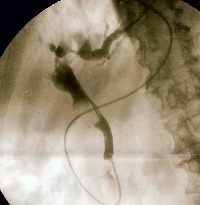
- ERCP - now principally therapeutic. It is both the confirmatory test and the treatment, and should not be used purely diagnostically because of its complication rate.1
Management
Immediate resuscitation
- A to E assessment with continuous monitoring and a NEWS2 score
- Blood cultures, then broad-spectrum intravenous antibiotics within one hour if the sepsis criteria are met, following local policy. Typical regimens cover enteric Gram-negatives and anaerobes - for example piperacillin-tazobactam, or a cephalosporin with metronidazole, with a carbapenem reserved for healthcare-associated or resistant infection.3
- Intravenous fluid resuscitation with balanced crystalloid, guided by lactate and urine output
- Nil by mouth in anticipation of ERCP, with analgesia and antiemetics
- Urinary catheter and hourly urine output
- Correct coagulopathy with vitamin K, and fresh frozen plasma or prothrombin complex concentrate if intervention is imminent and the INR is prolonged
- Early critical care referral in Grade III disease, and early discussion with gastroenterology or hepatobiliary surgery about drainage
Biliary decompression
This is the definitive treatment, and the timing is determined by severity:
| Grade | Definition | Approach |
|---|---|---|
| Grade I (mild) | No organ dysfunction, responds to initial treatment | Antibiotics and supportive care, with elective drainage if there is no response within 24 hours or a stone is confirmed |
| Grade II (moderate) | No organ dysfunction but risk features present | Early biliary drainage, typically within 24 to 48 hours |
| Grade III (severe) | Organ dysfunction - cardiovascular, neurological, respiratory, renal, hepatic or haematological | Organ support plus urgent drainage as soon as the patient can be stabilised, with the priority on decompression rather than on complete stone clearance |
- ERCP with sphincterotomy is the first-line method, allowing stone extraction with a basket or balloon, and stent placement where the duct cannot be cleared or the obstruction is malignant. In the sickest patients the aim is simply to place a stent and drain the duct, deferring definitive clearance to a second procedure.
- Percutaneous transhepatic cholangiography and drainage (PTC) is used where ERCP fails, is not technically possible (for example after Roux-en-Y reconstruction or gastric bypass), or is not available
- Endoscopic ultrasound-guided biliary drainage is an increasingly used alternative in specialist centres
- Open or laparoscopic bile duct exploration is now rarely needed as a primary emergency procedure, but remains an option when endoscopic and percutaneous routes fail
After the acute episode
- Laparoscopic cholecystectomy should follow in patients whose cholangitis was caused by gallstones, ideally during the same admission or soon afterwards, since leaving the gallbladder in place carries a substantial risk of recurrent biliary events
- Complete duct clearance must be confirmed, with a repeat ERCP or MRCP if a stent was placed as a temporary measure
- Stent surveillance and planned exchange where a stent has been left in situ, since blocked stents cause recurrent cholangitis
- Investigation and staging of a malignant cause, with referral to the hepatopancreatobiliary multidisciplinary team
- Address the underlying condition in primary sclerosing cholangitis or a benign stricture, with gastroenterology or specialist surgical input
Complications
- Septic shock and multi-organ failure, the principal cause of death
- Acute kidney injury, from sepsis, hypovolaemia and the direct tubular effect of conjugated bilirubin
- Hepatic abscesses, often multiple and small, from ascending infection through the biliary radicles
- Acute pancreatitis, either from the impacted stone itself or as a complication of ERCP
- Coagulopathy and bleeding, from vitamin K malabsorption and from disseminated intravascular coagulation in severe sepsis
- Complications of ERCP - post-ERCP pancreatitis in around 3 to 5%, bleeding after sphincterotomy, duodenal or duct perforation, and worsening of sepsis if the duct is instrumented but not drained
- Secondary biliary cirrhosis with repeated or chronic obstruction
- Recurrence, which is common where the gallbladder is left in place or a stent blocks
Red flags
Prognosis
With prompt antibiotics and timely biliary drainage, most patients with mild or moderate cholangitis recover fully, and overall mortality in contemporary series is around 5 to 10%. That figure is a dramatic improvement on the pre-endoscopic era and is attributable almost entirely to the availability of ERCP.
Severe Grade III disease is a different matter. Mortality in patients with organ dysfunction remains substantial, and the strongest predictors of death are the number of failing organ systems, older age, malignant obstruction, and - critically - delay to biliary drainage. Observational data consistently show worse outcomes when drainage is delayed beyond 24 to 48 hours in patients who need it.
The message to carry from the topic is therefore about sequencing rather than about drug choice. Resuscitate and give antibiotics immediately, but treat the antibiotics as a holding measure. The clock that matters is the one running until the biliary tree is decompressed.
References
- Williams E, Beckingham I, El Sayed G et al. Updated guideline on the management of common bile duct stones. Gut. 2017. Available here
- Kiriyama S, Kozaka K, Takada T et al. Tokyo Guidelines 2018: diagnostic criteria and severity grading of acute cholangitis. Journal of Hepato-Biliary-Pancreatic Sciences. 2018. Available here
- NICE NG51. Sepsis: recognition, diagnosis and early management. 2016, updated 2024. Available here
This article is written for revision and education. It is not clinical guidance and must not be used to make decisions about the care of a patient. Always check current NICE guidance and local protocols.