
Gallstones and Biliary Colic
Key points
- Prevalence: gallstones are present in 10 to 15% of UK adults. Around 80% never cause symptoms, and only 1 to 4% of silent stones become symptomatic each year.
- Stone types: cholesterol and mixed stones account for about 80%. Black pigment stones follow chronic haemolysis; brown pigment stones follow biliary infection and stasis.
- Admirand triangle: stones form when bile is supersaturated with cholesterol, nucleation is promoted, and the gallbladder empties poorly.
- Biliary colic: transient obstruction of the gallbladder neck or cystic duct by a stone. Severe constant right upper quadrant pain lasting 30 minutes to a few hours, with no fever and no peritonism.
- The key distinction: biliary colic has no inflammatory response. Fever, a raised CRP or a positive Murphy sign means acute cholecystitis, not colic.
- First-line imaging: transabdominal ultrasound, which is over 95% sensitive for gallbladder stones and also measures wall thickness and duct diameter.
- Duct stones: suspect if the bile duct is dilated above 6 mm or the LFTs show an obstructive pattern. MRCP is the non-invasive confirmatory test.
- Treatment: laparoscopic cholecystectomy is offered to people with symptomatic gallstones. Asymptomatic stones found incidentally are left alone.
Introduction
Gallstones are among the commonest findings in abdominal imaging, present in roughly 10 to 15% of UK adults and rising steeply with age and body mass index.3 The great majority cause nothing at all, and the central clinical skill in this topic is deciding whether the stones seen on a scan are actually responsible for the patient's symptoms.
That question matters because cholecystectomy is one of the commonest elective operations in the NHS, with around 70,000 performed each year, and because operating on a patient whose pain has another cause leaves them with the pain and the risks of surgery. A patient with typical biliary colic and stones on ultrasound should be offered an operation; a patient with vague chronic dyspepsia and incidental stones usually should not.
The other half of the topic is the spectrum of complications. Gallstones cause disease by lodging somewhere, and where they lodge determines the illness - the gallbladder neck causes colic and cholecystitis, the common bile duct causes jaundice and cholangitis, and the ampulla causes pancreatitis.
Types of stone and how they form
Bile is a solution held in delicate balance. Cholesterol is insoluble in water and is kept in solution by micelles formed with bile salts and phospholipids. When that balance fails, cholesterol crystallises.
Admirand triangle describes the three conditions that must combine for cholesterol stones to form:
- Supersaturation of bile with cholesterol, either because too much cholesterol is secreted (obesity, high-calorie diet, oestrogen) or because too few bile salts are available (terminal ileal disease or resection, which interrupts the enterohepatic circulation)
- Promotion of nucleation by mucin glycoproteins, calcium and bilirubin, which act as a scaffold for crystals to grow on
- Gallbladder hypomotility and stasis, which allows crystals time to aggregate. This is why pregnancy, prolonged fasting, total parenteral nutrition, rapid weight loss and octreotide all predispose to stones.
| Type | Share | Composition | Associations | Radio-opacity |
|---|---|---|---|---|
| Cholesterol and mixed | About 80% | Predominantly cholesterol, often with calcium salts and pigment | Obesity, female sex, increasing age, pregnancy, rapid weight loss, terminal ileal disease, oestrogen and fibrates | Usually radiolucent |
| Black pigment | About 15% | Calcium bilirubinate polymers | Chronic haemolysis - sickle cell disease, hereditary spherocytosis, thalassaemia, mechanical heart valves - and cirrhosis | Often radio-opaque |
| Brown pigment | About 5% | Calcium bilirubinate with bacterial debris and fatty acids | Biliary stasis and infection, biliary strictures, parasitic infestation. Often form within the ducts rather than the gallbladder. | Usually radiolucent |
Only around 10 to 15% of gallstones are radio-opaque, which is why a plain abdominal radiograph is useless for excluding them and ultrasound is the test of choice.
The spectrum of gallstone disease
Learn this as a map of where the stone is, rather than as a list of separate conditions. Almost every complication follows from a stone obstructing one of three places.
| Condition | Where the stone is | Key features |
|---|---|---|
| Biliary colic | Temporarily impacted at the gallbladder neck or in the cystic duct, then falls back | Severe constant right upper quadrant pain for 30 minutes to a few hours. No fever, normal inflammatory markers, normal LFTs. |
| Acute cholecystitis | Persistently impacted in the cystic duct | Continuous pain over 6 hours, fever, positive Murphy sign, raised white cell count and CRP |
| Chronic cholecystitis | Repeated episodes of impaction | Recurrent right upper quadrant pain, fat intolerance, a thick-walled contracted gallbladder on ultrasound |
| Choledocholithiasis | In the common bile duct | Obstructive jaundice, pale stools, dark urine, pruritus, raised ALP and bilirubin, dilated duct |
| Ascending cholangitis | Obstructing the common bile duct with infection above it | Charcot triad of fever, jaundice and right upper quadrant pain. A surgical emergency. |
| Gallstone pancreatitis | Impacted at the ampulla of Vater | Severe epigastric pain radiating to the back, amylase or lipase more than three times normal |
| Mirizzi syndrome | Impacted in the cystic duct or Hartmann pouch, compressing the common hepatic duct from outside | Obstructive jaundice without a stone in the duct itself |
| Gallstone ileus | Eroded through a cholecystoduodenal fistula and impacted at the ileocaecal valve | Small bowel obstruction with Rigler triad on CT - pneumobilia, obstruction and an ectopic gallstone |
| Gallbladder carcinoma | Chronic irritation, classically with a porcelain gallbladder | Rare, usually advanced at diagnosis, and a reason to remove a calcified gallbladder |
Clinical features of biliary colic
Despite its name, biliary colic is not colicky. The gallbladder contracts against an obstructed neck and stays contracted, so the pain builds to a plateau and then eases as the stone falls back, rather than coming in waves.
- Site - right upper quadrant or epigastrium, often described as deep and boring
- Character - severe and constant, rising to a plateau over about 15 minutes and lasting from 30 minutes to several hours before easing. Pain persisting beyond six hours suggests cholecystitis.
- Radiation - to the right scapula or interscapular region, through the shared T5 to T9 innervation
- Precipitants - classically a fatty meal, which stimulates cholecystokinin release and gallbladder contraction, though many episodes have no obvious trigger and nocturnal onset is common
- Associated features - nausea, vomiting and restlessness. The patient may pace or roll about, in contrast to peritonitis.
- Absent features - there should be no fever, no rigors, no jaundice and no peritonism. Their presence means the diagnosis is something further along the spectrum.
Examination
Between attacks the examination is normal. During an attack there is right upper quadrant tenderness without guarding, and Murphy sign should be negative - if it is positive, the gallbladder wall is inflamed and the diagnosis is cholecystitis. Check specifically for jaundice, scleral icterus and scratch marks, and examine for a palpable gallbladder.
Differential diagnosis
- Acute cholecystitis - the same pain, but lasting longer, with fever and a positive Murphy sign
- Peptic ulcer disease and gastro-oesophageal reflux - burning epigastric pain related to meals and posture, often responding to acid suppression
- Acute pancreatitis - epigastric pain radiating to the back, relieved by sitting forward, with a raised amylase or lipase
- Renal or ureteric colic - loin to groin pain with haematuria and genuine colic
- Right lower lobe pneumonia - pleuritic pain and referred upper abdominal tenderness
- Inferior myocardial infarction - epigastric pain with nausea and sweating; always consider an ECG
- Irritable bowel syndrome and functional dyspepsia - the important differential in a patient with incidental stones and atypical symptoms
- Sphincter of Oddi dysfunction - biliary-type pain persisting after cholecystectomy
Investigations
Blood tests
- FBC and CRP - normal in uncomplicated biliary colic. A leucocytosis or raised CRP shifts the diagnosis to cholecystitis or cholangitis.
- LFTs - normal in biliary colic. A rise in ALP and bilirubin with a proportionally smaller rise in transaminases indicates an obstructive picture and raises suspicion of a duct stone. A transient sharp rise in ALT can occur as a stone passes the ampulla.
- Amylase or lipase - to exclude gallstone pancreatitis, which is part of the same disease and must not be missed
- U&Es, clotting and group and save where surgery is anticipated
- Blood cultures if febrile
Imaging
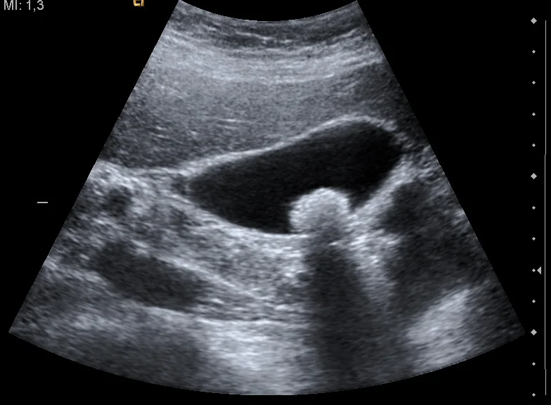
- Transabdominal ultrasound is first line and should be requested in anyone with suspected gallstone disease. It is over 95% sensitive for stones in the gallbladder, and it also reports gallbladder wall thickness (above 3 mm suggests inflammation), pericholecystic fluid, a sonographic Murphy sign, and the calibre of the common bile duct.1
- Common bile duct diameter - normally up to about 6 mm, rising by roughly 1 mm per decade after the age of 60, and up to 10 mm after cholecystectomy. A dilated duct on ultrasound is the main trigger for further imaging.
- MRCP - the non-invasive test of choice for suspected common bile duct stones where ultrasound has not visualised them. It is highly sensitive and avoids the risks of ERCP.2
- Endoscopic ultrasound - an alternative to MRCP with comparable accuracy, useful where MRCP is contraindicated or for small distal stones
- ERCP - now primarily a therapeutic procedure rather than a diagnostic one, used to remove duct stones by sphincterotomy and basket or balloon extraction
- CT - poor at detecting gallstones because most are radiolucent, but useful for complications such as perforation, gallstone ileus, abscess or a suspected malignancy
- HIDA (cholescintigraphy) - reserved for equivocal cases, where failure of the gallbladder to fill indicates cystic duct obstruction
Management
The acute episode
- Analgesia - a non-steroidal anti-inflammatory such as intramuscular diclofenac is effective and reduces progression to cholecystitis; add intravenous opioid if needed, with an antiemetic
- Antiemetics and intravenous fluid if vomiting
- Nil by mouth during severe pain, then a low-fat diet
- No antibiotics are needed for uncomplicated biliary colic, since there is no infection
- Reassess for complications - fever, jaundice, persistent pain beyond six hours or a rising CRP all mean the diagnosis has moved on
Definitive treatment
Laparoscopic cholecystectomy should be offered to people with symptomatic gallstones, because the natural history of symptomatic disease is recurrent attacks with a meaningful annual risk of a serious complication.1 Around 90% of cases are completed laparoscopically, with conversion to open surgery for adhesions, difficult anatomy or bleeding.
- The operation is performed as a day case in fit patients, with return to normal activity in one to two weeks
- Critical view of safety dissection within Calot triangle - bounded by the cystic duct, the common hepatic duct and the inferior border of the liver - is the technique used to avoid bile duct injury, and the cystic artery is typically found within it4
- In acute cholecystitis, surgery is best performed within the same admission, ideally within 72 hours of symptom onset, rather than deferred
- Where the duct also contains stones, options are pre-operative ERCP, intra-operative bile duct exploration, or post-operative ERCP, decided locally
Asymptomatic gallstones
Incidental gallstones in a patient with no biliary symptoms need no treatment and no follow-up. The annual risk of developing symptoms is low, most patients who do develop them present with colic rather than a complication, and prophylactic surgery does not benefit the population as a whole. Explaining this clearly is often the whole consultation.
There are a few situations where cholecystectomy is considered despite the absence of symptoms:
- Porcelain gallbladder - a diffusely calcified wall, historically associated with gallbladder carcinoma
- Gallbladder polyps above 10 mm, or smaller polyps that are growing or occur with primary sclerosing cholangitis
- Very large stones, conventionally above 3 cm, on the grounds of a higher cancer risk
- Sickle cell disease and other chronic haemolytic states, where a biliary complication is difficult to distinguish from a crisis
- Patients undergoing another upper abdominal operation, or awaiting organ transplantation, where a later biliary emergency would be particularly hazardous
Complications
Of the disease
These are the entities set out in the spectrum table above: acute and chronic cholecystitis, empyema and gangrene of the gallbladder, perforation, choledocholithiasis, ascending cholangitis, gallstone pancreatitis, Mirizzi syndrome, gallstone ileus and gallbladder carcinoma. Around a fifth of patients with symptomatic stones will develop one of these if left untreated over several years.
Of cholecystectomy
- Bile duct injury - uncommon but serious, as above
- Bile leak from the cystic duct stump or an accessory duct of Luschka, presenting with pain, ileus and a collection, and usually treated by ERCP with stenting plus drainage
- Retained common bile duct stone, presenting with jaundice or pancreatitis after surgery and treated by ERCP
- Bleeding, wound infection and port-site hernia
- Injury to bowel or vessels at port insertion, and conversion to open surgery
- Post-cholecystectomy syndrome - persistent right upper quadrant pain, dyspepsia or diarrhoea after surgery, affecting up to 10 to 15%. Causes include a retained stone, sphincter of Oddi dysfunction, bile acid malabsorption causing diarrhoea, and, commonly, a pre-existing functional disorder that was never due to the stones.
Red flags
Prognosis
The prognosis of asymptomatic gallstones is excellent. Most people carry them for life without ever knowing, and the annual conversion rate to symptomatic disease is only a few per cent, falling further with time since discovery.
Once symptoms begin, the picture changes. Around two thirds of patients who have had one episode of biliary colic have another within two years, and a significant minority present with a complication rather than a further attack of pain. This asymmetry - low risk while silent, appreciable risk once symptomatic - is the entire logic of the NICE recommendation to leave incidental stones alone and to offer surgery once they declare themselves.
Laparoscopic cholecystectomy resolves symptoms in the large majority, with a low complication rate and no long-term consequence from the absence of a gallbladder, since the bile duct dilates modestly and bile continues to reach the duodenum. The important caveat is patient selection: the more atypical the symptoms, the less likely the operation is to help, and the higher the chance of joining the 10 to 15% with post-cholecystectomy syndrome.
References
- NICE CG188. Gallstone disease: diagnosis and management. 2014. Available here
- Williams E, Beckingham I, El Sayed G et al. Updated guideline on the management of common bile duct stones. Gut. 2017. Available here
- NICE Clinical Knowledge Summaries. Gallstones. Available here
- Wakabayashi G, Iwashita Y, Hibi T et al. Tokyo Guidelines 2018: surgical management of acute cholecystitis and safe steps in laparoscopic cholecystectomy. Journal of Hepato-Biliary-Pancreatic Sciences. 2018. Available here
This article is written for revision and education. It is not clinical guidance and must not be used to make decisions about the care of a patient. Always check current NICE guidance and local protocols.