
Hiatus Hernia
Key points
- Hiatus hernia: protrusion of part of the stomach through the oesophageal hiatus of the diaphragm into the thorax.
- Type I - sliding: about 95% of cases. The gastro-oesophageal junction itself moves above the diaphragm, so the anti-reflux mechanism fails and the problem is reflux.
- Type II - rolling: the fundus herniates alongside a junction that stays below the diaphragm. Reflux is uncommon, but obstruction, volvulus and strangulation are not.
- Types III and IV: mixed sliding and paraoesophageal, and herniation of other viscera such as colon or spleen. Both behave like type II.
- Anti-reflux mechanism: the lower oesophageal sphincter, the crural diaphragm acting as an external pincer, the acute angle of His, and the intra-abdominal segment of oesophagus.
- Diagnosis: usually found incidentally at endoscopy. A barium swallow best demonstrates the anatomy of a paraoesophageal hernia.
- Medical treatment: weight loss, smaller meals, avoiding late eating, raising the head of the bed, and a proton pump inhibitor for reflux symptoms.
- Surgery: laparoscopic hiatal repair with fundoplication, for refractory reflux, intolerance of long-term acid suppression, or a symptomatic paraoesophageal hernia.
Introduction
A hiatus hernia is herniation of part of the stomach through the oesophageal hiatus of the diaphragm into the posterior mediastinum. It is extremely common, found in perhaps a third of people over 50, and in most of them it causes nothing at all. Its clinical importance is therefore not that it exists but that it is the anatomical basis of most gastro-oesophageal reflux disease, and that one uncommon variant can kill.
The confusion students have with this topic comes from treating it as a single condition. It is better understood as two very different diseases that happen to share a hole in the diaphragm. The sliding hernia is a chronic problem of reflux and its long-term mucosal consequences. The paraoesophageal hernia is a mechanical problem that can obstruct, twist and strangulate.
Anatomy and the anti-reflux mechanism
The oesophagus passes through the diaphragm at the level of T10, through a sling formed mainly by fibres of the right crus. It is anchored there by the phreno-oesophageal ligament, a condensation of fascia that permits the few centimetres of longitudinal movement needed for swallowing while preventing the stomach from ascending.
Continence at the gastro-oesophageal junction is not the work of a single sphincter but of four mechanisms acting together. A hiatus hernia disrupts all of them at once, which is why reflux follows.
- The lower oesophageal sphincter - a physiological rather than anatomical sphincter, a zone of tonically contracted smooth muscle in the distal 3 to 4 cm of oesophagus
- The crural diaphragm, which acts as an external pincer around the oesophagus and contracts during inspiration and straining, exactly when intra-abdominal pressure rises. Once the junction has slid upwards, this pinch acts on the wrong place.
- The angle of His, the acute angle at which the oesophagus meets the gastric fundus. Gastric distension pulls the fundus against the oesophageal opening, creating a flap valve. Herniation obliterates the angle.
- The intra-abdominal segment of oesophagus, normally 2 to 4 cm long, which is compressed by intra-abdominal pressure so that a rise in that pressure closes rather than opens it. In a sliding hernia this segment is lost into the chest.
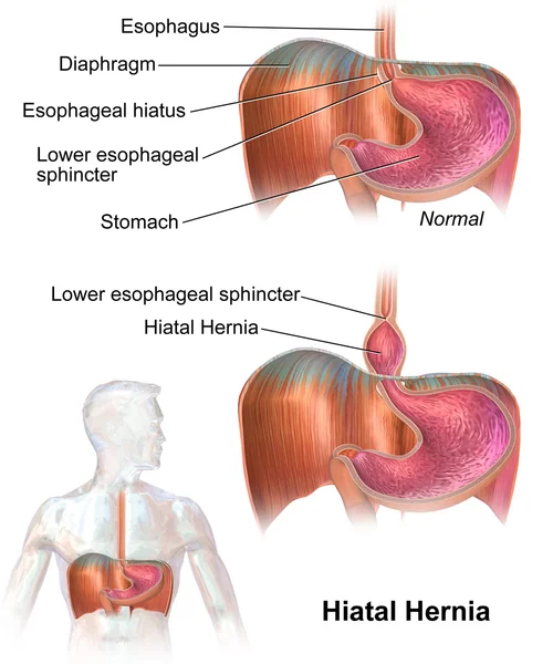
Classification
| Type | Name | Anatomy | Dominant problem |
|---|---|---|---|
| I | Sliding | The gastro-oesophageal junction and a portion of the cardia slide upwards through the hiatus into the chest | Reflux, oesophagitis, stricture and Barrett oesophagus |
| II | Rolling or paraoesophageal | The gastric fundus herniates alongside the oesophagus while the junction remains below the diaphragm | Mechanical - obstruction, incarceration, volvulus, strangulation, bleeding from Cameron lesions |
| III | Mixed | Both the junction and the fundus are displaced above the diaphragm | Mechanical and refluxive combined; behaves like type II |
| IV | Complex | Another organ - colon, spleen, pancreas or small bowel - is also in the sac | Mechanical, with a large defect and often a large hernia sac |
Around 95% of hiatus hernias are type I. Types II to IV together are often grouped as paraoesophageal hernias, and it is types III and IV rather than pure type II that make up most of them in practice. The distinction matters because paraoesophageal hernias are the ones considered for repair on anatomical grounds rather than for symptom control alone.
Risk factors
- Increasing age, through laxity of the phreno-oesophageal ligament and widening of the hiatus
- Obesity, which raises the abdominal-to-thoracic pressure gradient and is the strongest modifiable factor
- Pregnancy, both from raised intra-abdominal pressure and from progesterone-mediated relaxation of the lower oesophageal sphincter
- Chronically raised intra-abdominal pressure - chronic cough, constipation and straining, ascites, heavy lifting
- Connective tissue disorders such as Marfan and Ehlers-Danlos syndromes
- Previous surgery at the hiatus, including previous fundoplication or oesophagectomy
- Kyphoscoliosis and other skeletal deformity, which distorts the hiatus
Factors that worsen reflux without causing the hernia itself include smoking, alcohol, caffeine, chocolate, fatty meals, and drugs that relax the lower oesophageal sphincter such as nitrates, calcium channel blockers, anticholinergics and theophylline.
Clinical features
Sliding hernia
The symptoms are those of gastro-oesophageal reflux disease:
- Heartburn - retrosternal burning, worse after meals, on bending forwards and on lying flat
- Acid regurgitation into the throat or mouth, particularly at night
- Waterbrash - sudden filling of the mouth with saliva
- Nocturnal cough, hoarseness, sore throat and worsening asthma from microaspiration, the so-called extra-oesophageal manifestations
- Chest pain that can closely mimic angina and is a recognised cause of unnecessary cardiac investigation
- Dysphagia, which in this context suggests a peptic stricture and must be investigated rather than attributed to the hernia
Paraoesophageal hernia
Many are asymptomatic and found incidentally on a chest radiograph as a retrocardiac gas shadow, sometimes with a fluid level. When symptoms occur they are mechanical rather than refluxive:
- Postprandial fullness, early satiety and epigastric or chest discomfort after meals
- Dysphagia, from angulation at the hiatus
- Breathlessness where a large hernia compresses the lung, and palpitations from cardiac compression
- Iron deficiency anaemia from Cameron lesions - linear erosions on the gastric folds where they cross the diaphragm, caused by mechanical trauma and ischaemia. These are a genuinely important and easily missed cause of chronic occult blood loss.
- Recurrent chest infections from aspiration
- Acute severe symptoms if the hernia incarcerates or the stomach twists
Investigations
- Upper gastrointestinal endoscopy is the usual test. It confirms the hernia by measuring the distance from the diaphragmatic pinch to the squamocolumnar junction, grades any oesophagitis using the Los Angeles classification, identifies a peptic stricture, Cameron lesions or Barrett oesophagus, and allows biopsy to exclude malignancy.
- Barium swallow remains the best test for defining the anatomy of a large paraoesophageal hernia, showing how much stomach is in the chest and whether the junction is displaced. It is often requested before surgery.
- Chest radiograph may show a retrocardiac soft tissue shadow with an air-fluid level, which is sometimes the first clue in an asymptomatic patient
- CT of the chest and abdomen - defines large or complex hernias, identifies other herniated viscera, and is the investigation in suspected incarceration or volvulus
- Oesophageal manometry - performed before anti-reflux surgery to exclude a major motility disorder such as achalasia, which would be made worse by a fundoplication
- 24-hour ambulatory pH or pH-impedance monitoring - to confirm pathological acid exposure before surgery, particularly where symptoms have not responded to a proton pump inhibitor
- FBC and haematinics - to detect the iron deficiency anaemia of Cameron lesions
Management
Conservative and medical
Most sliding hernias are managed exactly as gastro-oesophageal reflux disease, and lifestyle measures should not be skipped over as a formality - weight loss in particular has a genuine effect.
- Weight loss where the patient is overweight
- Smaller, more frequent meals, and avoiding eating within three hours of lying down
- Raising the head of the bed by around 15 to 20 cm, using blocks rather than extra pillows, which only flex the neck
- Smoking cessation and reduction of alcohol, caffeine, chocolate and fatty or spicy foods where they are identified triggers
- Review the drug chart for nitrates, calcium channel blockers, anticholinergics, bisphosphonates and NSAIDs
- Antacids and alginates such as Gaviscon for breakthrough symptoms
- A proton pump inhibitor - a full-dose course for four to eight weeks, then stepping down to the lowest dose that controls symptoms. An H2 receptor antagonist is an alternative where a PPI is not tolerated.2
Surgery
The standard operation is laparoscopic hiatal repair with fundoplication: the hernia sac is reduced and excised, the crura are approximated to narrow the hiatus, and the fundus is wrapped around the distal oesophagus to recreate a valve. A Nissen fundoplication is a complete 360 degree wrap; a Toupet (posterior 270 degrees) or Dor (anterior 180 degrees) partial wrap causes less dysphagia and is preferred where oesophageal motility is impaired.
| Situation | Approach |
|---|---|
| Sliding hernia, symptoms controlled on a PPI | Continue medical treatment; surgery is not indicated |
| Sliding hernia, symptoms refractory to optimal medical therapy with proven acid reflux | Consider laparoscopic fundoplication after manometry and pH studies |
| Sliding hernia, patient unwilling to take lifelong acid suppression, or with volume regurgitation not helped by acid suppression | Reasonable indication for surgery after full workup |
| Symptomatic paraoesophageal hernia | Elective laparoscopic repair, with sac excision, crural repair and usually a fundoplication or gastropexy |
| Asymptomatic paraoesophageal hernia in a fit patient | Individualised. Routine repair of all asymptomatic hernias is no longer recommended, as the annual risk of an acute event is low and elective repair carries its own risk. |
| Incarceration, obstruction, gastric volvulus or ischaemia | Emergency surgery |
Complications
Of a sliding hernia, through reflux
- Reflux oesophagitis and ulceration
- Peptic stricture, presenting with progressive dysphagia to solids, and treated by endoscopic dilatation with long-term acid suppression
- Barrett oesophagus - metaplasia of the squamous lining to intestinal-type columnar epithelium, which is premalignant and requires endoscopic surveillance at an interval determined by segment length and dysplasia4
- Oesophageal adenocarcinoma, the endpoint of the metaplasia-dysplasia-carcinoma sequence, though the absolute annual risk from non-dysplastic Barrett is low
- Aspiration pneumonia, chronic cough, laryngitis and dental erosion
- Anaemia from chronic oesophagitis or ulceration
Of a paraoesophageal hernia, through mechanics
- Incarceration and obstruction
- Gastric volvulus with ischaemia, necrosis and perforation
- Bleeding from Cameron lesions, producing chronic iron deficiency anaemia or, less often, overt haemorrhage
- Respiratory compromise from a large intrathoracic stomach
- Perforation into the mediastinum or pleural cavity, which carries a very high mortality
Red flags
Prognosis
The great majority of hiatus hernias are asymptomatic and remain so, and the great majority of symptomatic sliding hernias are controlled by weight loss, lifestyle change and a proton pump inhibitor. Long-term PPI therapy is safe in the vast majority of patients, and the observational associations with fracture, renal disease and infection should be discussed honestly rather than used as a reason to withhold effective treatment from someone with erosive disease.
Laparoscopic fundoplication achieves good symptom control in around 85 to 90% of carefully selected patients at five years, with the caveat that a proportion resume acid suppression over time. Careful selection - proven acid reflux, normal motility, and symptoms that actually respond to a PPI - is the single strongest predictor of a good outcome, which is why the pre-operative workup is not a formality.
Paraoesophageal hernias are the exception to the generally benign picture. The annual risk of an acute event in an asymptomatic large hernia is low, but when it happens the mortality of emergency surgery is many times that of an elective repair. Balancing those two numbers, in a patient who is often elderly and comorbid, is the substance of the decision.
References
- NICE NG12. Suspected cancer: recognition and referral. 2015, updated 2023. Available here
- NICE CG184. Gastro-oesophageal reflux disease and dyspepsia in adults: investigation and management. 2014, updated 2019. Available here
- Society of American Gastrointestinal and Endoscopic Surgeons. Guidelines for the management of hiatal hernia. Available here
- Fitzgerald RC, di Pietro M, Ragunath K et al. British Society of Gastroenterology guidelines on the diagnosis and management of Barrett's oesophagus. Gut. 2014. Available here
This article is written for revision and education. It is not clinical guidance and must not be used to make decisions about the care of a patient. Always check current NICE guidance and local protocols.