
Barrett's Oesophagus: Diagnosis, Surveillance and Management
Key points
- Barrett's oesophagus: replacement of the normal squamous epithelium of the distal oesophagus with metaplastic columnar epithelium, driven by chronic gastro-oesophageal reflux.
- Significance: the main identifiable precursor of oesophageal adenocarcinoma, progressing through a metaplasia-dysplasia-carcinoma sequence.
- Presentation: usually asymptomatic and found incidentally at endoscopy for longstanding GORD symptoms.
- Diagnosis: endoscopic appearance of columnar-lined oesophagus confirmed by biopsy showing intestinal metaplasia.
- Risk factors: chronic GORD, male sex, obesity (central), smoking, older age, and a family history of Barrett's or oesophageal cancer.
- Surveillance: regular endoscopic surveillance with biopsies (Seattle protocol), with interval determined by segment length and grade of dysplasia.
- Management: long-term PPI therapy, and endoscopic therapy (radiofrequency ablation or resection) for dysplasia.
- Prognosis: annual cancer risk is low in non-dysplastic Barrett's, but surveillance detects dysplasia and early cancer while still curable.
Introduction
Barrett's oesophagus is a condition in which the normal squamous epithelium lining the distal oesophagus is replaced by metaplastic columnar epithelium, as an adaptive response to chronic acid and bile reflux.1 It is named after the surgeon Norman Barrett.
It is clinically important because it is the principal identifiable precursor lesion for oesophageal adenocarcinoma, one of the fastest-rising cancers in incidence in the UK.2 It is found in around 1-2% of the general population and up to 10-15% of patients undergoing endoscopy for chronic reflux symptoms.
Pathophysiology
Chronic exposure of the distal oesophageal mucosa to acid and bile from gastro-oesophageal reflux causes repeated injury and repair. Over time, the native squamous epithelium is replaced by a metaplastic columnar epithelium with goblet cells, known as intestinal metaplasia, which is more resistant to acid injury but carries malignant potential.1
Malignant transformation follows a recognised sequence: non-dysplastic Barrett's metaplasia progresses through low-grade dysplasia, then high-grade dysplasia, to invasive adenocarcinoma. This stepwise progression is the rationale for endoscopic surveillance, which aims to detect and treat dysplasia before invasive cancer develops.3
Risk factors
Risk factors overlap substantially with those for GORD and oesophageal adenocarcinoma:2
- Chronic or longstanding gastro-oesophageal reflux disease
- Male sex (around twice the risk of women)
- Central (visceral) obesity, independent of overall BMI
- Smoking
- Older age (typically diagnosed in the 50s-60s)
- White ethnicity
- Family history of Barrett's oesophagus or oesophageal adenocarcinoma
Clinical features
Barrett's oesophagus itself is asymptomatic; it does not have symptoms distinct from the underlying reflux disease. Most cases are found incidentally when a patient undergoes endoscopy for longstanding reflux symptoms, particularly if the patient is over 50 with a long history of heartburn.
Because it is silent, screening is not recommended for the general population, but endoscopy should be considered in patients with chronic GORD symptoms plus additional risk factors (male sex, over 50, obesity, smoking, family history).2 New dysphagia or weight loss in a patient with known Barrett's oesophagus raises concern for progression to malignancy and warrants urgent endoscopy.
Investigations
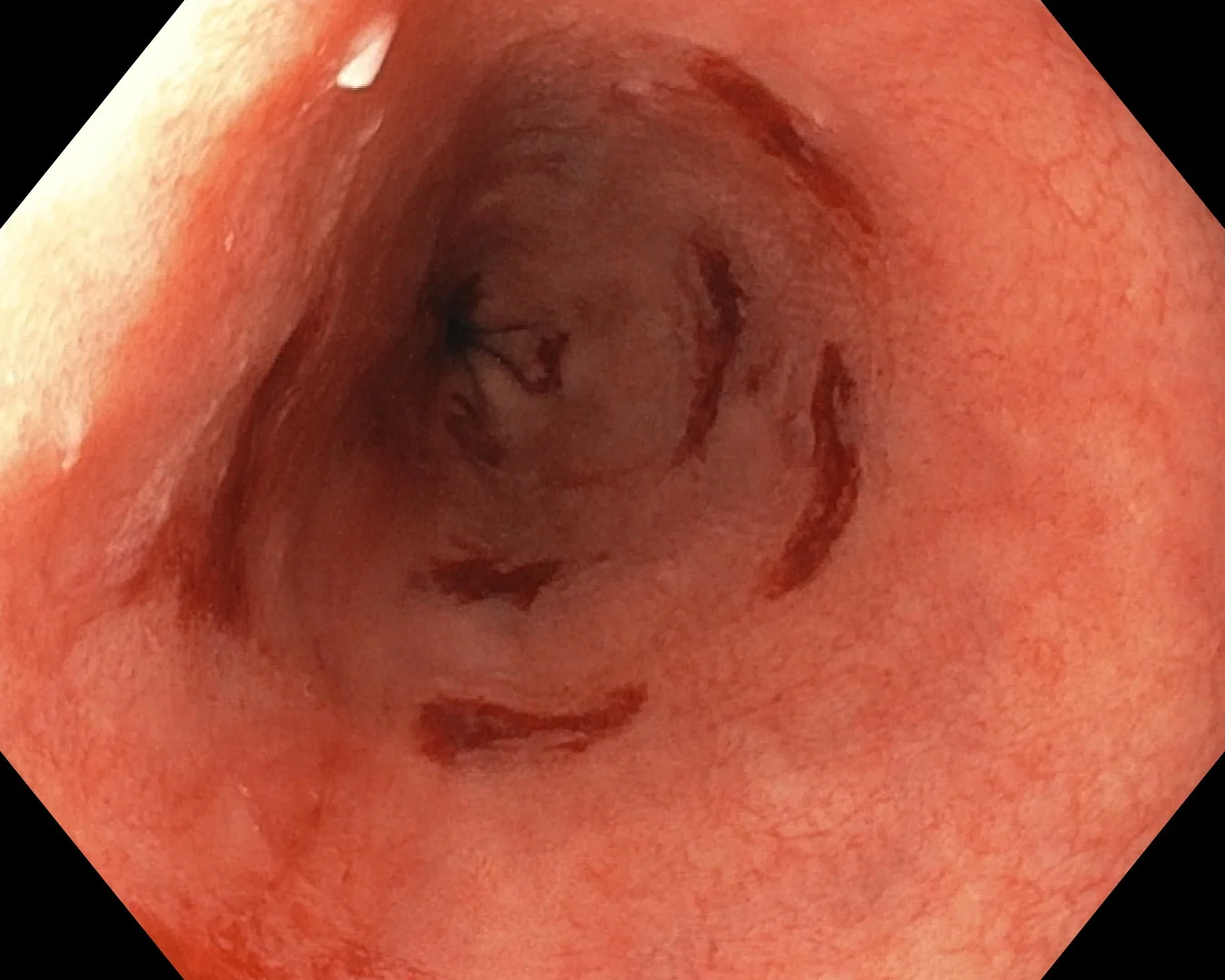
Diagnosis requires upper GI endoscopy with biopsy. At endoscopy, the squamocolumnar junction (Z-line) is seen to have shifted proximally, with salmon-pink columnar mucosa extending above the gastro-oesophageal junction into the tubular oesophagus, replacing the normal pale, glossy squamous mucosa.1
Biopsy protocol
Multiple biopsies are taken systematically from all four quadrants at 1-2 cm intervals along the length of the affected segment (the Seattle protocol), because dysplasia is patchy and can be missed with random or sparse sampling. Histology confirms intestinal metaplasia (goblet cells) and grades any dysplasia present.
Classification
The extent of columnar-lined oesophagus is recorded using the Prague classification, describing the circumferential (C) and maximum (M) extent of metaplasia in centimetres above the gastro-oesophageal junction. This, together with the presence and grade of dysplasia, determines the surveillance interval.
Differential diagnosis
The main diagnostic considerations relate to distinguishing Barrett's oesophagus from other causes of a columnar-appearing distal oesophagus or dysphagia:
- A large hiatus hernia, which can make the gastro-oesophageal junction difficult to define endoscopically
- Reflux oesophagitis without true intestinal metaplasia on biopsy
- Oesophageal or gastric adenocarcinoma, which must be excluded, particularly if there is a visible lesion
- Eosinophilic oesophagitis, in a patient presenting with dysphagia
Management
Management has two strands: controlling reflux, and surveillance or treatment based on the presence of dysplasia.
Acid suppression
Long-term proton pump inhibitor therapy is recommended for all patients with Barrett's oesophagus, both to control reflux symptoms and because reducing acid exposure may reduce progression to dysplasia.4
Surveillance
Patients with non-dysplastic Barrett's oesophagus undergo periodic surveillance endoscopy with Seattle protocol biopsies, typically every 2-5 years depending on segment length, to detect dysplasia early.2
| Finding | Typical approach |
|---|---|
| Non-dysplastic Barrett's | Long-term PPI plus surveillance endoscopy every 2-5 years |
| Indefinite for dysplasia | Optimise acid suppression and repeat endoscopy, often at 6 months |
| Low-grade dysplasia | Endoscopic therapy (RFA) or intensified surveillance, per specialist MDT |
| High-grade dysplasia or intramucosal cancer | Endoscopic resection and/or radiofrequency ablation |
| Invasive adenocarcinoma | Staging, with oesophagectomy or oncological treatment as appropriate |
Endoscopic therapy for dysplasia
Low-grade or high-grade dysplasia is managed endoscopically rather than surgically wherever possible. Endoscopic mucosal resection (EMR) removes visible nodular or irregular areas for staging and treatment, and radiofrequency ablation (RFA) is used to ablate the remaining flat Barrett's segment, reducing progression to cancer. Oesophagectomy is reserved for invasive adenocarcinoma not amenable to endoscopic therapy.3
Complications
The principal complication is progression to oesophageal adenocarcinoma, via low-grade then high-grade dysplasia. Other complications relate to the underlying reflux disease, including peptic stricture and oesophagitis. Endoscopic therapies themselves carry small risks of bleeding, perforation and post-ablation stricture.
Red flags
Prognosis
The annual risk of progression to adenocarcinoma in non-dysplastic Barrett's oesophagus is low, on the order of 0.1-0.3% per year, and most patients never progress.2 The risk rises substantially with high-grade dysplasia. Regular surveillance and, where indicated, endoscopic ablation of dysplastic segments substantially reduce the risk of a patient presenting with advanced, symptomatic cancer, and endoscopically detected early neoplasia has an excellent prognosis after treatment.
References
- NICE Clinical Knowledge Summaries (CKS). Barrett's oesophagus. 2023. Available here
- British Society of Gastroenterology. Guidelines on the diagnosis and management of Barrett's oesophagus. Gut. 2014. Available here
- Fitzgerald RC et al. British Society of Gastroenterology guidelines for surveillance and treatment of Barrett's oesophagus. Gut. 2014. Available here
- NICE CG184. Dyspepsia and gastro-oesophageal reflux disease in adults. 2014 (updated 2019). Available here
- Samir, CC BY-SA 4.0, via Wikimedia Commons. Available here
- Cancer Research UK. Oesophageal cancer risks and causes. Available here
- NHS. Barrett's oesophagus. 2021. Available here
This article is written for revision and education. It is not clinical guidance and must not be used to make decisions about the care of a patient. Always check current NICE guidance and local protocols.