
Peptic Ulcer Disease and Gastritis: Diagnosis and Management
Key points
- Peptic ulcer disease: a break in the mucosa of the stomach or duodenum extending through the muscularis mucosae, most often caused by Helicobacter pylori or NSAIDs.
- Gastritis: inflammation of the gastric mucosa without a discrete mucosal break, sharing the same main causes as peptic ulcer disease.
- Presentation: epigastric pain, classically related to meals; duodenal ulcer pain is relieved by eating, gastric ulcer pain is worsened by eating.
- Causes: H. pylori infection and NSAID use account for the large majority of cases; rarer causes include Zollinger-Ellison syndrome.
- Diagnosis: endoscopy with biopsy of any gastric ulcer to exclude malignancy, plus testing for H. pylori.
- Management: stop causative drugs, eradicate H. pylori if positive, and give a proton pump inhibitor; repeat endoscopy to confirm healing of gastric ulcers.
- Complications: upper GI bleeding, perforation, and gastric outlet obstruction from a pyloric channel or duodenal ulcer.
- Red flags: haematemesis, melaena, weight loss, dysphagia or an epigastric mass need urgent assessment and endoscopy.
Introduction
Peptic ulcer disease (PUD) is a break in the mucosal lining of the stomach or duodenum that extends through the muscularis mucosae, exposing deeper tissue layers to gastric acid and pepsin.1 Gastritis refers to mucosal inflammation without a discrete ulcer, and shares the same principal causes.
Duodenal ulcers are around four times more common than gastric ulcers and typically occur in a younger age group. Both are common presentations in primary care and gastroenterology, and are an important cause of upper GI bleeding.
Aetiology
Peptic ulceration results from an imbalance between mucosal protective factors (mucus and bicarbonate secretion, mucosal blood flow, prostaglandins) and damaging factors (acid, pepsin, H. pylori, NSAIDs).1 Two causes account for the great majority of cases.
Helicobacter pylori
H. pylori is a spiral, urease-producing, Gram-negative bacterium that colonises the gastric mucus layer. It disrupts the mucosal barrier and provokes chronic inflammation, which increases acid secretion in duodenal disease and causes mucosal atrophy in the stomach. It is present in the majority of duodenal ulcers and around 60-70% of gastric ulcers.2
NSAIDs and aspirin
NSAIDs inhibit cyclo-oxygenase (COX-1), reducing protective prostaglandin synthesis and impairing the mucosal defence, independent of H. pylori status. Risk is higher with older age, higher NSAID doses, concurrent corticosteroids or anticoagulants, and a past history of ulcer disease.
Other causes
Less common causes include Zollinger-Ellison syndrome (a gastrin-secreting tumour causing multiple or refractory ulcers), smoking, severe physiological stress (Cushing ulcers in raised intracranial pressure, Curling ulcers in major burns), and, rarely, malignancy presenting as an ulcer.
Clinical features
The cardinal symptom is epigastric pain, often gnawing or burning in character. The relationship to food can help distinguish gastric from duodenal ulcers, although this is not reliable enough to diagnose on history alone:
| Feature | Gastric ulcer | Duodenal ulcer |
|---|---|---|
| Relationship to food | Pain worsened by eating; may cause weight loss and food avoidance | Pain relieved by eating; may recur 2-3 hours later or wake the patient at night |
| Typical age | Older patients | Younger patients |
| Malignancy risk | Small but important risk - always biopsy | Very rarely malignant |
Other features include bloating, early satiety, nausea and dyspepsia. Many patients, particularly those on NSAIDs, are asymptomatic until a complication such as bleeding or perforation occurs.
Differential diagnosis
Epigastric pain has a broad differential:
- GORD: retrosternal burning, worse lying flat, without the discrete ulcer
- Gastric or oesophageal cancer: progressive symptoms, weight loss, dysphagia
- Biliary colic or cholecystitis: right upper quadrant pain, often after fatty food
- Acute pancreatitis: severe epigastric pain radiating to the back, raised lipase/amylase
- Acute coronary syndrome: cardiac chest pain can present as epigastric discomfort, particularly in inferior MI
- Functional (non-ulcer) dyspepsia: symptoms without a structural cause found at endoscopy
Investigations
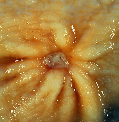
Upper GI endoscopy (OGD) is the investigation of choice when there are red-flag features or the patient is aged 55 or over with new dyspepsia, and confirms the diagnosis directly.3 Every gastric ulcer identified at endoscopy is biopsied from the ulcer margin to exclude malignancy, and a repeat endoscopy is arranged after 6-8 weeks of treatment to confirm healing, because a minority conceal an underlying gastric cancer.
Testing for H. pylori
All patients with peptic ulcer disease should be tested for H. pylori. The urea breath test and stool antigen test are non-invasive and most commonly used; rapid urease testing (CLO test) or histology can be performed on biopsies taken at endoscopy. PPIs should be stopped for around 2 weeks and antibiotics for 4 weeks before testing, as both cause false negatives.2
Other investigations
Full blood count may show iron-deficiency anaemia from chronic blood loss. Fasting gastrin levels are checked if Zollinger-Ellison syndrome is suspected (e.g. multiple or refractory ulcers, ulcers distal to the duodenum).
Management
Management addresses the underlying cause and promotes healing with acid suppression.
General measures
Stop NSAIDs and aspirin where possible; if they cannot be stopped (e.g. essential cardiovascular protection), co-prescribe a PPI for gastroprotection. Advise smoking cessation and reduced alcohol intake, both of which impair ulcer healing.
Helicobacter pylori eradication
Patients testing positive for H. pylori are offered first-line triple therapy: a PPI plus two antibiotics (typically amoxicillin with either clarithromycin or metronidazole) for 7 days.4 In penicillin allergy, clarithromycin and metronidazole are used together. Eradication is confirmed with a urea breath test if symptoms persist or the ulcer was complicated.
Acid suppression
A proton pump inhibitor is given for 4-8 weeks to allow healing, whether or not H. pylori is present. H2-receptor antagonists are an alternative where PPIs are not tolerated.
Refractory or complicated disease
Ulcers that fail to heal despite eradication and acid suppression need re-investigation for persistent H. pylori, ongoing NSAID use, Zollinger-Ellison syndrome, or an underlying malignancy. Surgery is now rarely needed except for complications such as perforation, uncontrolled bleeding, or gastric outlet obstruction.
Complications
- Upper GI bleeding: the most common complication, from erosion into a blood vessel; can present with haematemesis, melaena or haemodynamic instability
- Perforation: presents with sudden severe abdominal pain and peritonism, requiring urgent surgical assessment; erect chest X-ray may show free air under the diaphragm
- Gastric outlet obstruction: from scarring or oedema around a pyloric or duodenal ulcer, causing vomiting and early satiety
- Malignant transformation: a small proportion of apparently benign gastric ulcers are found to be malignant, hence the requirement for biopsy and healing confirmation
Red flags
Prognosis
Most peptic ulcers heal well with acid suppression and, where relevant, H. pylori eradication, and eradication substantially reduces the risk of recurrence.2 Ulcers that continue despite treatment, or that recur after eradication, need re-investigation for an alternative cause. Complicated ulcers, particularly those presenting with major bleeding or perforation, carry significant morbidity and mortality, especially in older patients with comorbidities.
References
- NICE Clinical Knowledge Summaries (CKS). Dyspepsia - unidentified cause. 2023. Available here
- NICE Clinical Knowledge Summaries (CKS). Helicobacter pylori infection. 2023. Available here
- NICE NG12. Suspected cancer: recognition and referral. 2015 (updated 2023). Available here
- BNF. Helicobacter pylori eradication. Available here
- Ed Uthman, MD, Public domain, via Wikimedia Commons. Available here
- NHS. Stomach ulcer. 2023. Available here
- Lanas A, Chan FKL. Peptic ulcer disease. Lancet. 2017. Available here
This article is written for revision and education. It is not clinical guidance and must not be used to make decisions about the care of a patient. Always check current NICE guidance and local protocols.