
Haemorrhoids
Key points
- Haemorrhoids: symptomatic enlargement and downward displacement of the normal anal cushions, which are vascular structures everyone has.
- Position: classically at the 3, 7 and 11 o'clock positions in the lithotomy position, corresponding to the terminal branches of the superior rectal artery.
- Internal versus external: the dentate line divides them. Above it the mucosa has visceral innervation and haemorrhoids are painless; below it the anoderm is somatically innervated and they hurt.
- Classic symptom: painless, bright red rectal bleeding, seen on the paper or dripping into the pan, separate from the stool.
- Grading: first degree do not prolapse, second reduce spontaneously, third need manual reduction, fourth are permanently prolapsed.
- Examination: internal haemorrhoids are not palpable on digital examination. Proctoscopy is required to see them.
- First-line treatment: increase dietary fibre and fluid, avoid straining and prolonged sitting on the toilet, and use a bulk-forming laxative.
- The rule that matters: never attribute rectal bleeding to haemorrhoids without excluding colorectal cancer in anyone with red flags or over the referral age threshold.
Introduction and anatomy
Everyone has anal cushions. They are three specialised vascular structures in the submucosa of the upper anal canal, containing arteriovenous communications supplied by the terminal branches of the superior rectal artery, held in place by a supporting framework of smooth muscle and elastic tissue known as the muscle of Treitz. They contribute perhaps 15 to 20% of resting anal canal pressure and are part of the mechanism of fine continence, sealing the canal against the passage of flatus and liquid.
Haemorrhoids are these normal cushions become symptomatic - engorged, enlarged, and displaced downwards as their supporting tissue degenerates. This is why the older description of haemorrhoids as varicose veins of the anus is wrong: the blood in them is arterial, which explains why haemorrhoidal bleeding is bright red rather than dark.
The cushions lie in the left lateral, right posterior and right anterior positions, described as 3, 7 and 11 o'clock with the patient in the lithotomy position. This is a favourite examination question and follows the branching pattern of the superior rectal artery.
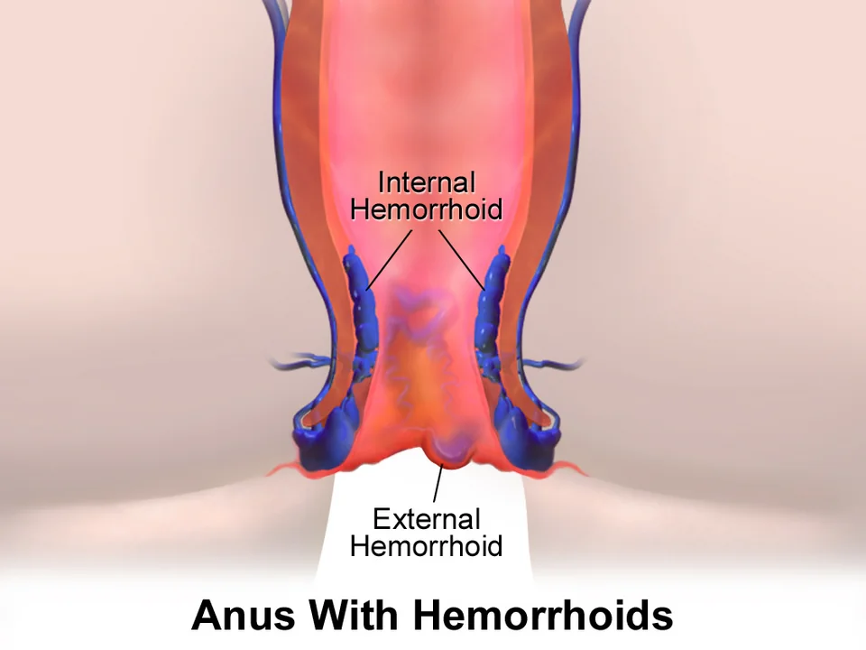
The dentate (pectinate) line is the critical landmark, marking the junction between endoderm-derived upper canal and ectoderm-derived lower canal. It determines everything about how a haemorrhoid presents:
| Above the dentate line (internal) | Below the dentate line (external) | |
|---|---|---|
| Epithelium | Columnar and transitional mucosa | Squamous anoderm and perianal skin |
| Innervation | Visceral, autonomic - insensitive to pain, touch and temperature | Somatic, via the inferior rectal nerve - exquisitely sensitive |
| Arterial supply | Superior rectal artery, from the inferior mesenteric artery | Inferior rectal artery, from the internal pudendal artery |
| Venous drainage | Superior rectal vein to the portal system | Inferior rectal vein to the systemic circulation |
| Lymphatic drainage | Internal iliac nodes | Superficial inguinal nodes |
| Clinical consequence | Painless bleeding and prolapse; can be banded in clinic without anaesthetic | Painful when thrombosed; banding is intolerable without anaesthetic |
Aetiology and risk factors
The mechanism is progressive degeneration of the supporting connective tissue of the cushions combined with repeated shearing forces, allowing them to engorge, slide downwards and eventually prolapse. Anything that raises intra-abdominal pressure or prolongs anal canal distension contributes.
- Constipation and straining at stool - the dominant modifiable factor, and the target of first-line treatment
- A low-fibre, low-fluid diet
- Prolonged sitting on the toilet, including reading or using a phone, which allows the cushions to engorge without support
- Pregnancy - from the pressure of the gravid uterus, progesterone-mediated venous relaxation and constipation. Haemorrhoids are extremely common in pregnancy and usually improve after delivery.
- Increasing age, through degeneration of the supporting tissue
- Chronic cough, heavy lifting and obesity
- Chronic diarrhoea, which is often forgotten but is a genuine cause
- A pelvic or rectal mass obstructing venous return - which is why a first presentation of haemorrhoids in an older patient deserves a moment of thought about what lies above them
Classification
| Degree | Definition | Usual treatment |
|---|---|---|
| First | Bleed but do not prolapse below the dentate line | Conservative measures; consider sclerotherapy or banding if persistent |
| Second | Prolapse on straining and reduce spontaneously | Conservative measures plus rubber band ligation |
| Third | Prolapse and require manual reduction | Rubber band ligation, or surgery if that fails |
| Fourth | Permanently prolapsed and cannot be reduced | Surgical haemorrhoidectomy |
The grading applies only to internal haemorrhoids. External haemorrhoids are dilated vessels below the dentate line covered by anoderm; they are typically asymptomatic until they thrombose. Interoexternal or mixed haemorrhoids span the dentate line and share features of both.
Clinical features
- Painless bright red rectal bleeding - the cardinal symptom. Typically seen on the toilet paper, dripping into the pan after defaecation, or streaking the outside of the stool. Blood mixed through the stool is not haemorrhoidal and points to a proximal source.
- Prolapse - a lump appearing on straining, which reduces spontaneously, needs pushing back, or stays out
- Pruritus ani and perianal soreness, from mucus discharge and faecal soiling around a prolapsed cushion
- Mucus discharge and a sensation of incomplete evacuation
- Pain is not a feature of uncomplicated internal haemorrhoids. Significant anal pain should prompt a search for a fissure, an abscess, a thrombosed external haemorrhoid or a strangulated prolapse.
- Iron deficiency anaemia is uncommon and should never be attributed to haemorrhoids without investigating the rest of the bowel
Examination
- Inspect the perianal area with the patient in the left lateral position, looking for prolapsed haemorrhoids, skin tags, fissures, fistula openings, excoriation and any suspicious lesion
- Ask the patient to strain, which may demonstrate prolapse not visible at rest
- Digital rectal examination - internal haemorrhoids are soft and compressible and are not palpable. The purpose of the examination is therefore to exclude a rectal mass, assess sphincter tone and detect tenderness, not to diagnose haemorrhoids.
- Proctoscopy - the definitive examination, allowing the cushions to be seen and graded as the instrument is withdrawn
- Rigid or flexible sigmoidoscopy to exclude a more proximal source of bleeding
- Abdominal examination for a mass, hepatomegaly or ascites
Differential diagnosis
- Colorectal carcinoma - the diagnosis that must never be missed. Look for change in bowel habit, weight loss, tenesmus, anaemia and blood mixed with stool.
- Anal fissure - severe pain during and after defaecation with a small amount of bright red blood, and often a visible tear at the posterior midline
- Anal fistula and perianal abscess - discharge, pain and a palpable indurated tract or a tender fluctuant swelling
- Rectal prolapse - full-thickness protrusion with concentric rather than radial mucosal folds, and often faecal incontinence
- Anal skin tags - the residue of previous thrombosed external haemorrhoids or associated with Crohn disease, and asymptomatic
- Anal carcinoma - an indurated, ulcerated or irregular lesion at the anal margin, with inguinal lymphadenopathy
- Inflammatory bowel disease - blood and mucus with diarrhoea, abdominal pain and systemic upset, and perianal disease in Crohn
- Anorectal varices in portal hypertension, and rectal polyps, both of which bleed
Investigations
- Proctoscopy in clinic to visualise and grade internal haemorrhoids
- FBC if there is significant or prolonged bleeding, or any suggestion of anaemia. Iron studies if the haemoglobin is low.
- Flexible sigmoidoscopy or colonoscopy where bleeding is atypical, the patient meets referral criteria for suspected colorectal cancer, or symptoms persist despite treatment
- Faecal immunochemical testing (FIT) is used in primary care to triage patients with lower gastrointestinal symptoms, but it does not replace a suspected cancer referral where clear red flags are present
- Coagulation screen in patients on anticoagulants or with liver disease, and review of any antiplatelet or anticoagulant therapy before intervention
Management
Conservative measures
These are effective for most first and second degree haemorrhoids and are the foundation of treatment at every grade, since procedures do not address the cause.2
- Increase dietary fibre to around 30 g daily and increase fluid intake, which softens the stool and reduces straining. This is the single most effective intervention.
- A bulk-forming laxative such as ispaghula husk if diet alone is insufficient, with an osmotic laxative such as macrogol if the stool remains hard
- Avoid straining and limit time on the toilet - go when the urge arises, and do not sit reading
- Topical preparations containing a local anaesthetic or a corticosteroid give short-term symptomatic relief. Steroid-containing preparations should be used for no more than about a week, since prolonged use causes skin atrophy and contact sensitisation.
- Simple analgesia with paracetamol; avoid opioids, which cause constipation, and use NSAIDs with care
- Good perianal hygiene, washing rather than vigorous wiping, and avoiding scented products that worsen pruritus
Outpatient procedures
| Procedure | How it works | Notes |
|---|---|---|
| Rubber band ligation | A band is applied to the base of the haemorrhoid above the dentate line, causing ischaemic necrosis and fixation of the cushion by scarring | The most effective outpatient treatment for second and third degree haemorrhoids. Painless if placed correctly above the dentate line - pain means the band is too low and must be removed. |
| Injection sclerotherapy | Phenol in almond oil is injected into the submucosa, causing fibrosis and fixation | Useful for first and second degree haemorrhoids and in patients on anticoagulants. Effects are less durable than banding. |
| Infrared coagulation | Infrared light causes coagulation and subsequent fibrosis | Comparable to sclerotherapy, with a low complication rate |
Surgery
- Milligan-Morgan open haemorrhoidectomy - excision of the three cushions leaving the wounds open. It is the most effective and most definitive treatment, but also the most painful, with a recovery of two to four weeks.
- Stapled haemorrhoidopexy - a circular stapler excises a ring of rectal mucosa above the dentate line, lifting the cushions back into position and interrupting their blood supply. Less immediate pain, but the eTHoS trial found worse long-term symptom control and more recurrence than conventional haemorrhoidectomy, so it is now used more selectively.3
- Haemorrhoidal artery ligation (HALO or THD) - Doppler-guided ligation of the feeding arteries, often with a mucopexy. Less painful than excision, with intermediate durability.
- Surgery is generally reserved for third and fourth degree haemorrhoids, for symptoms that fail outpatient treatment, and for large mixed haemorrhoids with a significant external component
Acute presentations
A thrombosed external haemorrhoid (perianal haematoma) presents as a sudden, exquisitely painful, tense blue lump at the anal margin, often after straining. Pain peaks at 48 to 72 hours and then subsides.
- Within 72 hours of onset, incision and evacuation of the clot under local anaesthetic gives rapid relief and speeds resolution
- Beyond 72 hours, the pain is already settling and conservative management is preferable - analgesia, stool softeners, ice packs and warm baths
- The lesion resolves over one to two weeks, often leaving a skin tag
Strangulated or thrombosed prolapsed internal haemorrhoids produce severe pain with irreducible, oedematous, dusky prolapsed cushions. Management is analgesia, stool softeners, ice and elevation, with urgent surgical review; some units perform an urgent haemorrhoidectomy, though this carries a higher complication rate than an elective one.
Complications
Of the condition
- Iron deficiency anaemia from chronic bleeding, though this always requires investigation of the whole bowel before it is accepted as the cause
- Thrombosis and strangulation, with severe pain and occasionally ulceration or gangrene of the prolapsed cushion
- Skin tags and pruritus ani from chronic prolapse and discharge
- Rarely, significant acute haemorrhage requiring transfusion, particularly in patients on anticoagulants
Of treatment
- Pain, particularly after excisional haemorrhoidectomy, which is genuinely severe and needs proper multimodal analgesia and laxatives rather than reassurance
- Secondary haemorrhage around day seven to ten after banding or haemorrhoidectomy, when the slough separates. It can be substantial and may need examination under anaesthetic.
- Urinary retention, common after haemorrhoidectomy
- Perianal sepsis, rarely progressing to a life-threatening pelvic sepsis after banding or stapling - fever and increasing pain after a procedure must be taken seriously
- Anal stenosis after over-enthusiastic excision, and faecal incontinence from sphincter injury or loss of the cushions' contribution to continence
- Recurrence, which is common after all non-excisional treatments
Red flags
Prognosis
Haemorrhoids are extremely common, with a lifetime prevalence commonly quoted around 50%, and the great majority are managed successfully with dietary and behavioural change alone. Symptoms in pregnancy usually resolve within weeks of delivery.
Rubber band ligation controls symptoms in around 70 to 80% of second and third degree haemorrhoids, but recurrence over several years is common and repeat banding is often needed. Excisional haemorrhoidectomy is the most durable option, with recurrence rates in low single figures, at the price of considerably more post-operative pain.
The most important prognostic point is not about the haemorrhoids at all. Recurrence after any procedure is driven by the same straining and constipation that produced the problem, so the dietary advice given at the first consultation matters more to the long-term outcome than the choice between banding and surgery. And the outcome that dominates everything else is whether a coexisting colorectal cancer was looked for in a patient who needed it to be.
References
- NICE NG12. Suspected cancer: recognition and referral. 2015, updated 2023. Available here
- NICE Clinical Knowledge Summaries. Haemorrhoids. Available here
- Watson AJM, Hudson J, Wood J et al. Comparison of stapled haemorrhoidopexy with traditional excisional surgery for haemorrhoidal disease (eTHoS): a pragmatic, multicentre, randomised controlled trial. The Lancet. 2016. Available here
This article is written for revision and education. It is not clinical guidance and must not be used to make decisions about the care of a patient. Always check current NICE guidance and local protocols.