
Pancreatic Cancer: Diagnosis, Staging and Management
Key points
- Pancreatic cancer: over 85% are ductal adenocarcinomas, and around 70% arise in the head of the pancreas, where they obstruct the bile duct.
- Classic presentation: painless obstructive jaundice with weight loss - dark urine, pale stools and pruritus, in a patient over 60.
- Courvoisier's law: a palpable, non-tender gallbladder with jaundice is unlikely to be gallstones - suspect malignant obstruction of the biliary tree.
- Other clues: new-onset diabetes over 60, epigastric pain radiating to the back relieved by sitting forward, and steatorrhoea.
- Trousseau sign: migratory thrombophlebitis - a paraneoplastic hypercoagulable state classically associated with pancreatic cancer.
- Investigation: urgent CT abdomen (pancreatic protocol) is first-line; NICE recommends CT within 2 weeks for anyone over 60 with weight loss and a suggestive symptom.
- Surgery: only around 15-20% are resectable at diagnosis; pancreaticoduodenectomy (Whipple procedure) is the operation for tumours of the head.
- Prognosis: the poorest of any common cancer - overall 5-year survival around 5-7%, and under 1 year median survival in metastatic disease.
Introduction
Pancreatic cancer is the tenth most common cancer in the UK but the fifth commonest cause of cancer death, a disparity that reflects its exceptionally poor prognosis.1 Around 10,500 people are diagnosed each year, and survival has improved only marginally over several decades, in contrast to most other major cancers.
Over 85% are ductal adenocarcinomas arising from the exocrine pancreas, and around 70% occur in the head of the gland. This anatomical distribution matters clinically: tumours of the head obstruct the common bile duct early and present with jaundice, sometimes while still resectable, whereas tumours of the body and tail grow silently and almost always present late with pain, weight loss or metastatic disease.
Less common tumour types include acinar cell carcinoma, and the biologically distinct pancreatic neuroendocrine tumours (PanNETs) - such as insulinoma, gastrinoma and glucagonoma - which are far rarer but carry a much better prognosis and are managed quite differently.
Risk factors
- Smoking - the strongest modifiable risk factor, roughly doubling risk and accounting for around 20-25% of cases
- Increasing age - incidence rises sharply after 60; it is uncommon under 45
- Chronic pancreatitis - increases risk around 15-16 fold; hereditary pancreatitis (PRSS1) carries a lifetime risk approaching 40%
- Diabetes mellitus - a complex relationship, being both a risk factor for and a consequence of pancreatic cancer. New-onset diabetes in a patient over 60, particularly with weight loss, is an important warning sign
- Obesity and a diet high in red and processed meat; physical inactivity
- Chronic alcohol excess, largely through chronic pancreatitis
- Family history - around 10% have a familial component
- Genetic syndromes: BRCA1 and BRCA2, Peutz-Jeghers syndrome (STK11), Lynch syndrome, familial atypical multiple mole melanoma (CDKN2A), familial adenomatous polyposis, and ataxia telangiectasia
- Cystic lesions: intraductal papillary mucinous neoplasms (IPMNs) and mucinous cystic neoplasms are premalignant
- Blood group non-O, Helicobacter pylori infection, and occupational exposure to chlorinated hydrocarbons
Molecularly, most ductal adenocarcinomas carry mutations in KRAS (over 90%), together with loss of CDKN2A, TP53 and SMAD4 - a sequence progressing from pancreatic intraepithelial neoplasia (PanIN) to invasive carcinoma. BRCA status matters clinically, since these tumours respond better to platinum-based chemotherapy and to PARP inhibitors.
Clinical features
Early pancreatic cancer is asymptomatic, and symptoms when they appear are often vague and non-specific - the principal reason for late diagnosis.
Tumours of the head
- Painless obstructive jaundice - the classic presentation. Yellow sclerae and skin, dark urine, pale, floating stools and pruritus (from bile salt deposition, which may precede visible jaundice)
- Weight loss and anorexia, often profound
- Steatorrhoea, from both biliary obstruction and pancreatic exocrine insufficiency
- Nausea and vomiting; gastric outlet obstruction if the duodenum is invaded
Tumours of the body and tail
- Epigastric pain radiating to the back, characteristically relieved by sitting forward, reflecting retroperitoneal and coeliac plexus invasion. Often severe and unremitting
- Weight loss and cachexia as the dominant feature
- Jaundice is a late feature, occurring only with liver metastases or extensive local spread
- Splenic vein thrombosis causing gastric varices
Signs and paraneoplastic features
| Sign | Significance |
|---|---|
| Courvoisier's law (sign) | A palpable, non-tender gallbladder in a jaundiced patient is unlikely to be due to gallstones - a chronically diseased, fibrotic gallbladder cannot distend. Suspect malignant obstruction of the biliary tree |
| Trousseau sign of malignancy | Migratory thrombophlebitis - successive episodes of superficial vein thrombosis at different sites, reflecting a paraneoplastic hypercoagulable state. Classically associated with pancreatic cancer |
| Virchow's node | Enlarged left supraclavicular lymph node from intra-abdominal malignancy |
| Sister Mary Joseph nodule | Metastatic umbilical nodule |
| New-onset diabetes | Particularly over 60 with weight loss; may precede the diagnosis by months |
| Hepatomegaly and ascites | Metastatic disease |
| Depression | Recognised as an early and sometimes presenting feature, occasionally preceding physical symptoms |
Patients are also at markedly increased risk of venous thromboembolism, and an unprovoked DVT or pulmonary embolism in an older patient with weight loss should prompt consideration of an occult malignancy.
Investigations
Referral criteria
NICE recommends:2
- Urgent direct-access CT abdomen within 2 weeks for people aged 60 and over with weight loss and any of: diarrhoea, back pain, abdominal pain, nausea, vomiting, constipation or new-onset diabetes
- Suspected cancer pathway referral (within 2 weeks) for people aged 40 and over with jaundice
- Consider urgent direct-access ultrasound if CT is not available
Imaging
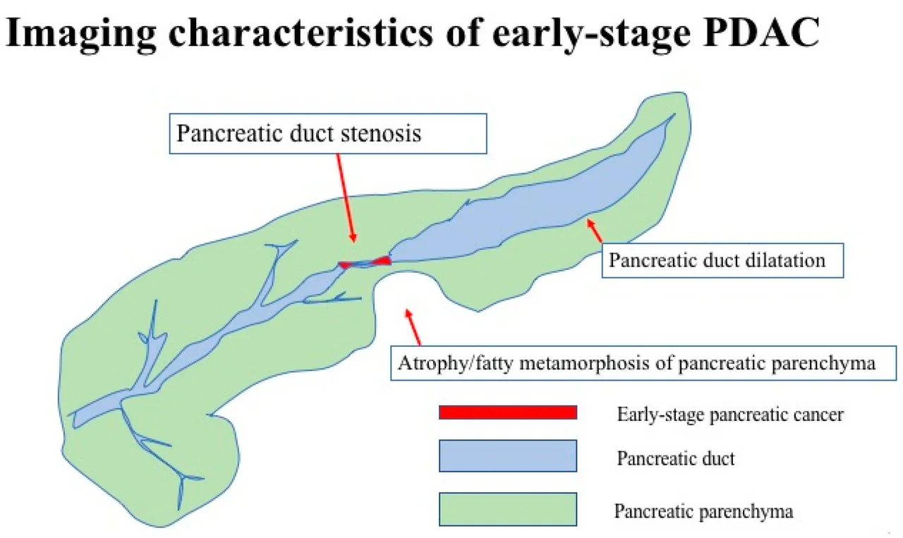
- Contrast-enhanced CT abdomen with a pancreatic protocol - the investigation of choice for both diagnosis and staging. It assesses the primary tumour, its relationship to the superior mesenteric artery, coeliac axis, superior mesenteric vein and portal vein (which determines resectability), nodal involvement and distant metastases
- Abdominal ultrasound - often the first test in a jaundiced patient, showing biliary dilatation and sometimes a pancreatic mass, but poor at visualising the pancreas fully
- Endoscopic ultrasound (EUS) - the most sensitive modality for small tumours, and allows fine-needle aspiration for histology, which is required before chemotherapy but not always before surgery
- MRI and MRCP - for characterising cystic lesions, and defining biliary and pancreatic ductal anatomy. The 'double duct sign' - simultaneous dilatation of the common bile duct and pancreatic duct - is highly suggestive of a periampullary tumour
- PET-CT - to detect occult metastatic disease in patients being considered for surgery
- Staging laparoscopy - to identify small-volume peritoneal disease not visible on CT
Blood tests
- Liver function tests - an obstructive (cholestatic) picture: markedly raised bilirubin and ALP with a proportionally smaller rise in transaminases, and raised gamma-GT
- Coagulation screen - obstructive jaundice causes vitamin K malabsorption and a prolonged INR, correctable with parenteral vitamin K
- Full blood count, urea and electrolytes, albumin, calcium and glucose/HbA1c
- CA 19-9 - a tumour marker used for prognosis and monitoring treatment response, not for diagnosis or screening. It is falsely raised in biliary obstruction, pancreatitis and other cancers, and is undetectable in the 5-10% of people who are Lewis antigen negative
- Histology by EUS-guided fine-needle aspiration or percutaneous biopsy - essential before chemotherapy, though patients with clearly resectable disease may proceed to surgery without it
Differential diagnosis
- Choledocholithiasis - obstructive jaundice, but usually painful and with a non-palpable gallbladder (Courvoisier's law)
- Chronic pancreatitis - shares symptoms, risk factors and imaging features, and can form an inflammatory mass indistinguishable from tumour without biopsy
- Autoimmune (IgG4-related) pancreatitis - a critical mimic, since it produces a mass and obstructive jaundice but resolves completely with corticosteroids. Check serum IgG4
- Cholangiocarcinoma and ampullary carcinoma - periampullary tumours presenting identically; ampullary tumours have a better prognosis
- Duodenal carcinoma
- Pancreatic neuroendocrine tumours - better prognosis, may have functional syndromes
- Pancreatic cystic neoplasms - IPMN, mucinous cystic neoplasm, serous cystadenoma
- Gastric cancer, lymphoma and metastases to the pancreas
- Drug-induced or viral cholestasis
Management
Management is determined by a specialist hepatobiliary multidisciplinary team, and the first question is always resectability, since surgery offers the only realistic chance of cure.3
| Category | Definition | Approach |
|---|---|---|
| Resectable | No arterial contact; no or limited venous contact | Upfront surgery, followed by adjuvant chemotherapy |
| Borderline resectable | Limited arterial contact or reconstructable venous involvement | Neoadjuvant chemotherapy, then reassess for surgery |
| Locally advanced (unresectable) | Encasement of the coeliac axis or superior mesenteric artery, or unreconstructable venous occlusion | Chemotherapy, sometimes with chemoradiotherapy |
| Metastatic | Distant spread - liver, peritoneum, lung | Palliative chemotherapy and best supportive care |
Surgery
- Pancreaticoduodenectomy (Whipple procedure) for tumours of the head. This removes the head of the pancreas, the duodenum, the gallbladder and common bile duct, the distal stomach (or pylorus is preserved in the modified version), and regional lymph nodes, with reconstruction by pancreaticojejunostomy, hepaticojejunostomy and gastrojejunostomy. It is major surgery with significant morbidity, though mortality in high-volume centres is now under 5%
- Distal pancreatectomy with splenectomy for tumours of the body and tail - patients require vaccination against encapsulated organisms and lifelong penicillin prophylaxis
- Total pancreatectomy occasionally, producing complete exocrine and endocrine failure
- Adjuvant chemotherapy after resection is standard and significantly improves survival - typically modified FOLFIRINOX in fit patients, or gemcitabine with capecitabine
Palliative management
Since around 80% of patients have unresectable disease, palliation is the mainstay of care and should be delivered actively rather than passively:
- Biliary obstruction: endoscopic stenting at ERCP (a self-expanding metal stent for longer patency) relieves jaundice and pruritus; percutaneous transhepatic drainage if ERCP fails, or a surgical bypass
- Gastric outlet obstruction: duodenal stenting or gastrojejunostomy
- Pain: the analgesic ladder including opioids, with coeliac plexus block or neurolysis for refractory back pain - highly effective in this cancer
- Chemotherapy: FOLFIRINOX in patients of good performance status, or gemcitabine with nab-paclitaxel; gemcitabine alone in frailer patients. PARP inhibitors (olaparib) for BRCA-mutated disease
- Pancreatic enzyme replacement therapy (PERT) - frequently forgotten, but important: exocrine insufficiency is very common and PERT improves weight, nutrition and quality of life. Prescribe it for anyone with steatorrhoea or weight loss
- Nutritional support with dietetic input, and management of cachexia
- Venous thromboembolism prophylaxis and treatment - the thrombotic risk is high
- Early palliative care involvement, which improves both quality of life and, in some cancers, survival
- Pruritus: relieved primarily by biliary drainage, with colestyramine as an adjunct
Complications
- Obstructive jaundice with pruritus, coagulopathy from vitamin K malabsorption, and cholangitis
- Gastric outlet and duodenal obstruction
- Pancreatic exocrine insufficiency with steatorrhoea and malnutrition
- Diabetes mellitus
- Venous thromboembolism and migratory thrombophlebitis
- Ascites and peritoneal carcinomatosis
- Severe pain from coeliac plexus and retroperitoneal invasion
- Cachexia, sarcopenia and profound malnutrition
- Depression and psychological distress, which are notably common
- Splenic vein thrombosis with gastric varices and bleeding
- Surgical complications: pancreatic fistula, delayed gastric emptying, anastomotic leak, haemorrhage and post-surgical diabetes
Red flags
Prognosis
Pancreatic cancer has the poorest survival of any common cancer, with overall UK 5-year survival of around 5-7%, a figure that has improved only slowly over decades.1 Around half of patients are dead within 6 months of diagnosis.
The reasons are threefold: the pancreas is retroperitoneal and clinically silent, so tumours grow undetected; symptoms are vague and non-specific, delaying presentation; and the tumour biology is aggressive, with early perineural, lymphatic and vascular invasion. As a result only around 15-20% of patients have resectable disease at diagnosis.
Prognosis stratifies sharply by stage. Patients undergoing successful resection with adjuvant chemotherapy achieve a median survival of roughly 2-3 years and 5-year survival of around 20-30% - and considerably better for small, node-negative tumours. Locally advanced disease has a median survival of around 12-15 months, and metastatic disease around 6-11 months depending on performance status and chemotherapy regimen.
Adverse prognostic factors include positive resection margins, nodal involvement, a high postoperative CA 19-9, poor performance status and weight loss. Because most patients will not be cured, early and active palliative care, effective pain control, biliary drainage and pancreatic enzyme replacement are as important to overall outcome as oncological treatment, and are the aspects most often neglected.
References
- Cancer Research UK. Pancreatic cancer statistics. Available here
- NICE NG12. Suspected cancer: recognition and referral. 2015 (updated 2023). Available here
- NICE NG85. Pancreatic cancer in adults: diagnosis and management. 2018. Available here
- Kanno A et al., CC BY 4.0, via Wikimedia Commons. Available here
- Neoptolemos JP et al. Therapeutic developments in pancreatic cancer. Nat Rev Gastroenterol Hepatol. 2018. Available here
- Conroy T et al. FOLFIRINOX or gemcitabine as adjuvant therapy for pancreatic cancer. N Engl J Med. 2018. Available here
- NHS. Pancreatic cancer. 2023. Available here
This article is written for revision and education. It is not clinical guidance and must not be used to make decisions about the care of a patient. Always check current NICE guidance and local protocols.