
Liver Function Test Interpretation
Key points
- The name is misleading: most 'liver function tests' measure hepatocyte injury or cholestasis, not function. True markers of function are albumin, prothrombin time/INR and bilirubin.
- First question: is the pattern hepatitic (ALT/AST dominant) or cholestatic (ALP/gamma-GT dominant)? This determines the entire subsequent workup.
- Hepatitic pattern: ALT-predominant rise - viral hepatitis, drugs, autoimmune hepatitis, alcohol, MASLD, ischaemia, Wilson disease and haemochromatosis.
- Cholestatic pattern: ALP-predominant rise - confirm hepatic origin with gamma-GT, then image to separate obstructive from intrahepatic causes.
- AST:ALT ratio: above 2 suggests alcohol; below 1 suggests MASLD; a ratio rising above 1 in known MASLD suggests advancing fibrosis.
- Isolated raised ALP: check gamma-GT. If normal, the ALP is likely bone in origin - think Paget disease, osteomalacia, fracture, malignancy or pregnancy (placental).
- Isolated raised bilirubin: if unconjugated with normal enzymes and no haemolysis, this is usually Gilbert syndrome - benign and needs no further action.
- Non-invasive liver screen: the standard panel for persistently abnormal LFTs: viral serology, autoantibodies with immunoglobulins, ferritin, caeruloplasmin, alpha-1 antitrypsin and coeliac serology.
Introduction
Abnormal liver blood tests are extremely common, found in around 10-20% of adults tested, and the great majority reflect either a benign finding or a treatable cause.1 The challenge is to work through them systematically rather than either over-investigating or, more dangerously, ignoring them - a substantial proportion of patients with cirrhosis are first identified through incidentally abnormal blood tests.
What each test means
| Test | What it reflects | Notes |
|---|---|---|
| ALT (alanine aminotransferase) | Hepatocyte injury | The most liver-specific transaminase. Located in the cytosol |
| AST (aspartate aminotransferase) | Hepatocyte injury | Less specific - also found in cardiac and skeletal muscle, red cells and kidney. Present in mitochondria, which is why alcohol (a mitochondrial toxin) raises it disproportionately |
| ALP (alkaline phosphatase) | Cholestasis, and bone turnover | Found in bile canaliculi, bone, placenta and intestine. A raised ALP is not necessarily hepatic |
| Gamma-GT (gamma-glutamyl transferase) | Cholestasis; enzyme induction | Used to confirm that a raised ALP is hepatic in origin. Also raised by alcohol and enzyme-inducing drugs, so a raised gamma-GT alone is non-specific |
| Bilirubin | Haem breakdown; excretory function | Fractionate into conjugated and unconjugated when isolated. Jaundice becomes clinically visible above roughly 50 µmol/L |
| Albumin | Synthetic function (and nutrition, inflammation) | Long half-life (~20 days), so falls in chronic disease. Also low in sepsis, nephrotic syndrome and malnutrition |
| Prothrombin time / INR | Synthetic function | The most sensitive marker of acute synthetic failure, as clotting factors have a short half-life (factor VII ~6 hours). Also prolonged by vitamin K deficiency and warfarin |
| Platelets | Indirect marker of portal hypertension | Not part of the LFT panel, but thrombocytopenia is often the earliest clue to cirrhosis |
Step 1: Identify the pattern
The single most useful step is to classify the abnormality as hepatitic, cholestatic or mixed, since this determines everything that follows.1
| Pattern | Biochemistry | Implication |
|---|---|---|
| Hepatitic (hepatocellular) | ALT and AST raised disproportionately to ALP | Hepatocyte injury - think viral, drug, autoimmune, metabolic or ischaemic causes |
| Cholestatic | ALP and gamma-GT raised disproportionately to ALT | Obstruction of bile flow, either mechanical (extrahepatic) or at the level of the small ducts (intrahepatic) |
| Mixed | Both raised proportionately | Common in drug-induced liver injury and in many chronic liver diseases |
| Isolated hyperbilirubinaemia | Bilirubin raised, enzymes normal | Fractionate: usually Gilbert syndrome if unconjugated, or haemolysis |
| Isolated abnormal synthetic function | Low albumin, prolonged INR, with near-normal enzymes | Suggests established cirrhosis or acute liver failure - potentially the most serious pattern of all |
The R factor formalises this: (ALT ÷ upper limit of normal) ÷ (ALP ÷ upper limit of normal). A value above 5 indicates a hepatitic pattern, below 2 a cholestatic pattern, and 2-5 a mixed picture. It is used particularly when classifying drug-induced liver injury.
The magnitude of the rise also helps
- ALT over 1000 U/L (a 'transaminitis in the thousands') has a short differential: acute viral hepatitis, drug or toxin-induced injury (especially paracetamol), ischaemic hepatitis ('shock liver'), acute autoimmune hepatitis, and acute biliary obstruction from a passing stone
- Modest rises (2-5 times normal) are far commoner and suggest MASLD, alcohol, chronic viral hepatitis, drugs or haemochromatosis
- A very high AST with a high LDH, falling rapidly over days in a patient who has been hypotensive suggests ischaemic hepatitis
- Remember that transaminase level does not reflect liver function or prognosis - a falling ALT with a rising bilirubin and INR indicates hepatocyte exhaustion, not recovery
Step 2: The hepatitic pattern
Where ALT and AST predominate, work through the causes systematically. The AST:ALT ratio is a useful early discriminator:
- AST:ALT above 2 - strongly suggests alcohol-related liver disease. Alcohol is a mitochondrial toxin and causes pyridoxine deficiency, both of which favour AST release. A ratio above 3 is highly suggestive
- AST:ALT below 1 - typical of MASLD and of chronic viral hepatitis
- A ratio that rises above 1 in known MASLD - suggests progression to advanced fibrosis or cirrhosis
- AST:ALT above 2.2 with a low ALP in acute liver failure - think Wilson disease
Causes of a hepatitic picture
- MASLD - by far the commonest cause of mildly abnormal LFTs in the UK
- Alcohol-related liver disease
- Viral hepatitis - A, B, C and E, plus EBV and CMV in acute presentations
- Drug-induced liver injury - paracetamol, antibiotics (co-amoxiclav, flucloxacillin, nitrofurantoin), anti-tuberculous drugs, statins, methotrexate, anti-epileptics, herbal and recreational drugs. Always take a full drug history including over-the-counter and herbal products
- Autoimmune hepatitis - raised IgG with positive ANA, ASMA or anti-LKM1
- Haemochromatosis - raised ferritin with a high transferrin saturation
- Wilson disease - essential to exclude in anyone under 40
- Alpha-1 antitrypsin deficiency
- Coeliac disease - a commonly overlooked cause of unexplained transaminitis
- Ischaemic hepatitis following hypotension, sepsis or cardiac arrest
- Non-hepatic causes: thyroid disease, muscle injury (check creatine kinase - a raised AST with normal ALT and high CK points to muscle rather than liver), and strenuous exercise
Step 3: The cholestatic pattern
Where ALP predominates, the first task is to confirm that the ALP is of hepatic origin by checking the gamma-GT. If gamma-GT is also raised, the ALP is hepatobiliary; if gamma-GT is normal, look for a bone source.
Having confirmed hepatic cholestasis, the next step is abdominal ultrasound to determine whether the biliary tree is dilated, which separates the two broad groups:
| Extrahepatic (obstructive - ducts dilated) | Intrahepatic (ducts not dilated) |
|---|---|
| Gallstones in the common bile duct (choledocholithiasis) | Primary biliary cholangitis - middle-aged women, pruritus, AMA positive, raised IgM |
| Pancreatic cancer and ampullary tumours | Primary sclerosing cholangitis - associated with ulcerative colitis, pANCA, beading on MRCP |
| Cholangiocarcinoma | Drug-induced cholestasis - co-amoxiclav, flucloxacillin, erythromycin, anabolic steroids, the combined oral contraceptive pill, chlorpromazine |
| Biliary strictures, benign or malignant | Sepsis and total parenteral nutrition |
| Chronic pancreatitis compressing the bile duct | Infiltrative disease - sarcoidosis, lymphoma, amyloidosis, liver metastases, tuberculosis |
| Extrinsic compression by nodes or tumour | Pregnancy - intrahepatic cholestasis of pregnancy; also viral hepatitis and alcoholic hepatitis can present cholestatically |
Isolated raised ALP with a normal gamma-GT
This indicates a non-hepatic source, most often bone:
- Paget disease of bone - a markedly raised ALP with normal calcium and phosphate
- Osteomalacia and vitamin D deficiency
- Bone metastases and primary bone tumours
- Healing fractures and recent orthopaedic surgery
- Growth spurts in children and adolescents - a physiological and common finding
- Pregnancy - placental ALP, particularly in the third trimester
- Hyperparathyroidism, hyperthyroidism and renal osteodystrophy
Step 4: Isolated hyperbilirubinaemia
Where bilirubin is raised but the enzymes are normal, the key step is to fractionate it into conjugated and unconjugated.
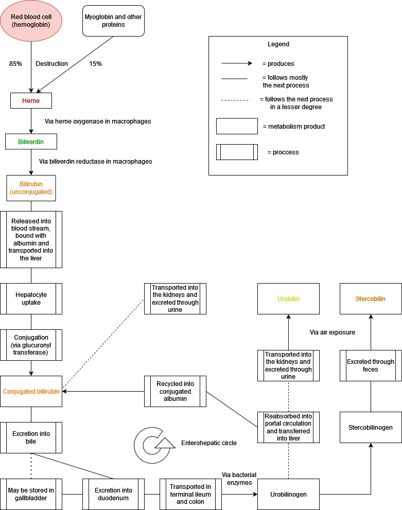
Unconjugated (indirect) hyperbilirubinaemia
Unconjugated bilirubin is water-insoluble and bound to albumin, so it is not excreted in urine - hence the classic description of jaundice with normal-coloured urine and stools.
- Gilbert syndrome - by far the commonest cause. An autosomal recessive reduction in UGT1A1 activity affecting around 5-10% of the population. Bilirubin rises with fasting, intercurrent illness, stress, exercise or dehydration, and typically stays below 100 µmol/L. It is entirely benign, requires no treatment or further investigation, and reassurance is the whole management. It is important to recognise so as to avoid unnecessary testing
- Haemolysis - check full blood count, reticulocytes, LDH (raised), haptoglobin (low) and a blood film and direct antiglobulin test
- Ineffective erythropoiesis - megaloblastic anaemia, thalassaemia
- Resorption of a large haematoma, or recent blood transfusion
- Crigler-Najjar syndrome - rare; type I is severe and presents in the neonate with kernicterus
Conjugated (direct) hyperbilirubinaemia
Conjugated bilirubin is water-soluble and is excreted renally, producing dark urine; if bile flow is obstructed, stools become pale. Causes include any hepatobiliary disease, plus the rare benign inherited disorders Dubin-Johnson syndrome (a characteristically black liver on biopsy) and Rotor syndrome, both of which are benign and require no treatment.
Investigating persistently abnormal LFTs
The British Society of Gastroenterology recommends that abnormal liver blood tests should not simply be repeated indefinitely - if an abnormality persists, or if there is any clinical concern, proceed to a non-invasive liver screen.1 Importantly, this should be done even for mild abnormalities, and the degree of enzyme elevation does not predict the severity of underlying fibrosis.
The non-invasive liver screen
- Hepatitis B surface antigen and hepatitis C antibody (with HCV RNA if positive)
- Autoantibodies: ANA, anti-smooth muscle antibody, anti-mitochondrial antibody, anti-LKM1
- Immunoglobulins: raised IgG suggests autoimmune hepatitis, raised IgM suggests primary biliary cholangitis, raised IgA suggests alcohol-related disease
- Ferritin and transferrin saturation for haemochromatosis - a raised ferritin alone is usually inflammation, not iron overload
- Caeruloplasmin in anyone under 40, for Wilson disease
- Alpha-1 antitrypsin level
- Coeliac serology (IgA tissue transglutaminase with total IgA)
- Thyroid function tests, and creatine kinase if a muscle source is possible
- Fasting glucose or HbA1c and lipid profile, for metabolic risk factors
- Abdominal ultrasound in all patients
Assessing fibrosis
Because enzymes correlate poorly with fibrosis, patients with risk factors should have fibrosis assessed directly using FIB-4 or the NAFLD fibrosis score as first-line triage, with transient elastography (FibroScan) or the enhanced liver fibrosis (ELF) test where these suggest intermediate or high risk. Liver biopsy is reserved for diagnostic uncertainty.
Throughout, take a careful history covering alcohol quantified in units, all medications including over-the-counter and herbal remedies, metabolic risk factors, travel, blood-borne virus risk factors, family history and autoimmune conditions.
Red flags
Summary of the approach
- Assess synthetic function first - check albumin, INR and bilirubin, and look at the platelet count. This tells you how sick the liver actually is, and whether this is urgent
- Classify the pattern - hepatitic, cholestatic, mixed, or isolated hyperbilirubinaemia
- If hepatitic: use the AST:ALT ratio, consider the magnitude of the rise, take a full drug and alcohol history, and send the non-invasive liver screen
- If cholestatic: check gamma-GT to confirm hepatic origin, then arrange ultrasound to distinguish dilated (obstructive) from non-dilated (intrahepatic) causes, and check AMA
- If isolated raised bilirubin: fractionate. Unconjugated with normal enzymes and no haemolysis is almost always Gilbert syndrome - reassure and stop
- If isolated raised ALP with a normal gamma-GT: look for a bone cause
- Assess fibrosis with FIB-4 and, where indicated, elastography - regardless of how mild the enzyme abnormality appears
- Refer urgently if there is synthetic failure, encephalopathy, suspected malignancy or cholangitis; and routinely if the screen identifies a specific liver disease or significant fibrosis
References
- Newsome PN et al. Guidelines on the management of abnormal liver blood tests. British Society of Gastroenterology. Gut. 2018. Available here
- NICE Clinical Knowledge Summaries (CKS). Abnormal liver blood tests. 2023. Available here
- NICE NG49. Non-alcoholic fatty liver disease (NAFLD): assessment and management. 2016 (updated 2025). Available here
- EvanWorse, CC BY-SA 4.0, via Wikimedia Commons. Available here
- NICE NG50. Cirrhosis in over 16s: assessment and management. 2016. Available here
- NICE NG12. Suspected cancer: recognition and referral. 2015 (updated 2023). Available here
- Kwo PY, Cohen SM, Lim JK. ACG clinical guideline: evaluation of abnormal liver chemistries. Am J Gastroenterol. 2017. Available here
This article is written for revision and education. It is not clinical guidance and must not be used to make decisions about the care of a patient. Always check current NICE guidance and local protocols.