
Thalassaemia
Key points
- Thalassaemia: an autosomal recessive group of inherited haemoglobin disorders caused by reduced or absent synthesis of one globin chain, causing an imbalance between alpha and beta chain production.
- Two families: alpha thalassaemia (deletion of one or more of the 4 alpha-globin genes) and beta thalassaemia (point mutations reducing or abolishing beta-globin production from the 2 beta-globin genes).
- Severity tracks gene dosage: in alpha thalassaemia, severity rises with the number of genes deleted (1 = silent carrier, 2 = trait, 3 = HbH disease, 4 = Hb Barts hydrops fetalis, incompatible with life). In beta thalassaemia: trait (one gene, asymptomatic), intermedia, and major (both genes, transfusion-dependent).
- Pathophysiology: unpaired globin chains precipitate within red cell precursors, causing ineffective erythropoiesis and haemolysis, which in turn drives massive marrow expansion and extramedullary haematopoiesis.
- Beta thalassaemia major: presents at 6-12 months as fetal haemoglobin (HbF) falls, with severe anaemia, failure to thrive, hepatosplenomegaly, and skeletal changes (frontal bossing, maxillary overgrowth) from marrow expansion.
- Diagnosis: microcytic anaemia disproportionate to the mild symptoms (unlike iron deficiency), blood film with target cells, and confirmation by haemoglobin electrophoresis/HPLC (raised HbA2 and/or HbF in beta thalassaemia trait).
- Management of major disease: lifelong regular blood transfusion to suppress ineffective erythropoiesis, combined with iron chelation to prevent transfusional iron overload. Allogeneic stem cell transplant is curative in selected patients.
- Leading cause of death: cardiac iron overload causing dilated cardiomyopathy and arrhythmia - chelation adherence is the single biggest determinant of survival.
Introduction
Thalassaemia is a group of inherited disorders of haemoglobin synthesis in which production of one or more globin chains is reduced or absent, in contrast to sickle cell disease and other haemoglobinopathies, where the globin chain is produced normally in quantity but is structurally abnormal.1 It is commonest in people of Mediterranean, Middle Eastern, South Asian and South-East Asian descent, mirroring historical malaria endemicity in the same way as sickle cell trait.
Normal adult haemoglobin (HbA) is a tetramer of two alpha chains and two beta chains. Thalassaemia is named for whichever chain is deficient, and severity depends on how completely production of that chain is lost.
Genetics and pathophysiology
Alpha thalassaemia
There are 4 alpha-globin genes (two on each copy of chromosome 16). Alpha thalassaemia usually results from gene deletions, and severity is proportional to how many of the four are lost:2
| Genes affected | Name | Clinical picture |
|---|---|---|
| 1 of 4 | Silent carrier | Asymptomatic, normal or near-normal blood count |
| 2 of 4 | Alpha thalassaemia trait | Mild microcytic anaemia, usually asymptomatic |
| 3 of 4 | HbH disease | Moderate-severe haemolytic anaemia; excess beta chains form unstable HbH (beta4) tetramers |
| 4 of 4 | Hb Barts hydrops fetalis | No functional alpha chains; incompatible with life - severe fetal anaemia, hydrops and intrauterine death or early neonatal death |
Beta thalassaemia
There are only 2 beta-globin genes (one on each copy of chromosome 11), and beta thalassaemia usually results from point mutations rather than deletions, reducing (beta+) or abolishing (beta0) production.
| Genes affected | Name | Clinical picture |
|---|---|---|
| 1 of 2 | Beta thalassaemia trait (minor) | Mild, often asymptomatic microcytic anaemia - frequently mistaken for iron deficiency |
| 2 of 2 (mixed severity) | Beta thalassaemia intermedia | Moderate anaemia not usually requiring regular transfusion, but with significant morbidity |
| 2 of 2 (severe) | Beta thalassaemia major (Cooley's anaemia) | Severe, transfusion-dependent anaemia presenting in infancy |
Pathophysiology
When one globin chain is deficient, the other is produced in relative excess. These unpaired chains are unstable and precipitate within red cell precursors in the bone marrow, causing premature destruction of erythroblasts (ineffective erythropoiesis) and shortened survival of the red cells that do reach the circulation (haemolysis).
The marrow responds to chronic anaemia by expanding dramatically - up to 15-30 times normal activity - which, if uncorrected by transfusion, causes skeletal deformity (marrow expansion in the skull produces a "hair-on-end" appearance on X-ray, and maxillary overgrowth produces "chipmunk facies") and extramedullary haematopoiesis in the liver and spleen, causing hepatosplenomegaly.
Clinical features
Alpha and beta thalassaemia trait are usually asymptomatic or cause only mild symptoms, often identified incidentally on a routine blood count.
Beta thalassaemia major
Typically presents at 6-12 months of age, once HbF (which does not require beta-globin) falls and the defect in HbA production becomes clinically apparent:
- Severe anaemia - pallor, lethargy, failure to thrive
- Hepatosplenomegaly from extramedullary haematopoiesis and increased red cell destruction
- Skeletal changes from marrow expansion - frontal bossing, maxillary hypertrophy ("chipmunk facies"), and the classic "hair-on-end" skull X-ray appearance
- Growth restriction and delayed puberty
- Jaundice from chronic haemolysis
HbH disease
A chronic haemolytic anaemia of variable severity, with splenomegaly and gallstones, generally milder than beta thalassaemia major but still clinically significant and sometimes transfusion-dependent.
Investigations
Full blood count and blood film
Microcytic, hypochromic anaemia, but characteristically disproportionately mild symptoms for the degree of microcytosis compared to iron deficiency - a useful bedside clue, since patients with thalassaemia trait can have a very low MCV with an only mildly reduced haemoglobin. The Mentzer index (MCV/red cell count) is typically below 13 in thalassaemia, versus above 13 in iron deficiency anaemia.
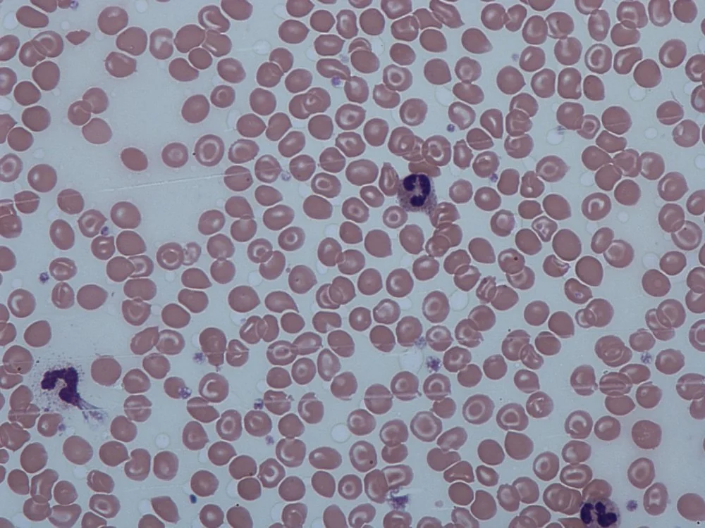
The blood film shows target cells, microcytes, and (in more severe disease) nucleated red blood cells and basophilic stippling.
Confirming the diagnosis
- Haemoglobin electrophoresis / HPLC - the key confirmatory test. Beta thalassaemia trait shows a raised HbA2 (and often mildly raised HbF); beta thalassaemia major shows absent or greatly reduced HbA with predominantly HbF
- Iron studies are normal - ferritin is not low, which is the crucial distinction from iron deficiency anaemia, since the two are frequently confused given the shared microcytosis
- Alpha thalassaemia often requires genetic testing for gene deletions, since electrophoresis can be normal in trait
- Skeletal X-ray in established beta thalassaemia major - "hair-on-end" skull appearance from marrow expansion
- Antenatal screening and genetic counselling for couples where both partners carry a thalassaemia trait, given the 1 in 4 risk of a severely affected child
Differential diagnosis
The key differential of any microcytic anaemia is iron deficiency anaemia and anaemia of chronic disease - both distinguished from thalassaemia by normal iron studies and a normal haemoglobin electrophoresis in thalassaemia. Sideroblastic anaemia is a rarer differential, distinguished by ring sideroblasts on bone marrow examination and a normal haemoglobin electrophoresis.
Management
Trait and mild disease
No treatment is required for silent carriers or thalassaemia trait beyond genetic counselling. Critically, iron supplementation must be avoided unless true concurrent iron deficiency is confirmed, since these patients are not iron deficient and inappropriate iron therapy risks iron overload.
Beta thalassaemia major
- Regular lifelong blood transfusion, typically every 2-4 weeks, to maintain haemoglobin above a target threshold (commonly around 95-105 g/L pre-transfusion) - this both corrects the anaemia and, importantly, suppresses the patient's own ineffective erythropoiesis, preventing skeletal deformity and marrow expansion3
- Iron chelation therapy is mandatory once transfusion begins, since each unit of blood adds iron that the body cannot excrete. Options include desferrioxamine (subcutaneous infusion), deferiprone and deferasirox (both oral) - chosen based on tolerability, organ involvement and adherence
- Folic acid supplementation, given increased erythropoietic turnover
- Splenectomy may be considered if hypersplenism causes excessive transfusion requirements, with the usual pre-operative vaccination and lifelong penicillin prophylaxis for hyposplenism
- Allogeneic haematopoietic stem cell transplant - the only curative option, most successful when performed early in childhood with a matched donor before iron overload and organ damage accumulate
- Emerging gene therapy is increasingly available for selected patients with severe transfusion-dependent thalassaemia
Complications
- Iron overload - from both increased intestinal absorption (driven by ineffective erythropoiesis) and repeated transfusion. Causes cardiomyopathy and arrhythmia (the leading cause of death), endocrine failure (diabetes, hypogonadism, hypothyroidism, growth failure), and liver fibrosis/cirrhosis
- Skeletal deformity if transfusion is inadequate or delayed
- Gallstones from chronic haemolysis
- Hypersplenism
- Osteoporosis
- Alloimmunisation and transfusion reactions from repeated transfusion
- Extramedullary haematopoietic masses, occasionally causing spinal cord compression in poorly transfused patients
- Infection risk related to hyposplenism (if splenectomised) and iron overload itself, which favours certain organisms (e.g. Yersinia)
Red flags
Prognosis
Thalassaemia trait carries a normal life expectancy and requires no treatment beyond genetic counselling. Alpha thalassaemia with 4 gene deletions (Hb Barts hydrops fetalis) is incompatible with life.
With modern transfusion and chelation programmes, patients with beta thalassaemia major now frequently survive into middle age and beyond, a dramatic improvement from a historical life expectancy of only a few years - but outcomes remain closely tied to chelation adherence, and cardiac iron overload remains the leading cause of premature death.3 Allogeneic stem cell transplant, where a suitable donor is available and performed early, offers the prospect of cure and normal life expectancy.
References
- Taher AT, Weatherall DJ, Cappellini MD. Thalassaemia. Lancet. 2018. Available here
- Thalassaemia International Federation. Guidelines for the management of transfusion dependent thalassaemia. Available here
- British Society for Haematology. Standards for the clinical care of children and adults with thalassaemia in the UK. Available here
- Dr Graham Beards, CC BY-SA 3.0, via Wikimedia Commons. Available here
- NHS. Thalassaemia. 2023. Available here
- Public Health England. NHS Sickle Cell and Thalassaemia Screening Programme. Available here
This article is written for revision and education. It is not clinical guidance and must not be used to make decisions about the care of a patient. Always check current NICE guidance and local protocols.