
Haemolytic Anaemia
Key points
- Haemolytic anaemia: anaemia caused by premature destruction of red blood cells, occurring faster than the bone marrow can compensate. Red cell lifespan falls from the normal ~120 days to as little as a few days in severe cases.
- Two broad mechanisms: intravascular haemolysis (destruction within the circulation, releasing free haemoglobin) and extravascular haemolysis (destruction by macrophages in the spleen and liver) - the pattern of results points to which is occurring.
- Causes split into two groups: inherited (membrane defects, enzyme deficiencies, haemoglobinopathies) and acquired (autoimmune, mechanical, infective, drug-induced).
- Proving haemolysis is occurring: raised reticulocyte count (marrow compensation), raised LDH and unconjugated bilirubin (breakdown products), and low haptoglobin (mopped up by free haemoglobin) - the core biochemical tetrad.
- Direct antiglobulin test (Coombs test): the pivotal investigation that separates autoimmune (Coombs-positive) from non-immune (Coombs-negative) causes.
- Clinical features: jaundice (unconjugated, so no dark urine or pale stools, unlike obstructive jaundice), pallor, splenomegaly, and a history of gallstones or leg ulcers in chronic cases.
- Management: treat the underlying cause; folic acid to support increased erythropoietic demand; corticosteroids for autoimmune haemolytic anaemia; transfusion for severe/symptomatic cases; splenectomy for selected refractory or hereditary causes.
- Complications: pigment gallstones, aplastic crisis (classically triggered by parvovirus B19), folate deficiency, and iron overload from repeated transfusion.
Introduction
Haemolytic anaemia is anaemia resulting from accelerated destruction of red blood cells, occurring at a rate that outstrips the bone marrow's considerable ability to compensate by increasing production up to sixfold. Normal red cell lifespan is around 120 days; in significant haemolysis this can fall to days.1
The topic is approached in two steps: first, confirm that haemolysis is actually happening, using a consistent set of laboratory markers; second, work out the mechanism and cause, which determines both prognosis and treatment.
Classification and aetiology
Haemolysis is classified along two axes: where it happens (intravascular vs extravascular) and why it happens (inherited vs acquired).
Intravascular vs extravascular
| Feature | Intravascular | Extravascular |
|---|---|---|
| Site | Within the blood vessels | Reticuloendothelial system (spleen, liver) |
| Mechanism | Complement-mediated lysis, mechanical shearing | Macrophage phagocytosis of opsonised/abnormal red cells |
| Free plasma haemoglobin | Raised | Normal |
| Haemoglobinuria/haemosiderinuria | Present | Absent |
| Haptoglobin | Markedly reduced | Reduced |
| Examples | ABO mismatch transfusion, PNH, G6PD deficiency crisis, mechanical heart valves, DIC, malaria | Hereditary spherocytosis, warm autoimmune haemolytic anaemia, thalassaemia |
Inherited causes
- Membrane defects: hereditary spherocytosis, hereditary elliptocytosis
- Enzyme deficiencies: glucose-6-phosphate dehydrogenase (G6PD) deficiency, pyruvate kinase deficiency
- Haemoglobinopathies: sickle cell disease, thalassaemia
Acquired causes
- Autoimmune haemolytic anaemia (AIHA) - warm (IgG-mediated, usually extravascular) or cold (IgM-mediated, complement-fixing, often triggered by Mycoplasma or EBV, exacerbated by cold)
- Alloimmune - haemolytic transfusion reactions, haemolytic disease of the newborn
- Mechanical (microangiopathic haemolytic anaemia, MAHA) - prosthetic heart valves, disseminated intravascular coagulation, thrombotic thrombocytopenic purpura, haemolytic uraemic syndrome, severe pre-eclampsia/HELLP syndrome - red cells are physically sheared, producing schistocytes
- Infective - malaria, Clostridium perfringens sepsis
- Drug-induced - e.g. penicillin (immune-mediated), or oxidative haemolysis precipitated by certain drugs in G6PD deficiency
- Paroxysmal nocturnal haemoglobinuria (PNH) - acquired clonal defect causing complement-mediated intravascular haemolysis, with a characteristic association with venous thrombosis at unusual sites
- Hypersplenism - any cause of splenomegaly can increase red cell destruction
Clinical features
Features reflect both the anaemia itself and the breakdown of haemoglobin:
- Generic anaemic symptoms - fatigue, dyspnoea, pallor
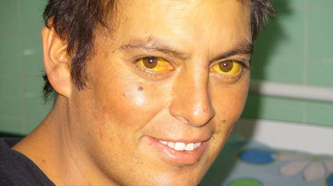
- Jaundice - from unconjugated hyperbilirubinaemia. Because this is a pre-hepatic process, urine and stool colour are normal (unconjugated bilirubin is not water-soluble and is not excreted in urine) - a key point that distinguishes it from obstructive (cholestatic) jaundice
- Splenomegaly - from increased reticuloendothelial workload, particularly in extravascular and chronic haemolysis
- Dark urine specifically in intravascular haemolysis, from haemoglobinuria - distinct from the bilirubinuria of obstructive jaundice
- History of gallstones (pigment stones from chronic excess bilirubin turnover) or leg ulcers in chronic haemolytic states
- Family history in inherited causes
Investigations
Step one: confirm haemolysis is occurring
- Reticulocyte count raised - the marrow's compensatory response; a low reticulocyte count in an anaemic, jaundiced patient should prompt reconsideration (e.g. an aplastic crisis, or a non-haemolytic cause of jaundice)
- LDH raised - released from destroyed red cells
- Unconjugated bilirubin raised
- Haptoglobin low - haptoglobin binds free haemoglobin and is cleared with it, so it falls, especially in intravascular haemolysis
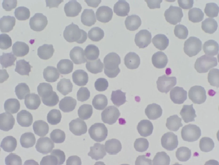
- Blood film - polychromasia (reflecting reticulocytosis) is seen in haemolysis of any cause; specific red cell morphology often points to the mechanism (see below)
Step two: find the mechanism and cause
- Direct antiglobulin test (Coombs test) - the pivotal test. Positive indicates an immune-mediated cause (autoimmune or alloimmune); negative points to an inherited or mechanical cause
- Blood film morphology - spherocytes (hereditary spherocytosis or warm AIHA), schistocytes/red cell fragments (MAHA - DIC, TTP, HUS, mechanical valve), bite and blister cells with Heinz bodies (G6PD deficiency, oxidative haemolysis), sickle cells (sickle cell disease)
- Haemoglobinuria/urinary haemosiderin - supports intravascular haemolysis
- For suspected inherited causes: osmotic fragility test or eosin-5-maleimide (EMA) binding test (hereditary spherocytosis), G6PD assay (ideally after the acute crisis, as levels can transiently normalise), haemoglobin electrophoresis (haemoglobinopathies)
- Flow cytometry (FLAER) for suspected PNH
- Coagulation screen, platelet count and renal function if MAHA is suspected, since this group includes medical emergencies (TTP, HUS, DIC)
Differential diagnosis
The main alternative to consider when a patient is anaemic and jaundiced is hepatobiliary disease causing conjugated hyperbilirubinaemia, which is distinguished by dark urine, pale stools, deranged liver enzymes and a normal reticulocyte count. Also consider ineffective erythropoiesis (e.g. severe B12/folate deficiency, myelodysplasia), which can mimic haemolysis biochemically (raised LDH and bilirubin from intramedullary cell death) but with a low, not raised, reticulocyte count.
Management
- Treat the underlying cause - stop an implicated drug, treat the triggering infection, manage the underlying autoimmune or malignant condition
- Folic acid supplementation, given the greatly increased erythropoietic demand depletes folate stores
- Warm AIHA: first-line oral corticosteroids; second line includes rituximab or splenectomy for refractory cases2
- Cold AIHA: avoid cold exposure; corticosteroids are typically less effective (the pathology is complement-mediated), so rituximab is often used instead
- Transfusion for severe or symptomatic anaemia - use with caution and cross-match carefully, since autoantibodies can make compatibility testing difficult
- Splenectomy for selected cases of hereditary spherocytosis (with pre-operative vaccination and lifelong penicillin prophylaxis given the hyposplenism risk) and for refractory autoimmune haemolysis
- MAHA emergencies (TTP, HUS) require urgent specialist management (e.g. plasma exchange in TTP) rather than routine haemolysis work-up
Complications
- Pigment gallstones, from chronically increased bilirubin turnover
- Aplastic crisis - a sudden, transient cessation of erythropoiesis, classically triggered by parvovirus B19 infection, causing a dangerous fall in haemoglobin in patients who depend on continuous high marrow output (e.g. hereditary spherocytosis, sickle cell disease)
- Folate deficiency, from ongoing depletion
- Iron overload in patients requiring recurrent transfusion
- Chronic leg ulcers in some inherited haemolytic anaemias
- Thromboembolism, notably in PNH and cold AIHA
Red flags
Prognosis
Prognosis depends entirely on the underlying cause. Drug-induced and infection-triggered haemolysis typically resolve once the precipitant is removed or treated. Warm AIHA responds to corticosteroids in the majority of patients, though relapse is common and some require second-line therapy.2 Inherited causes (hereditary spherocytosis, G6PD deficiency, haemoglobinopathies) are lifelong, with severity ranging from mild and well-compensated to transfusion-dependent, and management focuses on avoiding triggers and managing complications rather than cure - splenectomy aside.
References
- Barcellini W, Fattizzo B. The changing landscape of autoimmune hemolytic anemia. Front Immunol. 2020. Available here
- Jäger U, Barcellini W, Broome CM et al. Diagnosis and treatment of autoimmune hemolytic anemia: recommendations from the First International Consensus Meeting. Blood Rev. 2020. Available here
- British Society for Haematology. Guidelines on the management of drug-induced immune and secondary autoimmune haemolytic anaemia. Available here
- Sheila J. Toro, CC BY 4.0, via Wikimedia Commons. Available here
- Vives-Corrons JL, Krishnevskaya E. Rare anemias in adolescents. Acta Biomed. 2021. CC BY 4.0, via Wikimedia Commons. Available here
- NICE Clinical Knowledge Summaries (CKS). Anaemia - haemolytic. 2023. Available here
This article is written for revision and education. It is not clinical guidance and must not be used to make decisions about the care of a patient. Always check current NICE guidance and local protocols.